
 Worksheets
Sono week 3 powerpoint
Total questions: 100
Worksheet time: 50mins
The normal liver measurement is _____ when measured on the ____ lobe from diaphragm to inferior edge.
5 - 8cm, right
12 - 17cm, right
5 - 8cm, left
12 - 17cm, left
The _____ is the largest organ in the abdomen.
gallbladder
stomach
liver
pancreas
The liver is an acoustic cavity to view which organs?
gallbladder
pancreas
great vessels & bile duct
all answers are correct
The three lobes of the liver are:
right, left, caudate
anterior, posterior, caudate
medial, lateral, caudate
superior, inferior, caudate
What is the ligament that separates the right and left lobes?
ligamentum venosum
falciform
ligamentum teres
main lobar fissure

What ligament is this?
falciform
teres
triangular
coronary

What ligament is this?
teres
falciform
triangle
coronary

What ligament is this?
teres
falciform
triangle
coronary
What ligament attaches the liver to the diaphragm?
teres
falciform
triangle
coronary
What ligament attaches the liver to the anterior wall?
teres
falciform
triangle
coronary
What ligament is a remnant of the left umbilical vein?
teres
falciform
triangle
coronary
The peritoneum is ______ to the liver.
anterior
posterior
superior
inferior
Glisson's capsule is a ______.
serous membrane
thin membrane
fibrous membrane
thick membrane
Which of the following is not an acoustic window for scanning?
xiphoid
sternal
subcostal
intercostal
The groove on the posterior side of the liver lateral to the caudate lobe is where the ______ runs through the liver.
the aorta
the portal veins
the IVC
the gallbladder
Ligaments within the liver appear as ______ lines.
anechoic
hyperechoic
echopenic
echogenic
Use ______ as a landmark to find the left portal vein.
ligamentum teres
ligamentum venosum
main lobar fissure
falciform ligament
Use ______ as a landmark to find the left portal vein and caudate lobe.
ligamentum teres
ligamentum venosum
main lobar fissure
falciform ligament
Use ______ as a landmark to find the right portal vein and gallbladder.
ligamentum teres
ligamentum venosum
main lobar fissure
falciform ligament
Which of the following ligaments is the only one routinely seen on scans?
falciform ligament
teres ligament
coronary ligaments
triangular ligaments

What structure is indicated by the red line?
ligamentum venosum
ligamentum teres
main lobar fissure
falciform ligament
What plane is the image scanned in?
long
trans
sagittal
midclavicular
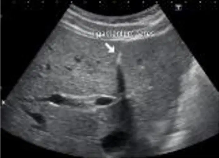
What structure is indicated by the white arrow?
ligamentum teres
ligamentum venosum
falciform ligament
main lobar fissure
What plane is the image scanned in?
long
trans
sagittal
midclavicular
The liver touches the ______ but not the ______.
aorta, IVC
IVC, portal veins
hepatic veins, portal veins
IVC, aorta
The ligamentum teres is a remnant of what structure?
ductus venosus
portal sinus
ductus sinus
portal venosus
The ligamentum venosum is a remnant of what structure?
ductus venosus
portal sinus
ductus sinus
portal venosus
During fetal circulation, the ductus venosus shunts blood from the _____ to the _____.
RPV to IVC
LPV to IVC
RHA to AO
LHA to AO
Ligamentum teres separates the ____ and ____ portions of the left lobe.
anterior and posterior
superior and inferior
medial and lateral
right and left
Ligamentum venosum separates the ____ and ____ lobes of the liver.
right and left
right and caudate
left and caudate
right, left and caudate
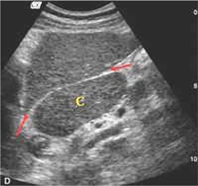
What structure is indicated by the red arrows?
ligamentum teres
main lobar fissure
falciform ligament
ligamentum venosum
What plane is the image scanned in?
long
trans
sagittal
midclavicular
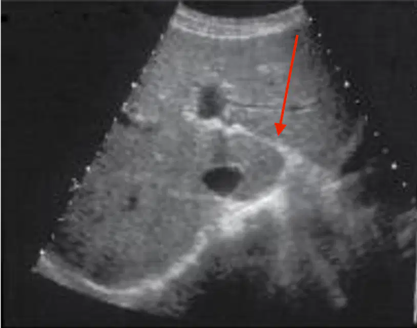
What structure is indicated by the red arrow?
ligamentum venosum
ligamentum teres
main lobar fissure
coronary ligament
What plane is the image scanned in?
long
trans
sagittal
midclavicular

What is 1?
right lobe
left lobe
caudate lobe
pancreas
What is 2?
right lobe
left lobe
caudate lobe
pancreas
What is 5?
right lobe
left lobe
caudate lobe
pancreas
What is 3?
aorta
IVC
right portal vein
left portal vein
What is 4?
aorta
IVC
right portal vein
left portal vein
What is 6?
aorta
IVC
right portal vein
left portal vein
Anatomically, the main lobar fissure separates the
right and caudate lobes
right and left lobes
left and caudate lobes
the caudate and axillary lobes
Sonographically, the main lobar fissure joins the
right and left lobes
gallbladder and left portal vein
gallbladder and right hepatic vein
gallbladder and right portal vein
The proper hepatic artery is a branch of the
gastroduodenal artery
splenic artery
common hepatic artery
right gastric artery
The _____ enters the liver at the porta hepatis.
proper hepatic artery
right hepatic artery
left hepatic artery
common hepatic artery
The porta hepatis is the _____ of the liver.
base
apex
hilum
fundus
The proper hepatic artery is seen adjacent to the
right portal vein
main portal vein
left portal vein
left hepatic vein
______ are almost always bigger than ______.
veins/arteries
arteries/veins
organs/vessels
vessels/organs
The portal vein is formed at the confluence of the ______ and the ______.
right and left portal vein
anterior and posterior portal vein
splenic vein and SMV
splenic vein and IMV

What is 1?
MPV
RPV
HA
IVC
What is 2?
MPV
RPV
HA
IVC
What is 3?
MPV
RPV
HA
IVC
What is 4?
MPV
RPV
HA
IVC
_______% of the volume of blood in the liver is supplied by the portal vein, and _____% of the volume of blood in the liver is supplied by the hepatic artery.
80/20
20/80
50/50
100/0
_______% of the oxygenated blood in the liver is supplied by the portal vein, and _____% of the oxygenated blood in the liver is supplied by the hepatic artery.
80/20
20/80
50/50
100/0
The left portal vein supplies the left lobe with blood. The left lobe is split into ____ and ____ segments.
superior/inferior
anterior/posterior
medial/lateral
left/right
The right portal vein supplies the right lobe with blood. The right lobe is split into ____ and ____ segments.
superior/inferior
anterior/posterior
medial/lateral
left/right
The liver is drained by the _____ veins, directly into the _____.
portal/IVC
hepatic/portal veins
portal/aorta
hepatic/IVC
Like the portal veins, the walls of the hepatic veins are echogenic
true
false
The hepatic veins increase in caliber as they drain towards the IVC.
true
false

What is 1?
right hepatic vein
middle hepatic vein
central hepatic vein
left hepatic vein
What is 2?
right hepatic vein
middle hepatic vein
central hepatic vein
left hepatic vein
What is 3?
right hepatic vein
middle hepatic vein
central hepatic vein
left hepatic vein
In Couinaud's System, the _____ veins divide the liver horizontally into superior and inferior regions while the _____ veins divide the liver vertically into left and right regions.
hepatic/renal
renal/portal
hepatic/portal
portal/hepatic
Couinaud's System divides the liver into how many segments?
5
6
8
9

What is 1?
left lateral superior
right anterior inferior
caudal
medial
What is 2?
left lateral superior
right anterior inferior
left lateral inferior
medial
What is 3?
right posterior inferior
right anterior inferior
left lateral inferior
medial
What is 4?
right posterior inferior
right anterior inferior
left lateral inferior
medial
What is 5?
right posterior inferior
right anterior inferior
right posterior superior
right anterior superior
What is 6?
right posterior inferior
right anterior inferior
right posterior superior
right anterior superior
What is 7?
right posterior inferior
right anterior inferior
right posterior superior
right anterior superior
What is 8?
right posterior inferior
right anterior inferior
right posterior superior
right anterior superior

What is 1?
splenic vein
splenic artery
gastroduodenal artery
left gastric artery
What is 2?
splenic vein
splenic artery
gastroduodenal artery
left gastric artery
What is 8?
splenic vein
splenic artery
gastroduodenal artery
left gastric artery
What is 3?
celiac axis
inferior mesenteric artery
superior mesenteric artery
left renal vein
What is 4?
celiac axis
inferior mesenteric artery
superior mesenteric artery
left renal vein
What is 6?
celiac axis
inferior mesenteric artery
superior mesenteric artery
left renal vein
What is 5?
IVC
aorta
diaphragm
esophagus
What is 7?
IVC
aorta
diaphragmatic crus
esophagus

What is 1?
right medial segment
right anterior segment
right lateral segment
right posterior segment
What is 2?
right medial segment
right anterior segment
right lateral segment
right posterior segment
What is 3?
left medial segment
left anterior segment
left lateral segment
left posterior segment
What is 4?
left medial segment
left anterior segment
left lateral segment
left posterior segment
What is 5?
aorta
main portal vein
IVC
SMA
What is 6?
left hepatic vein
middle hepatic vein
central hepatic vein
right hepatic vein
What is 7?
left hepatic vein
middle hepatic vein
central hepatic vein
right hepatic vein
What is 8?
left hepatic vein
middle hepatic vein
central hepatic vein
right hepatic vein
Hepatopedal means
blood flow away from liver
blood flow towards liver
blood flow around liver
blood flow through liver
Hepatofugal means
blood flow away from liver
blood flow towards liver
blood flow around liver
blood flow through liver
The portal triad consists of
portal vein, bile duct, hepatic vein
hepatic vein, IVC, SMA
portal vein, portal confluence, SMV
portal vein, bile duct, hepatic artery
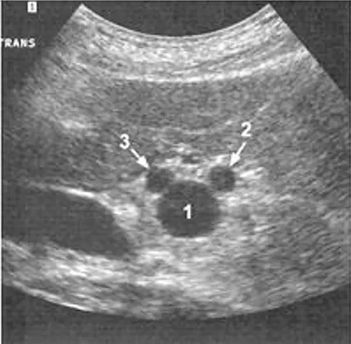
What is 1?
hepatic artery
ivc
portal vein
bile duct
What is 2?
hepatic artery
ivc
portal vein
bile duct
What is 3?
hepatic artery
ivc
portal vein
bile duct
In transverse, the portal triad forms a ______ but in longitudinal they form ______.
mickey mouse/shotgun
bunny ears/mickey mouse
shotgun/mickey mouse
mickey mouse/bunny ears
The diameter of the right and left hepatic ducts are
1-2 mm
< or = 4mm
< or = 7mm
greater than 8mm
The diameter of the common hepatic duct is
1-2 mm
< or = 4mm
< or = 7mm
greater than 8mm
The diameter of the common bile duct is
1-2 mm
< or = 4mm
< or = 7mm
greater than 8mm
The ______ ducts anastomose to become the _____ duct which becomes the _____ duct.
common bile, common hepatic, left and right hepatic
common hepatic, left and right hepatic, common bile
left and right hepatic, common hepatic, common bile
left and right hepatic, common bile, common hepatic
A liver protocol includes all of the following except
images taken in long and trans
measurements of specific structures
all images labeled with specific information
videos of structures in question

