
 Worksheets
CM - UE
Total questions: 65
Worksheet time: 33mins
You ALWAYS have to document if the pain involves the dominant side AND examine opposite side and the joint above/below the affected.
True
False
What is NOT a rotator cuff muscle?
Supraspinatus
Biceps
Subscapularis
Teres minor
Infraspinatus
Where do the rotator cuff tendons attach?
Acromion
Coracoid
Greater tuberosity
Lesser tuberosity
What rotator cuff tendon is the most common to tear?
Supraspinatus
Subscapularis
Teres minor
Infraspinatus
Pt presents with a deep sharp burning shoulder pain. Pt is able to sleep after finding a comfortable sleeping position and using NSAIDs/ice. ROM also improves with NSAIDs. Pt is able to bring arm overhead, but it is painful. PE shows pain over the lateral aspect of the shoulder (distal to insertion site of cuff tendon), pain with forced external rotation, + Neer test, + Hawkins test. Pt denies any popping feelings. What do you suspect?
Rotator Cuff Tear
Rotator Cuff Tendinitis
Subacromial Bursitis
Labral (SLAP) Tear
Glenohumeral Arthritis
Pt presents with shoulder pain. They recently had trauma to the shoulder where they felt a pop/pulling sensation. Pt reports that the pain is really bad at night. Pt doesn't get much improvment in strength/ROM with NSAIDs/ice. Pt cannot bring arm overhead. PE shows pain over the lateral aspect of the shoulder (distal to insertion site of cuff tendon), pain with forced external rotation, atrophy of supra/infraspinatus muscles, + Neer test, + Hawkins test. X-ray shows high riding humerus. What do you suspect?
Rotator Cuff Tear
Rotator Cuff Tendinitis
Subacromial Bursitis
Labral (SLAP) Tear
Glenohumeral Arthritis
Shoulder dislocation in younger individuals is associated with rotator cuff tear approximately 80% of the time.
True
False
Impingement signs PE special test that checks for compression of the greater tuberosity of the humerus against the acromion.
Neer
Hawkins
Empty can
Drop arm test
Impingement signs PE special test where you make the arm look like a wing, makes the patient sing.
Neer
Hawkins
Empty can
Drop arm test
Rotator cuff PE special test checking for weakness of the supraspinatus.
Neer
Hawkins
Empty can
Drop arm test
Rotator cuff PE special test checking for a tear. It is + if the pt cannot lift arm or loses control as lowering.
Neer
Hawkins
Empty can
Drop arm test
What is the best diagnostic/gold standard for rotator cuff tendinitis/tears?
CT
MRI
X-ray
PET
What is NOT part of the treatment/recovery for rotator cuff tears?
Full recovery in 6-12 mns
Kinesio-taping
Surgery w/in 6 wks
Sling 4-6 wks
What is NOT a MRI finding that indicates a need for referral to a surgeon?
Complete tear
Partial thickness rotator cuff tear involving greater than 10% of the width of the tendon
Retraction +/-
> 3.5 cm tear with high risk re-rupture
Pt with PMH significant for diabetes presents with dull, poorly localized ache in the shoulder. Their job involves a lot of repetitive overhead movements. PE shows intact ROM, except for loss of internal rotation. X-ray shows impingement/arthritis. What do you suspect?
Rotator Cuff Tear
Rotator Cuff Tendinitis
Subacromial Bursitis
Labral (SLAP) Tear
Glenohumeral Arthritis
Pt presents with a sharp, vague pain deep within the shoulder. They have also report clicking/popping deep within their shoulder, feelings of instability, numbness/tingling in the 4th and 5th fingers. Pt tried RICE, but there was no improvement. Of note, pt loves playing tennis and does it often. PE shows scapular dyskinesia, + Mayo Shear test, + active compression. X-ray shows impingement. MRI arthrogram shows dye leakage. What do you suspect?
Rotator Cuff Tear
Rotator Cuff Tendinitis
Subacromial Bursitis
Labral (SLAP) Tear
Glenohumeral Arthritis
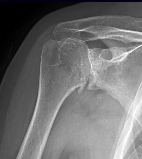
Pt presents with shoulder pain. PE shows crepitus. X-ray is done (image here). They deny specific trauma. What is NOT a possible treatment?
Restricted mobility
PT
Cortisone injection
Surgery
NSAIDs
Pt presents with pain in the shoulder that radiates to the deltoid and neck. PE shows pain with palpation over the AC joint and + adduction sign (crossover test). Of note, pt is a weightlifter. You suspect something that is caused by repetitive overhead use and cross chest activity (aka weightlifter). What do you suspect?
Rotator Cuff Tear
Rotator Cuff Tendinitis
Subacromial Bursitis
Labral (SLAP) Tear
Acromioclavicular osteoarthritis
What is NOT included in the diagnosis and treatment of Acromioclavicular joint osteoarthritis?
RICE + NSAIDs
Surgery (Distal clavicle excision)
Plain x-rays
Cortisone injection
Surgery (Tenotomy)
What type is related to POPEYE DEFORMITY?
Biceps tendon acute tear
Biceps tendon slow gradual tear
Biceps tendinitis
What two are correct regarding biceps tendon tears? (choose 2)
Distal: may or may not be able to supinate and pronate
Proximal: may or may not be able to supinate and pronate
Distal: able to supinate and pronate
Proximal: able to supinate and pronate
What is the imaging study of choice for biceps tendon rupture?
MRI
Plain x-rays
CT
US
What is NOT included in the treatment of biceps tendinitis?
RICE
Surgery (Distal clavicle excision)
PT
Cortisone injection
PO or topical NSAIDs
All of the following apply to tenotomy for bicep tendon rupture EXCEPT:
May result in mild weakness and popeye deformity
Arthroscopic release of the remaining attached tendon
Partial tears ONLY
Quick recovery
Generally done for young athletes or for aesthetic reasons
All of the following apply to tenodesis for bicep tendon rupture EXCEPT:
Usually done as part of more extensive surgery (RCR or SLAP repair)
Arthroscopic release of the remaining attached tendon & reattached to the proximal humerus
Longer recovery and more pain
Quick recovery
Generally done for young athletes or for aesthetic reasons
If a biceps tendon is completely torn, surgery is the ONLY option.
True
False
What is the MC pediatric fracture?
Epicondylar fx
Radial head
Clavicular fx
Supracondylar fx
Clavicular fracture classifications: middle 1/3 of bone; proximal fragment often displaced upward.
Class A
Class B Type 1
Class B Type 2
Class B Type 3
Class C
Clavicular fracture classifications: distal 1/3 of bone; non-displaced with intact ligaments.
Class A
Class B Type 1
Class B Type 2
Class B Type 3
Class C
Clavicular fracture classifications: distal 1/3 of bone; displaced, possible tear of coracoclavicular ligament; no AC joint involvement.
Class A
Class B Type 1
Class B Type 2
Class B Type 3
Class C
Clavicular fracture classifications: distal 1/3 of bone; displaced with AC joint involvement; ligament tears.
Class A
Class B Type 1
Class B Type 2
Class B Type 3
Class C
Clavicular fracture classifications: proximal 1/3
Class A
Class B Type 1
Class B Type 2
Class B Type 3
Class C
AC joint sprain/separation grades: AC joint intact; mild stretching of AC ligament; pain is mild; can usually move arm; "sprain"
Grade 1
Grade 2
Grade 3
AC joint sprain/separation grades: Disruption AC ligament; pain is moderate; limited movement
Grade 1
Grade 2
Grade 3
AC joint sprain/separation grades: Disruption AC and coracoclavicular ligaments; severe pain; distal clavicle is prominent; ROM limited
Grade 1
Grade 2
Grade 3
What is NOT included in treatment of GRADE 1 "SPRAIN" AC joint separation?
Full recovery in 4-6 wks
Gentle ROM
Sling until pain improves
RICE + NSAIDs
Refer to ortho for possible surgical consideration
What is NOT included in treatment of GRADE 2 and 3 AC joint separation?
Full recovery in 4-6 wks
Pain control
Sling
Refer to ortho for possible surgical consideration
What imaging should you consider for AC joint sprain/separation if x-rays are normal, but you have high suspicion?
CT
MRI
Sunrise view US
Weighted x-rays
What is the MC dislocated joint in the body?
Hip
Ankle
Shoulder
Elbow
What is the MC direction of shoulder dislocations?
Posterior
Inferior
Anterior
What is the MC nerve injured during shoulder dislocation?
Axillary
Radial
Ulnar
Median
What 2 are correct regarding shoulder dislocation direction and presentation?
Anterior: UE abducted and externally rotated
Anteiror: UE adducted and internally rotated
Posteior: UE adducted and internally rotated
Posterior: UE abducted and externally rotated
What is NOT part of the shoulder dislocation diagnosis and treatment?
Reduction
Post-reduction x-rays
Sling
X-ray
MRI
Pt presents with left elbow pain. Pt reports pain with picking up cups and driving. The elbow is stiff and painful in the mornings. PE reveals pain with resisted extension of the wrist and pain at lateral epicondyle. Of note, pt is an avid tennis player. What is NOT part of the diagnosis and treatment?
X-ray, MRI
Surgery
Tennis elbow band
Neoprene elbow sleeve
PRP or cortisone injections
Pt presents with left elbow pain. PE reveals pain with resisted flexion of the wrist and pain at medial epicondyle. Of note, pt is an avid golfer. What is NOT part of the diagnosis and treatment?
X-ray, MRI, arthrogram
Surgery
Tennis elbow band
Neoprene elbow sleeve
PRP or cortisone injections
Pt presents to clinic after FOOSH with pain at the elbow. PE reveals pain with supination and pronation and tenderness to palpation over radial head. X-ray shows an anterior sail sign. What is the treatment for occult, non-displaced, or minimally displaced?
Sling for 1 wk, then start PT
Radial head replacement
Posterior splint
ORIF
Pt presents to clinic after FOOSH with pain at the elbow. PE reveals pain with supination and pronation and tenderness to palpation over radial head. X-ray shows 3 mm depression of elbow joint and anterior sail sign. What is NOT a treatment option?
Sling for 1 wk, then start PT
Radial head replacement
ORIF
The most common complication after a radial head fx is permanent loss of full flexion.
True
False
What is NOT an indication for ORIF or radial head replacement if a pt has a displaced radial head fx?
> 1/3 of the radial head
3 mm of depression of joint
60 degree angulation
28 month-old child is brought in by their parent because they are having elbow pain after someone pulled the child up by their arm. The child is holding their arm in a flexed position. You plan to reduce it by holding UE in supination, placing thumb on radial head, and flex arm. What do you suspect?
Radial head subluxation
Supracondylar Fx
Epicondylar Fx
6 yo pt is brought in by their parent after a FOOSH. PE reveals parasthesias when pt does OK sign, bruising, tenderness, deformity, and edema of the elbow. X-ray shows posterior sail sign. What do you suspect?
Radial head subluxation
Supracondylar Fx
Epicondylar Fx
What is the correct order of frequency for pediatric traumatic fractures of the elbow?
1. Supracondylar
2. Lateral epicondyle
3. Medial epicondyle
1. Lateral epicondyle
2. Supracondylar
3. Medial epicondyle
1. Medial epicondyle
2. Lateral epicondyle
3. Supracondylar
1. Lateral epicondyle
2. Medial epicondyle
3. Supracondylar
1. Supracondylar
2. Medial epicondyle
3. Lateral epicondyle
What is the correct order of closure of elbow growth plates?
1. Capitellum (1 yr)
2. Trochlea (3 yr)
3. Int/Med epicondyle (5 yr)
4. Ext/lat epicondyle (7 yr)
5. Olecranon (9 yr)
6. Radial head (11 yr)
1. Capitellum (1 yr)
2. Radial head (3 yr)
3. Int/Med epicondyle (5 yr)
4. Trochlea (7 yr)
5. Olecranon (9 yr)
6. Ext/lat epicondyle (11 yr)
1. Olecranon (1 yr)
2. Capitellum (3 yr)
3. Int/Med epicondyle (5 yr)
4. Trochlea (7 yr)
5. Radial head (9 yr)
6. Ext/lat epicondyle (11 yr)
1. Int/Med epicondyle (1 yr)
2. Ext/lat epicondyle (3 yr)
3. Capitellum (5 yr)
4. Trochlea (7 yr)
5. Olecranon (9 yr)
6. Radial head (11 yr)
Pt presents with a painless soft mass on the posterior aspect of the right elbow. It is not hot or red and there is no purulent drainage. What is NOT part of the workup and treatment?
Aspiration (culture, cell count, gram stain, crystals)
Cortisone injection
Compression w/ neoprene sleeve
Send immediately to ortho
Pt presents with a soft mass on the posterior aspect of the right elbow. The area is warm, red, and painful. What is NOT part of the workup and treatment?
Aspiration (culture, cell count, gram stain, crystals)
Cortisone injection
Start empiric ABX after cultures obtained
Send immediately to ortho
Pt presents with numbness of the thumb, index, and long finger. There is also pain at the wrist that extends up the forearm. PE reveals + Tinel and Phalen signs. There is interosseous muscle and thenar eminence wasting. What is NOT included in the workup and treatment?
Night splint
Thumb spica splint
X-rays
Electromyograph (EMG) / Nerve conduction studies (NCS)
Carpal tunnel release
Pt presents with pain in the wrist. PE reveals pain to palpation of first doral compartment and + finkelstein test. What is NOT included in the treatment?
Thumb spica splint
Surgery to release fascia
NSAIDs
Cortisone injection
Night splint
What is the MC bone fractured in the hand?
Lunate
Capitate
Scaphoid
Trapezium
Pt presents with wrist pain after a FOOSH. PE reveals tenderness to palpation of anatomic snuff box. X-ray may or may not show fx. What is the treatment?
Thumb spica splint
Surgery to release fascia
Cortisone injection
Night splint
Pt presents with finger flexed at DIP joint. You suspect injury to the extensor tendon. Of note, pt plays baseball. What is the treatment?
Splint with PIP in full flexion
Surgery
NSAIDs
Splint holding finger in extention for 6 wks
Glucosamine injection
Pt presents with finger flexed at PIP joint and extended at DIP joint. You suspect injury to the extensor tendon where central slip tears/separates and the lateral band slips to the side. What are possible treatment opitons? (choose 2)
Splint with PIP in full extension
Surgery
NSAIDs
Glucosamine injection
Pt presents with finger flexed at DIP joint and extended at PIP joint. You suspect degeneration of a ligament on the palmar (volar) side of the PIP. What is the treatment?
Splint with PIP in full extension
Surgery
NSAIDs
Glucosamine injection
What is NOT included in the treatment of hand osteoarthritis?
Immobility
Surgery
NSAIDs
Glucosamine injection
Hand arthritis: What is NOT true about Heerden's Nodes?
Outgrowths/cysts on the DIP from calcified spurs of articular cartilage
Common
Outgrowths/cysts on the PIP from calcified spurs of articular cartilage
OA > RA
Hand arthritis: What is NOT true about Bouchard's Nodes?
May be seen with RA
Outgrowths/cysts on the PIP from calcified spurs of articular cartilage
OA > RA

