
 Worksheets
CM - LE
Total questions: 57
Worksheet time: 29mins
When getting history for hip pain what is the MOST IMPORTANT questions?
Onset
Provocative & palliating factors
Symptoms associated w/ pain?
Does the pain go to the groin?
The neck is the MC area of the hip to be fractured which is bad because it can easily disrupt blood flow to the head.
True
False
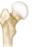
Garden Classification based upon AP x-rays: Incomplete
Type 1
Type 2
Type 3
Type 4

Garden Classification based upon AP x-rays: Complete Fx; nondisplaced
Type 1
Type 2
Type 3
Type 4
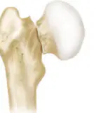
Garden Classification based upon AP x-rays: Complete; partially displaced
Type 1
Type 2
Type 3
Type 4
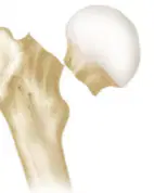
Garden Classification based upon AP x-rays: Complete; fully displaced
Type 1
Type 2
Type 3
Type 4
Pt presents with right leg pain and they cannot bear weight. The right leg is shortened, externally rotated, and abducted. What do you suspect?
Hip fracture
Hip dislocation
Pt presents with right leg pain. The right leg is shortened and internally rotated. What do you suspect?
Hip fracture
Hip dislocation
Loss of blood supply to femoral head that causes bone death and collapse. Most atraumatic cases are caused by idiopathic, corticosteroids, and alcohol. Imaging would show crescent sign. What is the best diagnostic tool and treatment? (choose 2)
X-ray
Hemiarhtroplasty
Total hip arthroplasty (THA)
US
CT/MRI
Pt presents with pain in the groin region, decreased ROM, pain with ambulating, and dull ache at night. X-ray shows joint space narrowing of the femoral acetabular joint, change in shape of femoral head, and osetophyte formation. What is NOT included in the treatment?
Shoe wear/ gait pattern
THA
Activity modification + weight loss
Casting
Joint injections
Pt presents with lateral hip pain that makes them unable to sleep on their side. PE reveals point tenderness over lateral hip. What do you suspect?
Trochanteric Bursitis
Iliotibial band tendonitis
Osteoarthritis of hip
Hip fracture
Hip dysplasia
What is NOT included in the treatment of trochanteric bursitis?
PT
Proper footwear
Steroid injection
THA
Gentle stretching
Pt presents with lateral hip pain. They can sometimes feel a snapping sensation. It feels like "hip is popping and and is unstable." PE reveals tenderness higher than the greater trochanter, and worse pain with active hip abduction. Evalute with MRI. Tx with PT, NSAID, injections, activity modification, and proper footwear. What do you suspect?
Trochanteric Bursitis
Iliotibial band tendonitis
Osteoarthritis of hip
Hip fracture
Hip dysplasia
What is the definitive treatment for hip dysplasia (anatomic abnormality in which the acetabulum does not completely cover the femoral head -> instability)?
Hemiarthroplasty
Immobilizing cast
THA
PT
The femoral head is the primary abnormality in hip dysplasia.
True
False
What direction is most likely for a hip dislocation?
Anterior
Posterior
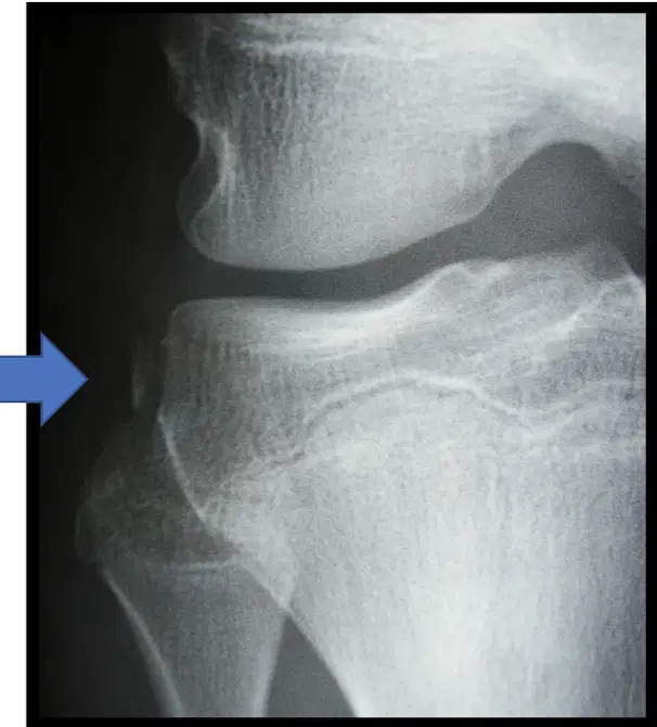
A Segond fx off lateral tibia plateau can be pathognomonic for what?
PCL tear
MCL tear
ACL tear
LCL tear
A Segond fx off lateral tibia plateau can be pathognomonic for what?
PCL tear
MCL tear
ACL tear
LCL tear
Meniscus tear
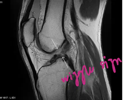
Wiggle sign on MRI is diagnostic for what?
PCL tear
MCL tear
ACL tear
LCL tear
Meniscus tear
What is the #1 indication for knee surgery?
PCL tear
MCL tear
ACL tear
LCL tear
Meniscus tear
Pt presents with sudden knee pain after a plant & pivot injury. PE reveals limited ROM, pain with weight-bearing, and + McMurry/Apley. What do you suspect?
PCL tear
MCL tear
ACL tear
LCL tear
Meniscus tear
MRI is unnecessary to diagnose a meniscal injury.
True
False
Pt presents with pain after a fall directly onto their knee. PE reveals swelling, ecchymosis, and inability to straighten the knee. What is NOT included in the diagnosis and treatment?
Surgery (Tension band)
Hinged locked brace
X-ray
Patella Tracking Orthosis
Weight bearing as tolerated (WBAT)
Pt presents with knee pain after a twisting injury where the pt felt a pop, then the knee immediately swelled. PE shows hemarthrosis, medial patella & lateral femoral condyle tender to palpation, + apprehension. What is NOT included in the diagnosis and treatment?
J brace
Medial patella-femoral ligament recon/realignment
X-ray
Patella Tracking Orthosis
Surgery (Tension band)
Pt presents with anterior knee pain. PE shows localized pain anterior & posterior-one finger test, + J sign, patella tracking laterally, weak VMO/core/hips with one leg squat, grind +/crepitus, and hypermobile patella. What is NOT included in your diagnostic workup and treatment?
Total patella replacement
Bracing
Inserts
X-ray
PT/taping
Teen pt presents with anterior knee pain. PE reveals enlarged tibial tubercle, pain w/ extension or kneeling, tenderness to palpation. Of note, pt participates in a jumping sport. XR shows enlarged and fragmented tibial tubercle. What is NOT included in the treatment?
PT
Activity modification or immobilization
Brace
Ossicle excision
TKA
Pt presents with knee swelling, painful medial joint line, and increased pain with varus/valgus stress. Xrays and MRI are completed. What is NOT part of the tx?
NSAIDs
Custom MCL brace
RICE
PT
ACL repair
Pt reports progressive functionally limiting knee pain, night pain, feelings on instability, swelling, stiffness, and mechanical sx. Pt notes this pain impacts their QOL and ADL. What is the imaging of choice?
MRI
CT
Weight-bearing x-ray
US
Pt reports progressive functionally limiting knee pain, night pain, feelings on instability, swelling, stiffness, and mechanical sx. Pt notes this pain impacts their QOL and ADL. All of the following are possible treatments, but what is the definitive treatment?
PRP or hyaluronic acid injections
Glucosamine Chondroitin sulfate
Unicompartmental, TKA, or PFA
Stem cells
Matrix-induced autologous chondrocyte implantation (MACI)
What is NOT included in the terrible triad?
MCL
Medial meniscus
ACL
LCL
Patella dislocation can be related to all of the following EXCEPT
Patella alta (kneecap sits too high in groove)
Trochlea dysplasia (shallow groove)
Patella bajo (kneecap sits too low in groove)
Tilt
35 yo pt presents with mild discomfort in the back of the ankle. The pt was playing basketball when they heard a pop. PE reveals limp, loss of push-off strength, weak ankle, pt is still able to plantar flex ankle. PE also shows ecchymosis, swelling, + Thompson test, and palpable defect. All of the following are treatments, but what is the gold standard?
Long-leg CAM boot w/ 2 in heel lift or plantar flexed posterior splint & crutches
Surgical repair
PT
RICE
What is NOT included in the treatment of achilles tendonitis?
RICE + NSAIDs
Brief immobilization in CAM boot
PT
Shoe lift, lifestyle mods
Surgery
What is the MC ankle ligament to tear?
Calcaneal fibular ligament (CFL)
Posterior talofibular ligament (PTFL)
Anterior talofibular ligament (ATFL)
Pt presents with lateral ankle pain, swelling, and ecchymosis. PE reveals anterior drawer test with > 10 mm translation. What is NOT included in the diagnostic workup and treatment?
MRI
X-ray
WBAT in CAM boot or lace-up ankle sleeve
RICE + NSAIDs
PT
Which of the following doesn't describe grade I ankle sprain?
Mild swelling
Ligament length normal
Partial ligament tear
Negative anterior drawer
ATFL
Which of the following doesn't describe grade II ankle sprain?
Moderate swelling
Mildly positive anterior drawer
Partial ligament tear
ATFL, CFL
ATFL
Which of the following doesn't describe grade III ankle sprain?
Severe swelling
Grossly positive anterior drawer
Complete ligament tear
ATFL, CFL, PTFL
ATFL, CFL
Pt presents with ankle pain. Stress view x-ray shows widening of the tibiotalar joint (mortise) and/or widening of syndesmotic space. What is NOT part of the treatment?
Splint
Self-limited
Surgery
Crutches
Bimalleolar fx includes fibular fx with a deltoid ligament tear.
True
False
The Ottawa Ankle Rules are meant to reduce x-ray imaging by ruling out significant ankle fractures. What is NOT one of hte criteria?
Pain near malleoli
Inability to weight bear immediately after & in ED (4 steps)
Tenderness at posterior edge or tip of lateral malleolus
Tenderness at posterior edge or tip of medial malleolus
Gross swelling at either malleloi
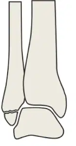
Weber classification of ankle fracture: fibular fx is distal to ankle joint (probably won't need surgery)
Weber A
Weber B
Weber C
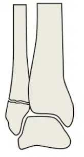
Weber classification of ankle fracture: spiral fx of fibular at distal tibiofibular joint (may need surgery)
Weber A
Weber B
Weber C
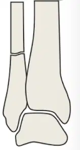
Weber classification of ankle fracture: fx above the level of the tibiofibular joint (suspect disruption of the synesmotic and ankle ligaments)
Weber A
Weber B
Weber C
Pt with ankle injury went to ER and was placed in a CAM boot, but they returned today because their pain is worsening. You suspect a combination of a spiral fx of the proximal 1/3 of the fibula with an unstable ankle injury (widening of the ankle mortise on x-ray). What do you suspect?
Weber A Fx
Pes Cavus Deformity
Maisonneuve Fx
Segond Fx
Pt presents with onset of pain, swelling, limited ROM, decreased ADL, shoes don't fit. You suspect narrowing of tibiotalar joint space. All of the following are treatment options, but what is the gold standard for end-stage OA?
Ankle fusion
NSAIDs
Glucosamine
Total ankle arthroplasty
Bracing
The Ottawa Foot Rules are meant to reduce x-ray imaging by ruling out significant foot fractures. What is NOT one of the criteria?
Pain in the midfoot
Inability to weight bear both immediately & in the ED (4 steps)
Tenderness at the navicular
Tenderness at the base of the 5th metatarsal
Gross swelling at calcaneous
High arch, walk on lateral border of foot, callous under 5th MT, claw toes, peek-a-boo heel sign from front
Cavovarus/Pes Cavus Deformity
Pes Planus Deformity
Forefoot rolls inward, can be 2/t posterior tibial tendon dysfunction. Acquired, congenital. Valgus achilles tendon. "Too many toes" or "peek-a-boo" toe sign from the back.
Cavovarus/Pes Cavus Deformity
Pes Planus Deformity
Pt presents with medial hindfoot & ankle pain, loss of strength, foot "rolls" over, "too many toes" sign, unable to tiptoe. MRI was done to assess integrity of tendon. Your treatment would include rest, NSAIDs, boot, AFO or arizone face. What do you suspect?
Posterior tibial tendon dysfunction
Pes cavus deformity
Metatarsal stress fx
Pes planus deformity
Plantar fasciitis
Pt presents with insidious onset of foot pain. The pain is worse during and after activity. PE shows swelling and point tenderness. What imaging modality will show a metatarsal stress fracture the earliest?
X-rays
Bone scan
US
MRI
What is the MC type of foot fx?
March
Jones
Maisonneuve Fx
Segond Fx
Transverse fx b/w base and shaft of 5th metatarsal (Zone 2/Transition Zone). Mostly surgical tx.
March
Jones
Maisonneuve Fx
Segond Fx
Obese pt presents with bilateral, atraumatic, gradual onset of heel pain. Plantar medial heel pain is worse in the morning, when barefoot, and on "start-up." Pt notes that achilles is tight. You suspect the MCC of heel pain. What is NOT included in the tx?
Stretching
Heel cushion, orthotic, proper footwear, custom orthotics
Surgery
Avoid high impact activity
Arizona brace
Deformity of the 1st MTP with medial prominence, increased metatarsal angle, and lateral capsule contracture. Most common deformity of forefoot and toes.
Phalanx Fx
Charcot Joint
Hallux Valgus/ Bunion
Lisfranc Deformity
Pt with DM presents with warm, red, swollen foot with little to no pain. PE shows pulses present, ulceration, and "rocker bottom" deformity (plantar surface prominence). You suspect a chronic, noninfective, painless joint destruction. What disease does this describe?
Phalanx Fx
Charcot Joint - Neuropathic osteoarthropathy
Hallux Valgus/ Bunion
Lisfranc Deformity
Pt presents with midfoot pain, inability to weight-bear, swelling, and medical plantar ecchymosis. Of note, pt plays a lot of baseball. You suspect tarsalmetatarsal (TMT) fx dislocation characterized by disruption of the ligament b/w the second metatarsal and the medial cuneiform.
Phalanx Fx
Charcot Joint - Neuropathic osteoarthropathy
Hallux Valgus/ Bunion
Lisfranc Fracture

