
 Worksheets
IB Bio 6.2 and D4
Total questions: 206
Worksheet time: 4hrs 10mins

When blood leaves the left atrium it passes through which valve?
mitral
aortic
pulmonary
tricuspid

Blood passes through this valve after leaving the right ventricle:
tricuspid valve
mitral valve
aortic valve
pulmonary valve

Which vessels transport de-oxygenated blood from the heart to the lungs?
pulmonary veins
pulmonary aorta
pulmonary arteries
pulmonary capillaries
A complete heartbeat
cardiac systole
cardiac diastole
cardiac cycle
cardiac arrest
Pacemaker of the heart
SA node
Right atrium
Left atrium
AV node
Place the following in the correct order:
1. atria contract
2. AV node
3. Bundle of His
4. Purkinje fibers
5. SA node
6. ventricles contract
5, 1, 2, 3, 4, 6
2, 1, 5, 3, 4, 6
5, 6, 2, 3, 4, 1
2, 6, 5, 3, 4, 1
What type of muscle is found in the heart?
skeletal
smooth
voluntary
cardiac

In the section of cardiac muscle shown, structures A and B respectively are
mitochondrion, intercalated disc
mitochondrion, myosin/actin filaments
nucleus, intercalated disc
nucleus, myosin/actin filaments
What is a similarity between cardiac and skeletal muscle?
both are striated
both fatigue
both multinucleate
both under voluntary control
Which is correct about the SA node?
located in left atrium
a collection of cardiac cells initiating action potentials
referred to as the artificial pacemaker
it is the equivalent of the AV node in birds
In general, what is systole?
contraction
the same as diastole
relaxation
the system of blood vessels in the heart
Where is the AV node?
in the left atrium wall
junction between the atria and ventricles
in the right atrium wall
in the ventricles
What is the correct sequence of electrical signals spreading throughout the heart?
SA node→bundle of His→AV node→Purkinje fibres
SA node→AV node→Purkinje fibres→bundle of His
SA node→AV node→bundle of His→Purkinje fibres
AV node→SA node→bundle of His→Purkinje fibres
What causes the "lub" sound?
AV valves closing
AV valves opening
semilunar valves closing
semilunar valves opening
An artificial pacemaker would be beneficial for which one of the following conditions?
obesity
inability of brain to send rhythmic signals to the hear
malfunctioning SA node
narrowing of coronary arteries
During diastole which one of the following is not occurring?
semilunar valves are closed
both atria and ventricles are relaxed
"dub" sound is made
only ventricles are relaxed, atria are contracting
Why is there a delay in the conduction of a signal from the AV node?
So that right atrium contracts before the left atrium
So that right ventricle contracts before the left ventricle
So the both atria have contracted and emptied all the blood into the ventricles
So that the AV valves are closed before the ventricles contract
What chamber of the heart has the thickest walls?
right atrium
left atrium
right ventricle
left ventricle
The human heart is referred to as myogenic because
nerve impulses stimulate the heart muscle tissue to contract
an individual has an SA node that is not functioning
a signal originates within the heart muscle tissue to contract
the size of the heart is genetically determined based on the myocyte gene
Read the following statements.
1.cells are self-excitatory
2.cells have a long linear shape for better contraction
3.cells contain many lysosomes
4.cell joined end to end by intercalated discs
5.cells have a large central nucleus
6.cells contain many mitochondria
7.arteries supply the cells with oxygenated blood
8.a branched structure allows for coordinated contraction throughout the heart
Which set of statements will get you the most marks if you were asked to describe the unique properties of cardiac muscle cells on an IB Biology exam?
1, 4, 6
1, 2, 4, 7
2, 3, 5, 6, 8
2, 4, 5, 8
The two causes of normal heart sounds are
AV valves closing and semilunar valves opening
AV valves opening and semilunar valves closing
AV valves closing and semilunar valves closing
AV valves opening and semilunar valves opening
What type of muscle is found in the heart?
skeletal
smooth
voluntary
cardiac
In the section of cardiac muscle shown, structures A and B respectively are
mitochondrion, intercalated disc
mitochondrion, myosin/actin filaments
nucleus, intercalated disc
nucleus, myosin/actin filaments
What is a similarity between cardiac and skeletal muscle?
both are striated
both fatigue
both multinucleate
both under voluntary control
Which is correct about the SA node?
located in left atrium
a collection of cardiac cells initiating action potentials
referred to as the artificial pacemaker
it is the equivalent of the AV node in birds
In general, what is systole?
contraction
the same as diastole
relaxation
the system of blood vessels in the heart
Where is the AV node?
in the left atrium wall
junction between the atria and ventricles
in the right atrium wall
in the ventricles
What is the correct sequence of electrical signals spreading throughout the heart?
SA node→bundle of His→AV node→Purkinje fibres
SA node→AV node→Purkinje fibres→bundle of His
SA node→AV node→bundle of His→Purkinje fibres
AV node→SA node→bundle of His→Purkinje fibres
What causes the "lub" sound?
AV valves closing
AV valves opening
semilunar valves closing
semilunar valves opening
An artificial pacemaker would be beneficial for which one of the following conditions?
obesity
inability of brain to send rhythmic signals to the hear
malfunctioning SA node
narrowing of coronary arteries
During diastole which one of the following is not occurring?
semilunar valves are closed
both atria and ventricles are relaxed
"dub" sound is made
only ventricles are relaxed, atria are contracting
Why is there a delay in the conduction of a signal from the AV node?
So that right atrium contracts before the left atrium
So that right ventricle contracts before the left ventricle
So the both atria have contracted and emptied all the blood into the ventricles
So that the AV valves are closed before the ventricles contract
The human heart is referred to as myogenic because
nerve impulses stimulate the heart muscle tissue to contract
an individual has an SA node that is not functioning
a signal originates within the heart muscle tissue to contract
the size of the heart is genetically determined based on the myocyte gene
Read the following statements.
1.cells are self-excitatory
2.cells have a long linear shape for better contraction
3.cells contain many lysosomes
4.cell joined end to end by intercalated discs
5.cells have a large central nucleus
6.cells contain many mitochondria
7.arteries supply the cells with oxygenated blood
8.a branched structure allows for coordinated contraction throughout the heart
Which set of statements will get you the most marks if you were asked to describe the unique properties of cardiac muscle cells on an IB Biology exam?
1, 4, 6
1, 2, 4, 7
2, 3, 5, 6, 8
2, 4, 5, 8
The two causes of normal heart sounds are
AV valves closing and semilunar valves opening
AV valves opening and semilunar valves closing
AV valves closing and semilunar valves closing
AV valves opening and semilunar valves opening

The p wave corresponds to what event in the heart?
repolarization of the atria
depolarization of the atria
repolarization of the ventricles
depolarization of the ventricles
The T wave corresponds to what event in the heart?
repolarization of the atria
depolarization of the atria
repolarization of the ventricles
depolarization of the ventricles
ECG is a reading that shows
the activity of heart
the electrical activity of cardiac muscle
the systole and diastole of cardiac muscle
the initiation of heartbeat
The correct sequence of the three events in ECG
P wave , T wave , QRS complex
QRS complex , P wave , T wave
T wave , QRS complex , P wave
P wave, QRS complex, T wave

Which one is represent T wave
A
B
C
E
By relating events in ECG with phases in cardiac cycle, at which event both atria and ventricles will be relaxed?
A
B
C
E
By relating events in ECG with phases in cardiac cycle, at which event the second sound of heart "dup" will be produced?
A
B
C
E

Identify the ECG
injured person
Dead person
Infant
Toodler
ECG stand for
Electrocardiograph
Electrocardiogram
Electrocardiopraphy
Electronicardiogram
Bradycardia .........
Shows irregular rhythm
Heart rate is more than 60bpm
Heart rate is less than 60bpm
Tachycardia.......
Shows irregular rhythm
Occurs due to suppression of SA
Heart rate is less than 100bpm
Heart rate is more than 100bpm
IA Assessment criteria:- Personal Engagement:
Assesses evidence of independent and creative thinking.
Assesses awareness of safety, environmental, and ethical considerations.
Assesses how well the investigation is presented in regards to the focus, process, and outcomes.
IA Assessment criteria:- Exploration:
Assesses evidence of evaluation of the investigation.
Assesses how well the investigation is presented in regards to the focus, process, and outcomes.
Assesses scientific context, a clear and focused question, on level with the Diploma Program.
IA Assessment criteria:- Analysis:
Assesses evidence of evaluation of the investigation.
Assesses how the student collected and *interpreted* the data.
Assesses scientific context, a clear and focused question, on level with the Diploma Program.
IA Assessment criteria:- Evaluation:
Assesses the extent to which the student’s report provides evidence of evaluation of the investigation and the results.
Assesses scientific context, a clear and focused question, on level with the Diploma Program.
Assesses how the student collected and *interpreted* the data.
IA Assessment criteria:- Communication:
Assesses evidence of evaluation of the investigation.
Assesses how well the investigation is presented in regards to the focus, process, and outcomes.
Assesses scientific context, a clear and focused question, on level with the Diploma Program.


What is the most muscular chamber of the heart?
left ventricle
left atrium
right atrium
right ventricle

Valve that blood is forced through from right ventricle into pulmonary arteries.
pulmonary semi lunar valve
aortic valve
mitral valve
In the walls of the heart, there are two thin layers around a thick layer of muscle called the
epithelial tissue.
pericardium.
connective tissue.
myocardium.


The heart layer that keeps blood from sticking to the inside of the heart made up of squamous epithelial cells
endocardium
pericardium
myocardium
epicardium
Pericardium is a serous membrane it has two layers. The parietal layers of the membranes line the walls of the body cavity. The inner visceral layer covers organ (viscera).
True
False
Aorta
The largest arterial trunk that carries blood from the heart to be distributed by branch arteries through the body
a vessel that returns blood to the heart
a body structure that is temporarily closes a passage or orifice, or permits movement of fluid in only one direction.
An integrated group of cells with a common function, structure, or both
Artery
A valve in the heart that guards the opening between the left atrium and the left ventricle; prevents the blood in the ventricle from returning to the atrium. Alternative name is bicuspid valve.
A vein that is the largest vein in the human body and returns blood to the right atrium of the heart from bodily parts below the diaphragm.
The body structure that temporarily closes a passage or orifice, or permits movement of fluid in only one direction.
Any of the tubular, branching, muscular, and elastic-walled vessels that carry blood from the heart through the body.
Mitral Valve
The semilunar valve separating the aorta from the left ventricle that prevents blood from flowing back into the left ventricle.
A vein that is the largest vein in the human body and returns blood to the right atrium of the heart from bodily parts below the diaphragm.
An anatomical cavity or passage especially a chamber of the heart that receives blood from the veins and forces it into a ventricle or ventricles
A valve in the heart that guards the opening between the left atrium and the left ventricle; prevents the blood in the ventricle from returning to the atrium. Alternative name is bicuspid valve.


What is the size of an adult’s heart?
two fists together
the size of a fist
the size of a ball
the size of an apple
How many chambers does the heart consist of?
2
3
4
6
What do the lungs provide the blood with?
oxygen
blood
hydrogen
carbon dioxide
What vessels carry the blood away from the heart?
arteries
veins
an aorta
capillaries
What vessels carry the blood back to the heart?
arteries
veins
an aorta
capillaries
What are the thinnest blood vessels called?
arteries
veins
capillaries
an aorta
What is biggest artery in a human body?
arteries
veins
capillaries
an aorta
How much blood if there in an adult human body?
3 liters
5 liters
10 liters
20 liters
How many times does a human heart beat a day?
100 times
1000 times
100,000 times
1,000,000 times
What is the total length of blood vessels in a human body?
6 miles
60 miles
6,000 miles
60,000 miles

when looking at heart diagrams the veins are usually ____, and the arteries are ____
red, blue
blue, red
pink, purple
blue, maroon

The first cavity of the heart that deoxygenated blood enters is
(a)
Draw a heart (the one inside, doesn't have to be perfect)

what is O
inferior vena cava
inferior cava vena
superior vena cava
superior cava vena

what is A
Aorta
vena cava
left atrium
artery

what is M
bicuspid valve
pulmonary valve
mitral valve
tricuspid valve

what is J
superior cava vena
inferior vena cava
superior vena cava
inferior cava vena
the heart has how many cavities?
2
3
4
5

what is H
Right Ventricle
Right atrium
Left Atrium
Left Ventricle
how many steps of the bloodflow to the heart
4
18
12
11
the LUBB of the heartbeat is the
Ventricular contraction
Ventricular Relaxation
the DUBB of the heartbeat is the
Ventricular Contraction
Ventricular Relaxation
The Atria has/does
has thin walls
receives blood that's returning to the heart
has thick wall
expels blood from the heart
The Ventricle....(pick two)
is the upper two quadrants of the heart
contract to force blood out of the heart
receives blood from the Atria
are connected to each other (right and left)
the heart has three layers, what are they?
Epicardium
myocardium
Endocardium
sarkicartium
The Atria ____ while ventricles relax, then _____ contract while atria relax.
contract, ventricles
contract, atria
relax, ventricles
relax, atria

What is B
Left plumonary artery
right pulmonary artery
left pulmonary veins
right pulmonary veins

what is e.
(a)

what is D
left pulmonary artery
right pulmonary artery
left pulmonary veins
right pulmonary veins


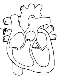

Which side of the heart pumps oxygen-rich blood to the rest of the body?
Right
Left
Top
Bottom
Which side of the heart pumps blood into the lungs?
Right
Left
Top
Bottom


Blood travels TO the heart through the ______.
lungs
veins
arteries
oxygen


Valve that blood is forced through from right ventricle into pulmonary arteries.
Pulmonary valve
Aortic valve
mitral valve
Tricuspid
Name of the valve that separates left atrium from left ventricle.
Mitral valve
Tricuspid Valve
Pulmonary valve
Aortic Valve
What portion of the heart is supposed to be the most muscular?
Atriums
Ventricles
Septum
Which valve is found between the left ventricle and the aorta?
Tricuspid Valve
Bicuspid Valve
Aortic Valve
Pulmonary Valve
What is the function of the Atrium?
Receive Blood
Send out Blood
Oxygenate Blood
Filter Blood
Atria pumps blood to the:
Arteries
Body cavities
Veins
Ventricles
The only artery in humans that carries deoxygenated blood is the:
Pulmonary artery
Renal artery
Aorta
Carotid artery
The direction of blood flow through the human body is maintained by:
The elasticity of the vein walls
The pump action of the heart
Muscular contractions
A system of valves
Blood:
Is pumped from the heart into the veins
Enters the right atrium from the lungs
Is not involved in temperature control
Can pass round the body in less than a minute
Blood leaves the heart in two vessels, namely the:
Aorta and pulmonary vein
Pulmonary artery and aorta
Aorta and venae cavae
Pulmonary artery and pulmonary vein
A boy is bitten by a venomous snake on his left leg. Poison gets to his heart through the:
Pulmonary vein
Left subclavian artery
Inferior vena cava
Hepatic portal vein
Which of the following contain deoxygenated blood?
Left ventricle
Pulmonary vein
Right atrium
Coronary artery
The phase in the cardiac cycle when blood is pumped out of the ventricles is called:
Atrial systole
Ventricular diastole
Atrial systole
Ventricular systole
A blood vessel is called an artery when it:
Transports oxygenated blood to the heart
Transports blood to the heart
Contains many valves
Transports blood away from the heart
One of the three functions of the circulatory system is:
Carries waste away from our cells
Helps regulate body temperature
Delivers needed substances to the cells
All of the above
Type of circulation that refers to blood flow from the heart to the lungs and back to the heart again?
Systemic circulation
Renal circulation
Portal circulation
Pulmonary circulation
Type of circulation that refers to the blood flowing through the entire body?
Systemic circulation
Renal circulation
Portal circulation
Pulmonary circulation
The contraction/emptying phase of the cardiac cycle?
Systole
Diastole
The relaxing/filling stage of the cardiac cycle?
Systole
Diastole
Coronary arteries...
supply the heart muscle cells with a supply of blood.
supply the brain cells with a supply of blood.
contain deoxygenated blood.
when clogged, cause a stroke.
The type of blood vessel that normally carries de-oxygenated blood back to the heart from body cells?
arteries
capillaries
veins
The type of blood vessel that normally carries oxygenated blood from the heart to the body cells?
arteries
capillaries
veins
The type of blood vessel where exchange of gases, nutrients, and wastes occurs?
arteries
capillaries
veins

An abnormal widening/ballooning of a portion of an artery.
aneurysm
embolism
thrombosis
atherosclerosis
A blood clot that travels through the circulatory system and eventually becomes lodged within a blood vessel?
aneurysm
embolism
thrombosis
atherosclerosis
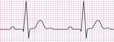
Type of device that measures the electrical currents created by the SA and AV nodes?
AED
MRI
ECG
CAT Scan

The main vein(s) that brings blood to the right atrium?
Aorta
Pulmonary veins
Superior and Inferior vena cava
Jugular vein

Which choice below correctly identifies the layers of the heart from the most outer layer to the most inner layer?
epicardium, myocardium, endocardium
endocardium, myocardium, epicardium
myocardium, endocardium, epicardium
myocardium, epicardium, endocardium
The sac that surrounds and protects the heart?
endocardium
epicardium
myocardium
pericardium

Instrument used to measure blood pressure?
AED
ECG
Sphygmomanometer
ultrasound

Name part N.
Right atrium
left atrium
right ventricle
left ventricle

Name part I.
pericardium
mediastinum
septum
unknownum

Name part A.
Pulmonary artery
Pulmonary vein
Superior vena cava
Aorta

Name part B.
Pulmonary arteries
Pulmonary veins
Superior Vena cava
Aorta

Name part M.
Tricuspid valve
Semilunar valve
Bicuspid valve
Mitral valve

