
Font size
 Worksheets
AP04 Cell Structures
Total questions: 82
Worksheet time: 1hrs 7mins
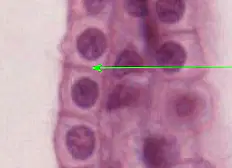
What cell structure is the green arrow pointing at?
(a)
What cell structure is the green arrow pointing at?
Rough ER
cell wall
What cell structure is the green arrow pointing at?
(a)
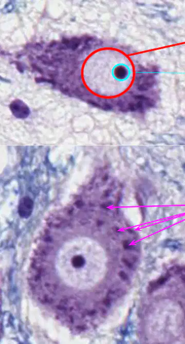
What cell structure has a light BLUE CIRCLE around it?
(Cerebellum, Monkey, Cresyl Violet)
(neurons)
(a)
What cell structure has a RED CIRCLE around it?
(Cerebellum, Monkey, Cresyl Violet)
(neurons)
(a)
What cell structure has a MAGENTA ARROWS pointing at it?
(Cerebellum, Monkey, Cresyl Violet)
(neurons)
(a)
Label the indicated structures.
(Cerebellum, Monkey, Cresyl Violet)
cell membrane
smooth ER
rough ER
nucleus
nucleolus
What kind of cells are shown here?
(a)
What kind of cells are shown here?
muscle
epithelial
blood
connective
What cell structure has a light BLUE CIRCLE around it?
(Cerebellum, Monkey, Cresyl Violet)
(neurons)
smooth ER
rough ER
nucleolus
What cell structure has a MAGENTA ARROWS pointed at it?
(Cerebellum, Monkey, Cresyl Violet)
(neurons)
smooth ER
rough ER
nucleolus
What cell structure has a RED CIRCLE around it?
(Cerebellum, Monkey, Cresyl Violet)
(neurons)
smooth ER
rough ER
nucleolus
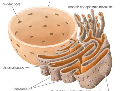
The main purpose of the Rough ER is to produce and package

Membrane bound polyribosomes are found where?
nucleus
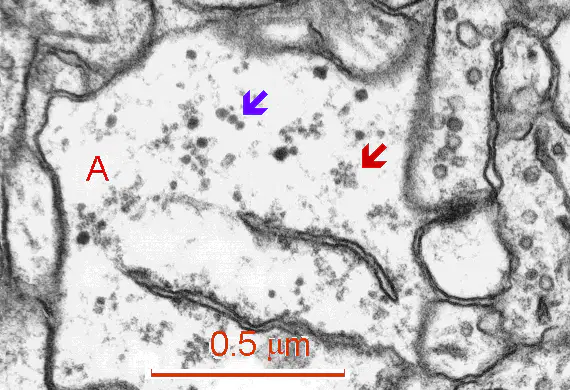
Where are free polyribosomes found in the cell?
golgi appartatus
What organelle can be embedded in the membranes of rough ER and the nucleus and is the site of polypeptide (protein) synthesis?
(a)
What organelle can be found embedded in the membranes of rough ER and the nucleus and is the site of polypeptide (protein) synthesis?
mitochondria
chloroplast
smooth ER
proteinosome
Label the structures indicated.
Pancreas (H&E)
cell membrane
smooth ER
nucleolus
nucleus
Rough ER
Eosinophilic
Label the structures indicated.
Kidney, Pelvis, & Ureter (Unilobular)
cell membrane
smooth ER
nucleolus
nucleus
Rough ER
Eosinophilic

One of the most important roles of smooth ER (sER) is (a) synthesis
One of the most important roles of smooth ER (sER) is _______ synthesis
protein
nucleic acid
carbohydrate
Smooth ER (sER) tends to stain poorly making it appear (a)
Smooth ER (sER) tends to stain (a) making it appear empty.
Label this section of an adrenal gland.
Remember: Steroid producing cells tend to have a significant amount of smooth ER (sER).
(Adrenal, Monkey, H&E)
less eosinophilic
more eosinophilic
Label this section of an adrenal gland.
Remember: Steroid producing cells tend to have a significant amount of smooth ER (sER).
(Adrenal, Monkey, H&E)
more sER
less sER

In this section of the adrenal gland, which cells would most like have the most smoothER (sER)?
(Adrenal, Monkey, H&E)
cells above the blue line
cells below the green line
cells between the blue and green lines
none of these cells have sER
all of these cells would have about the same amount of sER
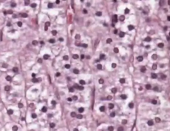
The lack of staining in these cells of the adrenal gland indicates that they contain an abundant amount of _____
(Adrenal, Monkey, H&E)
rough ER (rER)
nuclei
smooth ER (sER)
DNA
Lysosomes


______ are glycosylated or otherwise modified in the Golgi Complex.
Polypeptides (Proteins)
DNA
RNA
Polypeptides (proteins) are glycosylated in the ____
nucleus
nucleolus
Rough ER
Smooth ER
Golgi Appartus
Glycosylation is the process of attaching a(n) ______ to another biomolecule.
carbohydrate
DNA
RNA
Match the following
ribosome
lysosome
chloroplast
mitochondia
rough ER
nucleus
nucleolus
golgi complex
smooth ER
Match the following
produce energy
protein synthesis
control center
ribosome synthesis
modification and transport
lipid synthesis

Match the following
A
smooth ER
B
nucleolus
C
nucleus
D
Golgi complex
E
rough ER

Smooth Endoplasmic Reticulum (sER)
A
B
C
D
E
Nucleolus
A
B
C
D
E
Nucleus
A
B
C
D
E
Golgi complex
A
B
C
D
E
Rough Endoplasmic Reticulum (rER)
A
B
C
D
E
At the “maturing” face of the Golgi, new _____ pinch off and are sorted to different destinations according to their contents.
vessicles
ribosomes
centrioles
proteins
carbohydrates
At the “maturing” face of the Golgi, new ____ ____ ____...(plural) (3 words)
(a)
At the “maturing” face of the (a) , new vesicles pinch off.
At the “maturing” face of the Golgi, new vesicles pinch off and are (a) to different destinations according to their contents.
Most Golgi vesicles either condense to form secretory granules that release their contents by ____...
diffusion
facillitated diffusion
active transport via a protein pump
Most Golgi vesicles either condense to form ____ that release their contents by exocytosis...
secretory granuales
lysosomes
peroxisomes
ribosomes
carrier proteins
Golgi vesicles that remain in the cytoplasm, remain as ____ or ____.
secretory granules
lysosomes
peroxisomes
ribosomes
carrier proteins
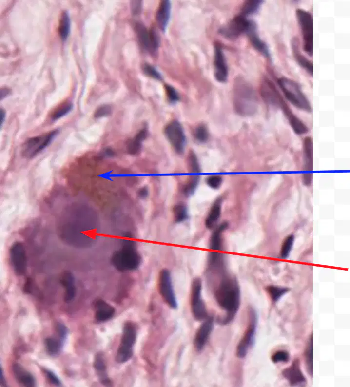
In long lived cells like a neuron, the brown staining material indicates the presence of ____
Sympathetic Ganglion, Human H&E
lysosomes
ribosome
peroxisomes
smooth ER
rough ER


Label the indicated structures of the neuron.
Sympathetic Ganglion, Human H & E
Lipofuscin granules
(residual bodies)
nucleus
nucleolus
rough ER
smooth ER
The red arrow marks the ____.
The blue arrow indicates the presence of ____.
Sympathetic Ganglion, Human H & E
ribosomes
peroxisomes
liposomes
lysosomes
nucleus
nucleolus
rough ER
smooth ER
Label the indicated structures.
Mitochondria
matrix
cristae

What organelle is shown in the picture?
mitochondria
nucleus
peroxisome
lysosome
chloroplast
The enzymes for Beta oxidation of fatty acids and the citric acid cycle are found in/on the structure labeled as
Label A
Label B
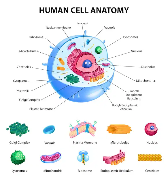
The enzymes for Beta oxidation of fatty acids and the citric acid cycle are found in/on the
mitochondrial matrix
mitochondrial cristae
cell cytoplasm
chloroplast thylakoid membrane
rough ER
What structure increases the surface area for ATP synthesis during cellular respiration?
mitochondrial matrix
mitochondrial cristae
cell cytoplasm
chloroplast thylakoid membrane
rough ER
The structure that increases surface area for ATP synthesis.
Label A
Label B

Mitochondria have their own (a) .
The genome that mitochondria have is
mDNA
rRNA
tRNA
mRNA

mDNA stands for
mitochondrial DNA
mitosis DNA
messenger DNA
membrane DNA
meiosis DNA
Label the structures.
nucleolus
euchromatic nucleus
heterochromatic nucleus
Label the structures.
ribosome synthesis
high levels of protein synthesis
low levels of protein synthesis
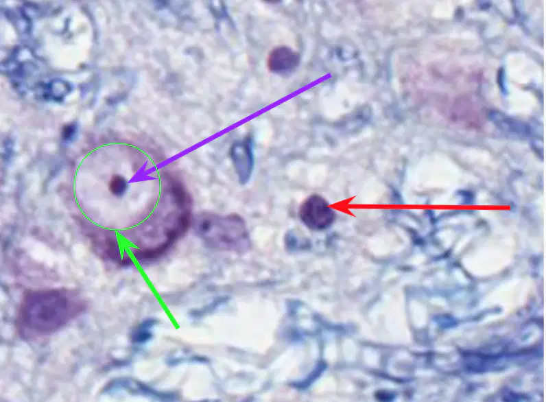
The purple arrow marks
euchromatic nucleus
heterochromatic nucleus
nucleolus
mitochondria
The red arrow marks
euchromatic nucleus
heterochromatic nucleus
nucleolus
mitochondria
The green arrow marks
euchromatic nucleus
heterochromatic nucleus
nucleolus
mitochondria
site of ribosome synthesis
green arrow
red arrow
purple arrow
high level of protein synthesis
green arrow
red arrow
purple arrow
low level of protein synthesis
green arrow
red arrow
purple arrow
Label the steps of mitosis (cell division)
(Please Make Another Taco)
anaphase
prophase
metaphase
telophase
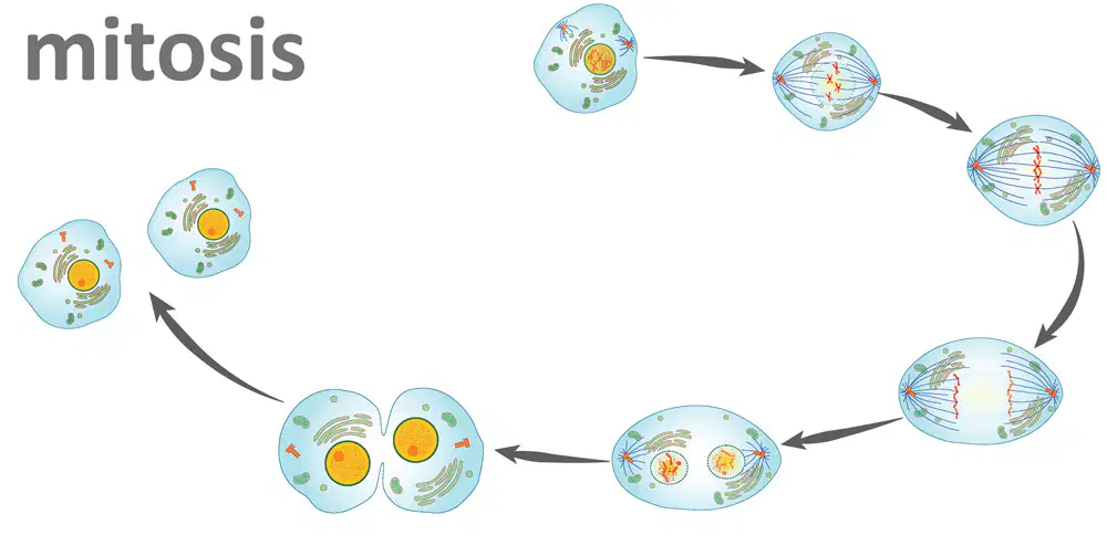
Put the stages of mitosis in order starting with Interphase.
(Please Make Another Taco)
Interphase
Prophase
Metaphase
Anaphase
Telophase
Put the stages of mitosis in order starting with Interphase.
Interphase
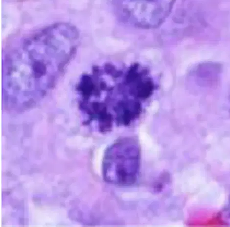
Prophase
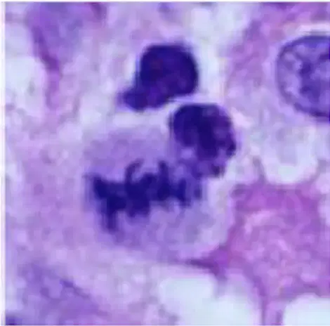
Metaphase
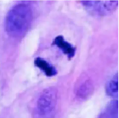
Anaphase
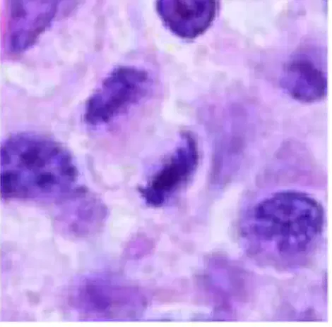
Telophase
Put the stages of mitosis in order starting with Interphase.
Interphase
Match the following Stages of Mitosis
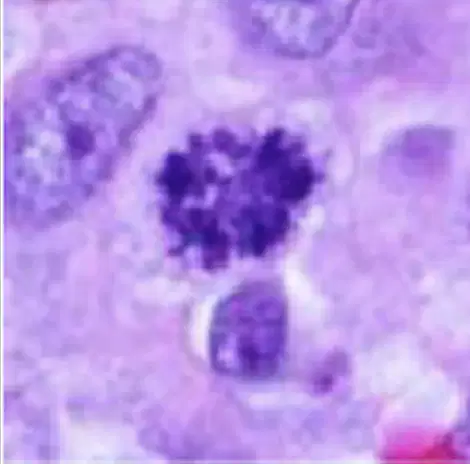
Prophase

Metaphase
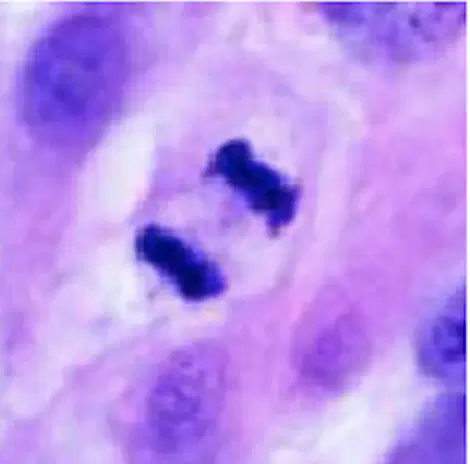
Anaphase
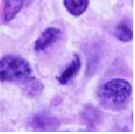
Telophase

What stage of mitosis?
Prophase
Telophase

What stage of mitosis?
Prophase
Telophase

What stage of mitosis?
Prophase
Telophase
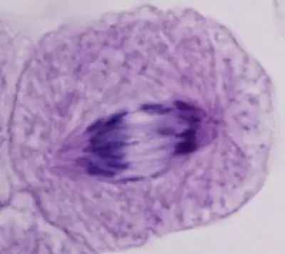
What stage of mitosis?
Prophase
Telophase

What stage of mitosis?
Prophase
Telophase

What stage of mitosis?
Prophase
Telophase

What stage of mitosis?
Prophase
Telophase

