
 Worksheets
A&P 1 Unit 4
Total questions: 162
Worksheet time: 3hrs 32mins
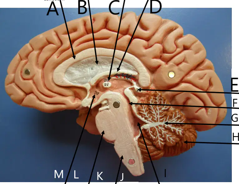
Name the brain structure labeled A:
(a)
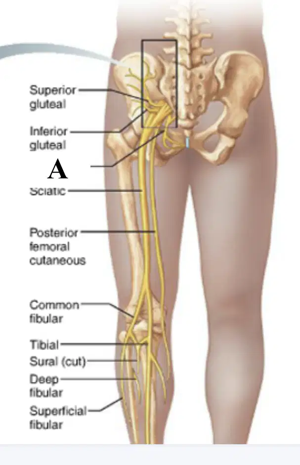
Name the missing nerve structure labeled A:
(a)

Name the missing nerve structure labeled ?:
(a)
Name the missing nerve structure labeled ?:
(a)

Name the sheep brain structure labeled A:
(a)

Name the sheep brain structure labeled A:
(a)
Name the sheep brain structure labeled B:
(a)
Name the sheep brain structure labeled C:
(a)
Name the sheep brain structure labeled D:
(a)
Name the sheep brain structure labeled E:
(a)
Name the sheep brain structure labeled B:
(a)
Name the sheep brain structure labeled C:
(a)
Name the sheep brain structure labeled D:
(a)
Name the sheep brain structure labeled E:
(a)
Name the sheep brain structure labeled F:
(a)
Name the sheep brain structure labeled G:
(a)
Name the sheep brain structure labeled H:
(a)
Name the sheep brain structure labeled I:
(a)
Name the sheep brain structure labeled J:
(a)
Name the sheep brain structure labeled K:
(a)
Name the sheep brain structure labeled L:
(a)
Name the sheep brain structure labeled E:
(a)
Name the sheep brain structure labeled B:
(a)
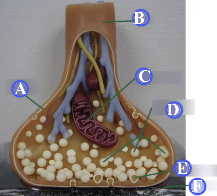
Name the synapse structure labeled A:
(a)

Name the nerve structure labeled A:
(a)
Name the nerve structure labeled B:
(a)
Name the nerve structure labeled C:
(a)
Name the nerve structure labeled D:
(a)
Name the nerve structure labeled E:
(a)
Name the nerve structure labeled F:
(a)
Name the nerve structure labeled G:
(a)
Name the nerve structure labeled H:
(a)
Name the nerve structure labeled I:
(a)
Name the nerve structure labeled J:
(a)
Name the nerve structure labeled K:
(a)
Name the synapse structure labeled B:
(a)
Name the synapse structure labeled C:
(a)
Name the synapse structure labeled D:
(a)
Name the synapse structure labeled E:
(a)
Name the synapse structure labeled F:
(a)
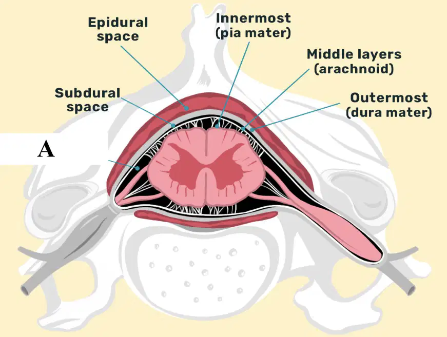
Name the meninges structure labeled A:
(a)
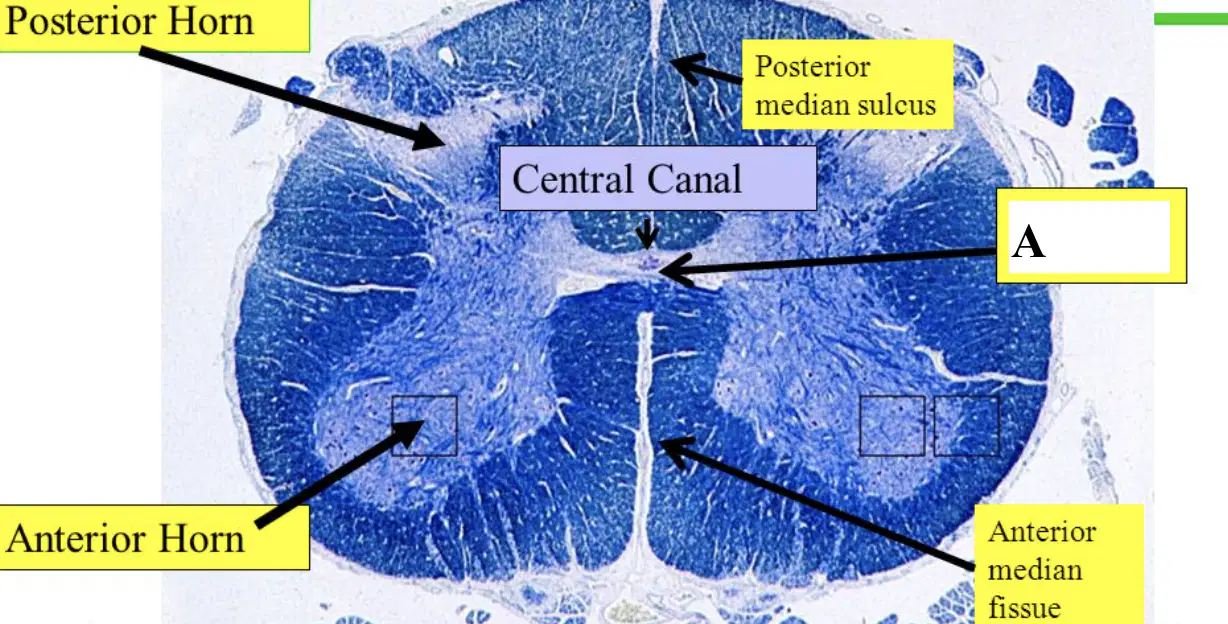
Name the meninges structure labeled A:
(a)
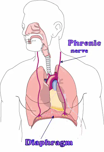
Which ventral rami give rise to the labeled nerve on this figure?
(a)
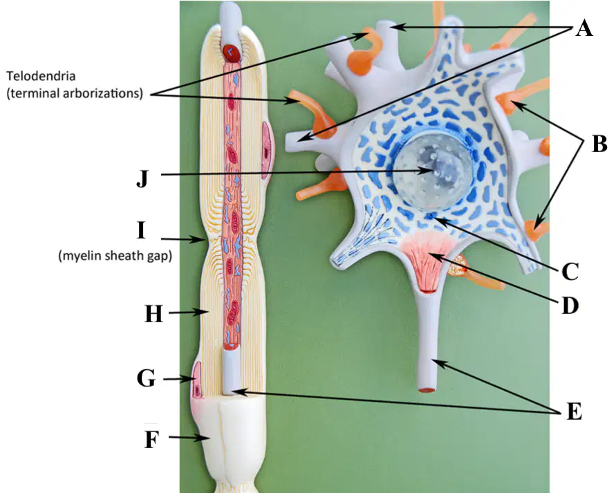
Name the neuron structure labeled A:
(a)
Name the neuron structure labeled B:
(a)
Name the neuron structure labeled C:
(a)
Name the neuron structure labeled D:
(a)
Name the neuron structure labeled E:
(a)
Name the neuron structure labeled F:
(a)
Name the neuron structure labeled G:
(a)
Name the neuron structure labeled H:
(a)
Name the neuron structure labeled I:
(a)
Name the neuron structure labeled J:
(a)
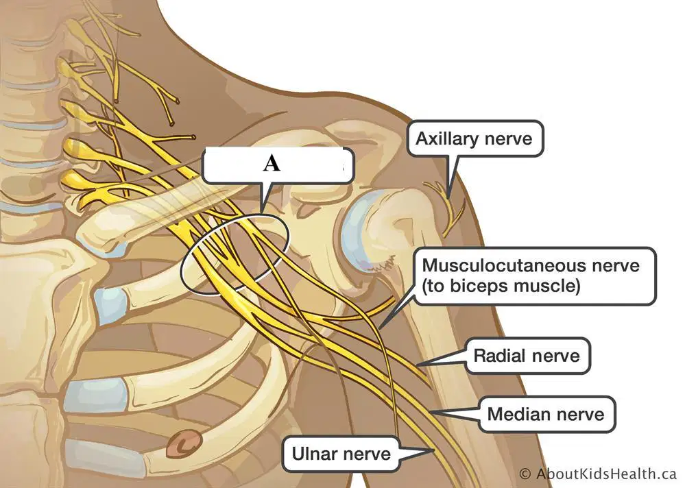
Label A is a stucture comprised of a set of nerves. It is called the:
(a)
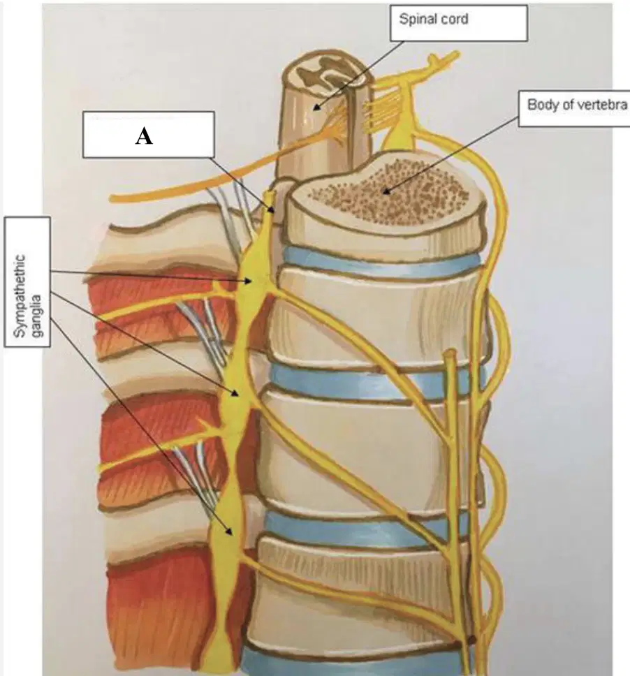
Label A is s called the:
(a)
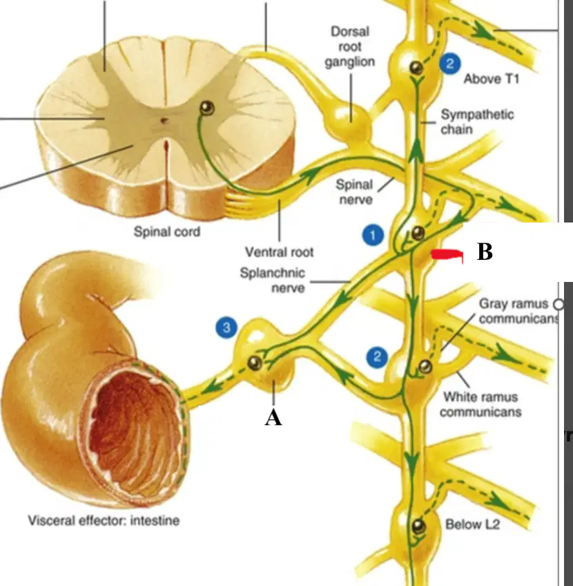
Label A is s called the:
(a)
Label A is a stucture comprised of a set of nerves. It is called the:
(a)
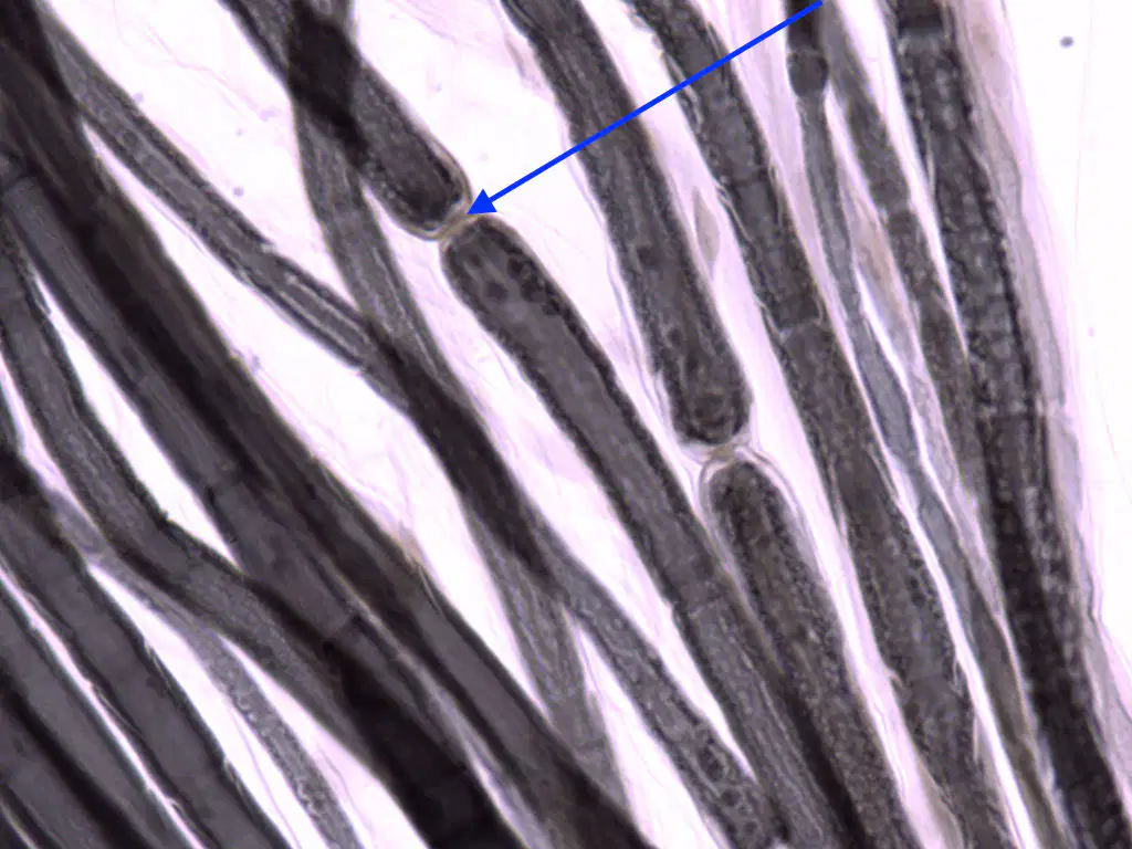
The pointer of the microscope is depicting what structure?
(a)
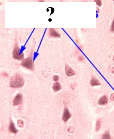
The pointer of the microscope is depicting what structure?
(a)

What type of neurons are being displayed?
(a)

What structures are being depicted by the purple clusters in the slide?
(a)
What is the function of a glial cell?
secrete & circulate CSF in the PNS
Send electrical impulses from the spine to the brain
secrete & circulate CSF in the CNS
Send electrical impulses from the brain to the spine
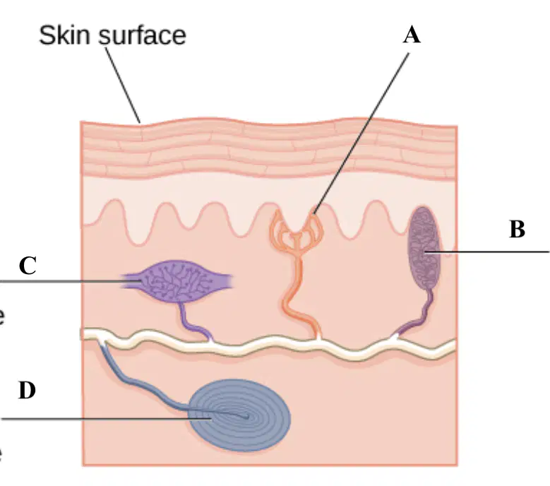
What is the sensory receptor that responds to light touch and allows for two-point discrimination, also labeled A?
(a)
What is the sensory receptor that relates to continuous touch or pressure or stretch of the skin, also labeled C?
(a)
What is the sensory receptor that relates to Deep cutaneous pressure/vibration, also labeled D?
(a)

The blue arrow is pointing at what brain structure?
(a)
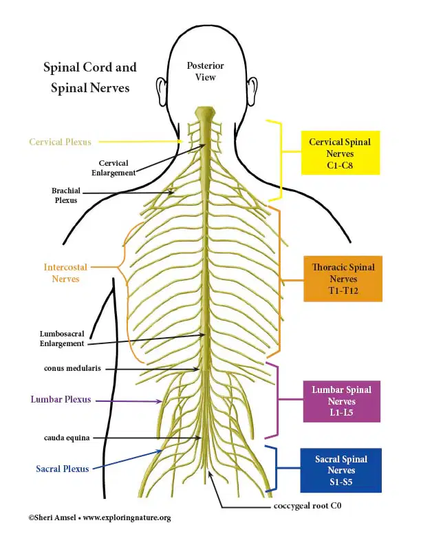
Be able to identify and count the spinal nerve if asked. For example (Cervical Nerve #3 could be an answer)
Thanks Pham
I will write out the name and number
I better get this right
I can count 1-10
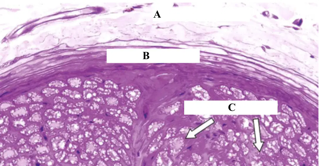
The slide shown is of a peripheral nerve. Which of the following correctly identifies each label?
A = epineurium
B = endoneurium
c = perineurium
A = perineurium
B = endoneurium
c = epineurium
A = epineurium
B = perineurium
c = endoneurium
A = endoneurium
B = perineurium
c = epineurium

Choose the answer that has each of the arm nerves correctly labeled:
A = Axillary Nerve
B = Musculocutaneous Nerve
c = Radial Nerve
d = Median Nerve
E = Ulnar Nerve
A = Axillary Nerve
B = Radial Nerve
C = Musculocutaneous Nerve
d = Median Nerve
E = Ulnar Nerve
A = Radial Nerve
B = Axillary Nerve
C = Musculocutaneous Nerve
d = Median Nerve
E = Ulnar Nerve
A = Axillary Nerve
B = Musculocutaneous Nerve
c = Median Nerve
d = Radial Nerve
E = Ulnar Nerve
A = Axillary Nerve
B = Musculocutaneous Nerve
c = Median Nerve
d = Ulnar Nerve
E = Radial Nerve
Be able to identify and count the spinal nerve if asked. For example (Cervical Nerve #3 could be an answer)
Thanks Pham
I will write out the name and number
I better get this right
I can count 1-10
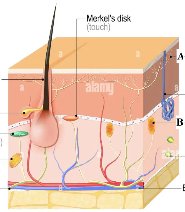
What is the function of the structure labeled A?
pain
light touch
pressure
skin stretching

For Label J, what is the function of the nerve associated?
Taste
Smell
Sight
Chewing
What is the function of the structure labeled A?
pain
light touch
pressure
skin stretching

Which of the following correctly labels the slide shown in the microscope:
A = Posterior Gray Horn
B = Anterior Median Fissure
C= White Columns/lateral funiculus
D = Central Canal
A = Anterior Gray Horn
B = Anterior Median Fissure
C= White Columns/lateral funiculus
D = Central Canal
A = Posterior Gray Horn
B = White Columns/lateral funiculus
C= Anterior Median Fissure
D = Central Canal
A =Anterior Gray Horn
B = White Columns/lateral funiculus
C= Anterior Median Fissure
D = Central Canal
A = Posterior Gray Horn
B = White Columns/lateral funiculus
C= Central Canal
D = Anterior Median Fissure
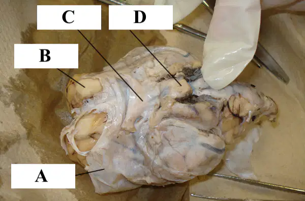
Name the brain structure labeled A:
(a)

Name the missing red structure:
(a)
Name the brain structure labeled B:
(a)
Name the brain structure labeled C:
(a)
Name the brain structure labeled D:
(a)
For Label A, name the correct nerve with its ,CN #:
(a)
For Label A, the following is correct that it is what type of nerve?
Sensory
Motor
Both
For Label B, name the correct nerve with its ,CN #:
(a)
For Label C, name the correct nerve with its ,CN #:
(a)
For Label C, name the correct nerve with its ,CN #:
(a)
For Label D, name the correct nerve with its ,CN #:
(a)
For Label D, is this a motor or sensory nerve?
Motor
Sensory
For Label E, name the correct nerve with its ,CN #:
(a)
For Label F, name the correct nerve with its ,CN #:
(a)
For Label G, name the correct nerve with its ,CN #:
(a)
For Label L, name the correct nerve with its ,CN #:
(a)
For Label K, name the correct nerve with its ,CN #:
(a)
For Label J, name the correct nerve with its ,CN #:
(a)
For Label J, name the correct nerve with its ,CN #:
(a)
For Label I, name the correct nerve with its ,CN #:
(a)
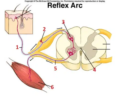
For the reflex arc, identify label 6
(a)
For Label H, name the correct nerve with its ,CN #:
(a)
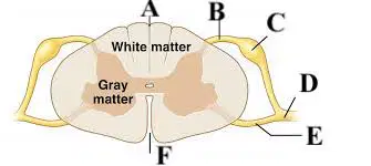
Name the brain structure labeled A:
(a)
Name the brain structure labeled B:
(a)
Name the brain structure labeled C:
(a)
Name the brain structure labeled D:
(a)
Name the brain structure labeled E:
(a)
Name the brain structure labeled F:
(a)
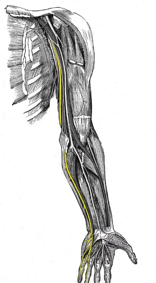
The highlighted yellow nerve is exhibiting which arm nerve?
(a)
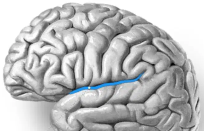
The highlighted blue brain feature is known as the
(a)

Name the brain structure colored yellow:
(a)
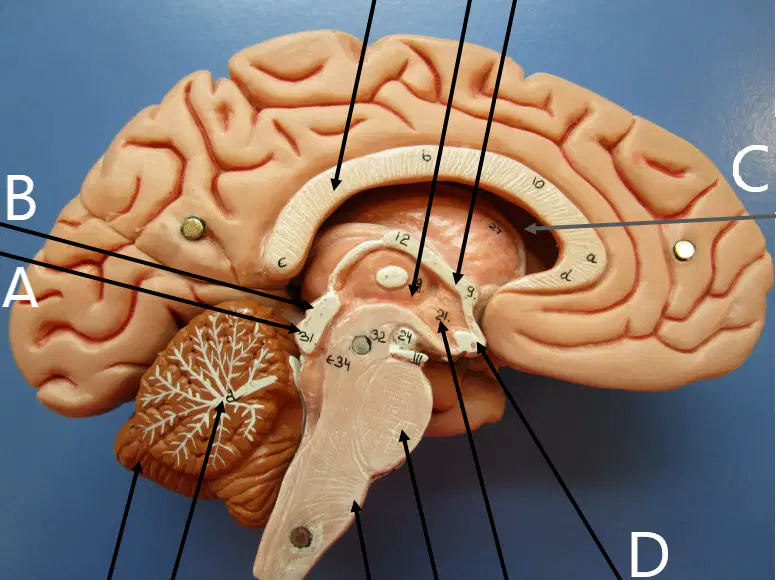
Name the brain structure labeled A:
(a)
Name the brain structure labeled C:
(a)
Name the brain structure labeled B:
(a)
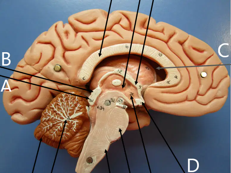
Name the brain structure labeled D:
(a)
Name the brain structure labeled C:
(a)
Name the brain structure labeled D:
(a)
Name the brain structure labeled E:
(a)
Name the brain structure labeled F:
(a)
Name the brain structure labeled G:
(a)
Name the brain structure labeled H:
(a)
Name the brain structure labeled I:
(a)

Name the missing brain structure labeled ?:
(a)

Name the missing nerve structure labeled ?:
(a)
Name the muscle(s) that is/are innervated by the missing nerve structure labeled ?:
biceps femoris
brachialis
brachioradialis
triceps brachii
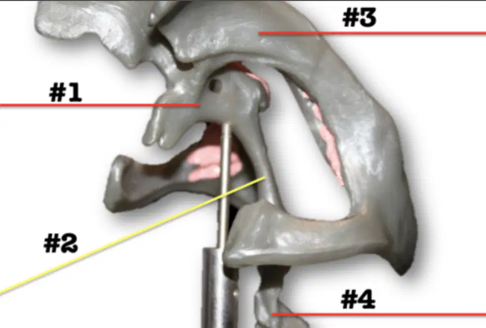
Where did the CSF in #1 come from directly before?
interventricular foramen
cerebral aqueduct
3rd ventricle
lateral ventricle
Which of the following correctly identifies label 2?
interventricular foramen
cerebral aqueduct
3rd ventricle
lateral ventricle
Name a skin region innervated by the missing nerve structure labeled ?:
dorsum of foot
posterior leg
anterior leg
plantar side of foot
Name the missing brain structure labeled ?:
(a)
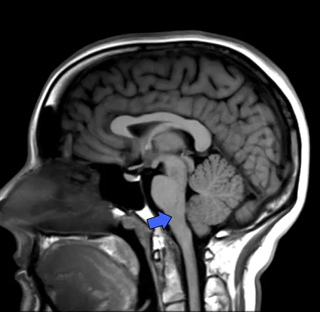
Name the structure in the MRI:
(a)
Name the brain structure labeled J:
(a)
Name the brain structure labeled K:
(a)
Name the brain structure labeled L:
(a)
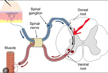
The red arrow is pointing to what structure
interneuron
motor neuron
synaptic neuron
sensory neuron
Reorder the following meninge layers from superficial to deep (1= superficial, 3 = deep)
1 = Dura Mater
2 = Arachnoid Mater
3 = Pia Mater
1 = Arachnoid Mater
2 = Dura Mater
3 = Pia Mater
1 = Arachnoid Mater
2 = Pia Mater
3 = Dura Mater
1 = Pia Mater
2 = Arachnoid Mater
3 = Dura Mater
The decreased ability to create facial expressions and abduct the eyeball indicates damage to which CN's?
V, III
VII, III
VII, VI
V, VI
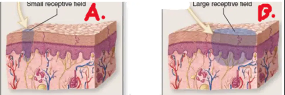
Which receptive field is located in your fingertips?
A) Small Receptive Field
B) Large Receptive Field
Neither
Both
A receptive field that has very few mechanoreceptors with very large receptive fields. You would expect to find this type of receptive field in the:
Fingertips
Back
Lips
Hand

Which of the following correctly corresponds to the colored brain?
orange - frontal lobe
red - precentral gyrus
blue (right) - postcentral gyrus
blue (left) - parietal lobe
green - occipital lobe
orange - frontal lobe
red - precentral gyrus
blue (left) - postcentral gyrus
blue (right) - parietal lobe
green - occipital lobe
orange - frontal lobe
red - precentral gyrus
blue (left) - postcentral gyrus
blue (right) - temporal lobe
green - occipital lobe
orange - frontal lobe
red - postcentral gyrus
blue (left) - precentral gyrus
blue (right) - parietal lobe
green - occipital lobe

In infants, the normal response for the reflex shown here is called the (a) .

Which of the following nerves are involved in this reflex?
CN 4
CN 2
CN 5
CN 7
CN 2
The effector of the reflex shown is the
retina
smooth muscle of the iris
optic nerve
extrinsic ocular muscles
Whiat is the name of reflex being tested?
(a)

Which nerves are involved in the shown reflex?
CN V
CN IV
CN VI
CN VII
CN IX
What is the name of the reflex being tested in the image, which causes you to blink?
(a)

What is the name of the reflex being tested in the image, which causes plantar flexion?
(a)
Which nerve(s) can be tested by the shown reflex test?
(a)

What is the name of the reflex being tested in the image, which causes leg extension?
(a)
What is the name of the nerve being tested in the image, which causes leg extension?
(a)
what is the name of the nerve involved in the image's test?
(a)
What is the name of the reflex being tested in the image, which causes you to blink?
(a)

Which of the following identifies the correct reflex shown and the corresponding cranial nerves involved?
Consensual reflex
CN 2 and 3
Gag Reflex
CN IX and X
Gag Reflex
CN V and X
Salivary Reflex
VII and IX
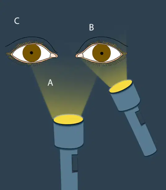
The (a) reflex is the change in pupil size in the eye opposite to the eye to which the light is directed
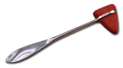
Which of the following reflexes can be tested using this device?
Patellar
Calcaneal
Crossed Extensor
Plantar

Which peripheral nerve is involved in the labeled reflex?
median nerve
axillary nerve
musculocutaneous nerve
radial nerve

You used calipers to test and measure which tactile phenomenon?
(a)
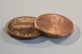
Pennies can be used to test and measure which tactile phenomenon?
(a)
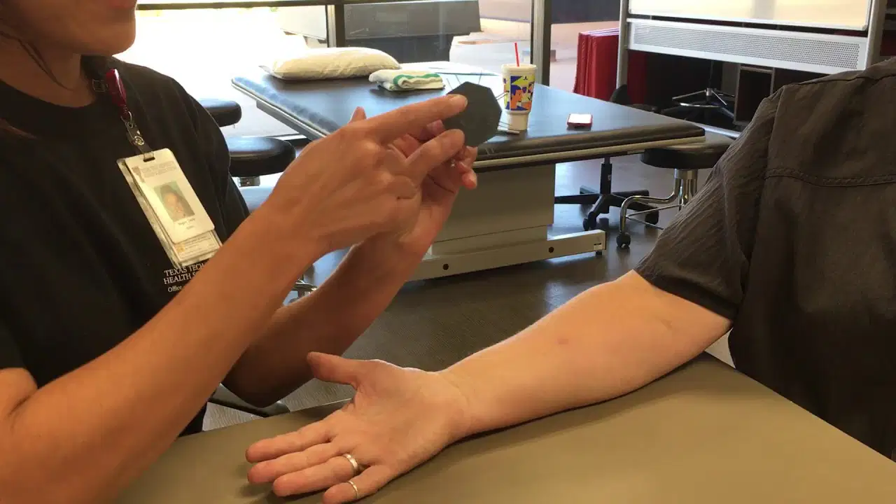
You used fine-point markers to test and measure which sensory phenomenon?
(a)
You used calipers to test and measure which tactile phenomenon?
(a)

Placing your hands in bowls with hot, cold, and room temperature water tested what?
(a)
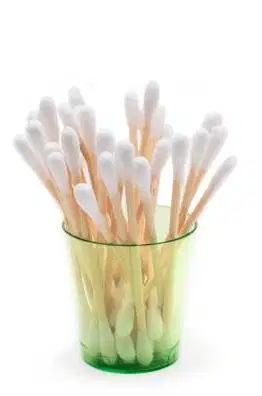
Which of the following reflex can be tested using this device?
Patellar
Consensual
Plantar
Crossed Extensor
Corneal
The normal effect of the sympathetic nervous system on the bronchioles of the lungs is:
Dilation of the bronchioles
Constriction of the bronchioles
No effect
The normal effect of the parasympathetic nervous system on heart rate is:
increase
decrease
no effect
The normal effect of the parasympathetic nervous system on the iris of the eye is:
Blinking
Dilation of pupil
Constriction of pupil
The normal effect of the sympathetic nervous system on the iris of the eye is:
Blinking
Dilation of pupil
Constriction of pupil
When a provider asks the patient to stick out their tongue, which cranial nerve is being tested?
Facial Nerve, CN VII
Hypoglossal, CN XII
Trigeminal, V
Vagal Nerve, CN X
What is the origin/course for label C?
facial canal to stylomastoid
midbrain to eye muscle
forebrain to nares
midbrain to mammillary bodies
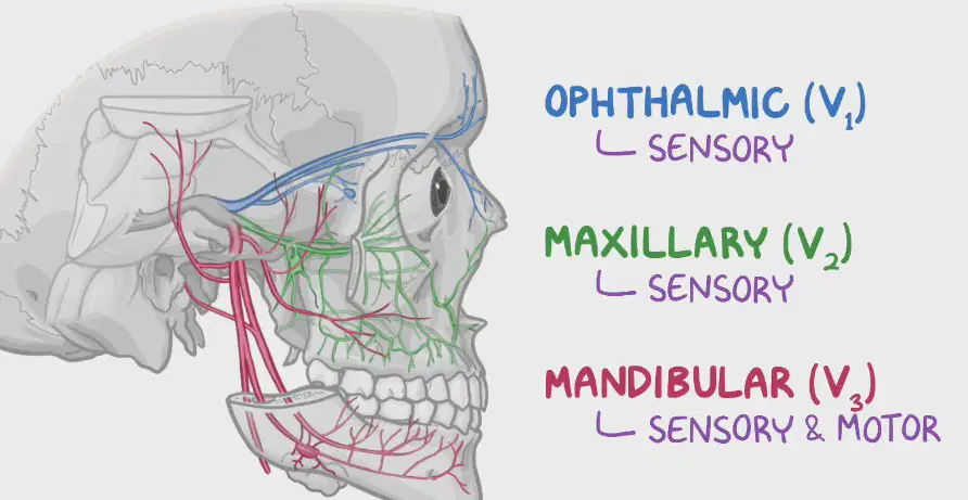
Identify the correct nerve name and its motor function:
Trigeminal Nerve
Motor Fxn = Mastication
Trigeminal Nerve
Motor Fxn = Pupil Constriction
Hypoglossal Nerve
Motor Fxn = Pupil Dilation
Hypoglossal Nerve
Motor Fxn = Mastication

