
 Worksheets
Science microscope
Total questions: 43
Worksheet time: 25mins

It controls the passing of light in the opening of the stage.
diaphragm
base
eyepiece/ocular lens
body tube

It supports the tube and connects it to the base.
eyepiece / ocular lens
arm
mirror
objective lenses

It holds the eyepiece and the revolving nosepiece in place.
pillar
draw tube
body tube
inclination joint

It allows you to look through and observe the enlarged image of the objective.
stage
ocular lens / eyepiece
condenser
mirror

It magnifies the image.
mirror
iris diaphragm
objective lenses
stage clip

It is used to provide light on the object and help focuses it.
coarse adjustment knob
mirror
base
fine adjustment knob

These are used to support and hold the slide in place.
arm and base
stage and stage clip
body tube
objective lenses

It moves the stage up and down and brings the image of the object into fine focus producing a clearer and more detailed view of the object.
coarse adjustment knob
fine adjustment knob
revolving / rotating nosepiece
objective lenses

It allows the proper position of the objectives lenses and the body tube holds the eyepiece.
dust shield
coarse adjustment knob
revolving nosepiece
body tube

The part above the base supports the other parts of the microscope.
arm
pillar
eyepiece
coarse adjustment knob

What is the name of this part?
base
leg
arm
eyepiece

What is the name of this part?
stageclips
leg
body tube
coarse adjustment knob

What is the name of this part?
objective lens
base
diaphragm
coarse adjustment knob

What is the name of this part?
arm
eyepiece/ocular lens
nosepiece
objective lens

What is the name of this part?
arm
eyepiece/ocular lens
nosepiece
objective lens

What is the name of this part?
stageclips
stage
nosepiece
slide

What is the name of this part?
stageclips
stage
objective lens
slide

What is the name of this part?
stageclips
stage
nosepiece
slide
What part of the microscope moves the stage up and down?
fine adjustment knob
coarse adjustment know
arm
Which of the following is the part of the microscope that illuminates the specimen?
Light Source
Diaphragm
Stage
Ocular
This flat platform holds the microscope slide up.
Arm
Diaphragm
Stage
Nose piece
This part of the microscope should be turned to clarify the image seen in the microscope when it is slightly out of focus.
Coarse adjustment knob
Fine adjustment knob
Stage clamp
Nose piece
This part of the microscope adjusts the amount of light shining up through the specimen to improve the image that you see.
Condenser
Diaphragm
Scanner
Low power objective lens
Allows you to change the objective that you are viewing the specimen through.
Body tube
Stage clamp
Stage
Nose piece
When switching to the 100X lens, what should you use?
few drops of immersion oil
low light intensity
wet cloth to clean the lens for clarity
use it with a 15X eyepiece only
A 100X lens is also known as _________.
scanning lens
low power lens
high power lens
immersion oil lens
When cleaning immersion oil from a 100X lens, what should be used?
damp, soft cloth
tissue paper
dry paper towel
specialized lens paper
Calculate the following total magnification: 5X eyepiece with a 10X objective.
50X total magnification
5X total magnification
100X total magnification
15X total magnification
The __________________ lens of the microscope would show you the MOST zoomed-in image of the specimen.
low power objective
medium power objective
high power objective
scanning
usually has 4x magnifications and can be identified by a red strip band around its perimeter of the lens it is designed for getting your bearings right before moving into low power lens
high power lens
scanning lens
low power lens
oil immersion lens
usually has 10x or 20 x magnifications and can be identified by a yellow strip band around its perimeter of the lens .the objective is useful for viewing prepared specimens on slides, it will achieve somewhere around 100 to 200x for a 10x 20 x eyepiece respectively .the sort magnification is great in viewing small specimens like fly eggs, onion skin brine shrimps hair
low power lens
high pawer lens
oil immersion lens
scanning lens
it is ideal for observing the small details within a specimen sample. The total magnification for this lens is equal to 400x magnification (10x eyepiece lens x the 40x objective equals 400).identifiable by a blue strip,it is used in looking at small details like germs moulids cell
low power lens
oil immersion lens
scanning lens
high power lens
This objective lens will achieve the greatest magnification and has a total magnification of 1000x (10x eyepiece lens x the 100x objective equals 1000). However, since the refractive index of air and the glass slide are slightly different, a special oil must be used to help fill the gap between the two. Without a drop of oil, the objective lens will not work properly and you will not achieve the desired magnification and resolution.it is identifiable with a white strip on its lens
high power lens
low power lens
oil immersion
scanning lens
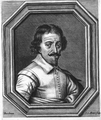
In 1590 he is the one who produced the first compound microscope with the help of his father Hans Janssen
(a)

The Jansen Microscope could magnify images up to..
(a)

in 1625 he invented one of the first compound microscope. His microscope composed of more than one lens
(a)
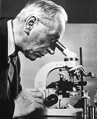
1932 he invented the phase
-contrast microscope that allowed for the study of colorless and transparent biological materials
(a)
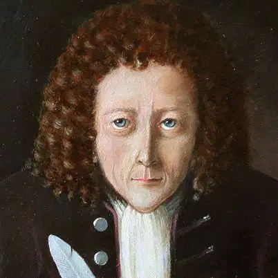
He discovered the cell using a microscope in the year 1665.He Examined a very Thin slice of cork, he thought the close up look resembled small, empty rooms
(a)

Name this Microscope that was invented in year 1670
(a)
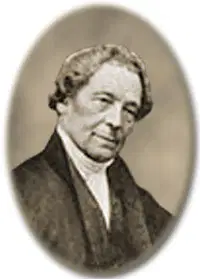
In 1830 He was credited for his prototype of the compound microscope
(a)
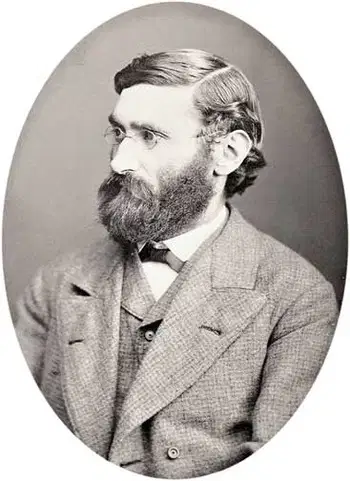
In 1872 he provided the calculations that allowed for the maximum resolution in microscope possible.
(a)
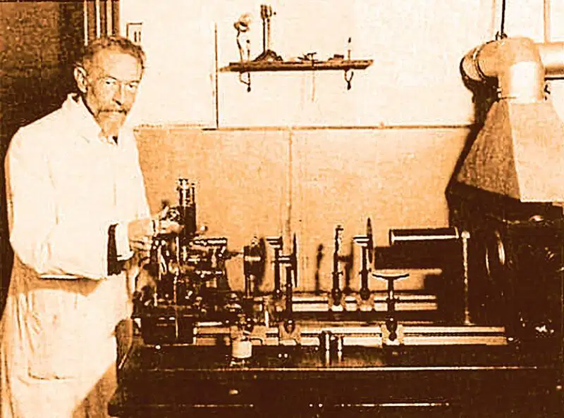
In 1903, he developed the ultra- microscope that could study objects below wavelength of light
(a)

In 1674 He build a simple microscope with only one lens to examine blood, yeast insects and other tiny objects .
He is called "the Father of Microbiology."
(a)

