
 Worksheets
Human Anatomy Fall Semester Review
Total questions: 176
Worksheet time: 2hrs 6mins
Basic units of life
Cells
Tissues
Organ
Cell Differentiation
A group of similar cells that perform a specific function.
Cells
Tissues
Organ
Cell Differentiation
A group of similar tissues that perform a specific function.
Cells
Tissues
Organ
Cell Differentiation
A group of organs that work together to perform vital body functions.
Cells
Tissues
Organ
Organ System
An individual living thing, made up of one or many cells, and possibly tissues, organs and organ systems, that is capable of growing and reproducing.
Cells
Tissues
Organ
Organism
Match the following
skeletal
framework of bones that protects organs
respiratory
organs that bring oxygen and remove CO2
circulatory
heart, vessels circulate blood
cells
smallest unit of life
tissue
group similar cells - perform function
Match the following
digestive
breaks down food
brain
processes information
stimulus
change in environment NS reacts
respiratory
oxygen
circulatory
delivers blood
Match the following
excretory
rid of waste
cross section
thin slice of organ or tissue
nerves
bundle of cells
sense receptor
neurons that detect stimuli
respiratory
oxygen
What are the major parts of the circulatory system? (a)
What are their functions? (b)
Which system of the human body provides protection to most of the major organs?
A. skeletal system
B. circulatory system
C. respiratory system
D. muscular system
Which are parts of the digestive system?
A. mouth, lungs
B. mouth, teeth
C. mouth, brain
In which system of the human body does the heart belong?
A. skeletal system
B. respiratory system
C. cardiovascular system
Which body system’s main purpose is to help the body move?
A. digestive system
B. muscular system
C. respiratory system
Which body system sends messages to the brain?
A. nervous
B. muscular
C. circulatory
Which body system includes the lungs?
A. respiratory system
B. digestive system
C. skeletal system
is the maintenance of a relatively stable internal environment
is the maintenance of a relatively stable external environment
During recess, Renee scraped her knee, so the nurse put a bandage on her skin. Which of the functions below that are normally provided by skin is now being provided by the bandage?
Preventing dehydration
Regulating body temperature
Keeping bacteria out of the body
Sensing when her skin has been touched
.The study of body structure is _______________.
anatomy
biology
physiology
pathophysiology
The study of body function is _______________.
anatomy
physiology
biology
pathology
The ________ separates the thoracic cavity from the abdominal cavity.
A. diaphragm
A. pericardial cavity
A. mediastinum
A. pelvic cavity
The cavity below the abdominal cavity is the __________.
A. dorsal cavity
A. thoracic cavity
A. ventral
A. pelvic cavity
What is the term for standing straight with palms forward and feet together?
Anterior
Posterior
Superior
Anatomical Position
Which term refers to the direction towards the front?
Anterior
Posterior
Superior
Inferior
What is the term for the direction towards the back?
Anterior
Posterior
Superior
Inferior
Which term describes the direction towards the head?
Superior
Inferior
Medial
Lateral
What is the term for the direction away from the head?
Superior
Inferior
Medial
Lateral
Which term refers to the direction towards the midline?
Medial
Lateral
Proximal
Distal
What is the term for the direction away from the midline?
Medial
Lateral
Proximal
Distal
Which term describes being closer to the point of attachment?
Proximal
Distal
Superficial
Deep
What is the term for being away from the point of attachment?
Proximal
Distal
Superficial
Deep
Which term refers to being near the surface?
Proximal
Distal
Superficial
Deep
What is the term for being away from the surface?
Proximal
Distal
Superficial
Deep
An anatomical section that separates the body into right and left portions is a __________ section.
frontal
transverse
coronal
sagittal

This plane divides the body into superior and inferior parts.
coronal plane
frontal plane
transverse plane
sagittal plane
_______ means towards the side of the body.
distal
proximal
medial
lateral

Cranial Cavity
Pleural Cavity
Pericardial Cavity
Vertebral Canal

The cavity surrounded by a box is?
Abdominal Cavity
Pelvic Cavity
Pleural Cavity
Cranial Cavity

The cavity surrounded by a box is?
Cranial Cavity
Abdominal Cavity
Pelvic Cavity
Abdominopelvic Cavity

The cavity surrounded by a box is?
Abdominal Cavity
Pelvic Cavity
Thoracic Cavity
Pericardial Cavity

The cavity surrounded by a box is?
Pericardial Cavity
Pleural Cavity
Abdominal Cavity
Pelvic Cavity

The cavity surrounded by a box is?
Thoracic Cavity
Abdominal Cavity
Pelvic Cavity
Vertebral Canal


How do we classify epithelial tissue?
Shape
Shape and number of layers
Counting the number of individual cells
Squamous
What are the four types of tissues in the human body? (Select all that apply)
Epithelial
Connective
Muscle
Nervous
What does pseudostratified mean?
Many layers of cells
Single layer that looks like many layers of cells
Single layer of cells
No layer of cells
What are the three types of muscle tissue?
cardiac
smooth
skeletal
biceps femoris

Select the correct tissue type.
simple columnar epithelium
stratified squamous epithelium
stratified columnar epithelium
transitional epithelium

Select the correct tissue type.
simple columnar epithelium
stratified cuboidal epithelium
stratified columnar epithelium
transitional epithelium

Which type of muscle tissue is shown here?
cardiac
skeletal
smooth
What is Histology mean?
Study of cells
Study of life and living organisms
Study of tissues
The four types of tissue are
Cardiac, transitional, epithelial, connective
Muscle, Nervous, Epithelial, Connective
Connective, Epithelial, Transitional, Muscle
Epithelial, Connective, Muscle, Squamous
This type of tissue is composed of neurons and neuroglia.
muscular
nervous
connective
epithelial
Which tissue is a continuous sheet of tightly packed cells, covers body surfaces, lines organs, and forms glands?
muscular
nervous
connective
epithelial

Squamous

Cuboidal

Columnar
This tissue has a free apical surface and is attached to a basement membrane.
muscular
nervous
connective
epithelial
I am a single layer of cube-like cells that are found in the tubules of the kidneys; my job includes excretion and absorption.
Simple columnar epithelium
Simple cuboidal epithelium
Stratified squamous epithelium
Stratified columnar epithelium
I am a single layer of tall cells with round or oval nuclei; I can be found in the linking of the digestive tract and my job includes absorption and secretion of enzymes
Simple Columnar Epithelium
Stratified Columnar Epithelium
Simple Squamous Epithelium
Pseudostratified Columnar Epithelium
I am arranged in several layers like stacks of cubes. I can be found in the sweat and salivary ducts of humans and my purpose is to protect!
Simple Cuboidal
Simple Columnar
Stratified Columnar
Stratified Cuboidal
I am a solid calcified matrix containing many collagen fibers. I support and protect! I provide levers for the muscles to act on; I store calcium and other minerals and fat. I have marrow inside for blood formation.
blood
loose connective tissue
dense connective tissue; tendons
bone
Parallel collagen fibers that are densely packed together. I am great for attaching muscles to bone. I also attach bone to bone. I can work under stressful conditions when pulling and pushing for movement.
Bone
loose connective tissue
dense connective tissue
blood
Long, cylindrical multinucleated cells; obvious striations. Voluntary movement locomotion. I am attached to bones and occasionally skin
Smooth muscle
Skeletal muscle
Cardiac muscle
Neurons
Branching, striated, uninucleated cells that are specialized. As I contract I propel blood into the circulatory system.
Smooth muscle
Skeletal muscle
Cardiac muscle
Neurons
What are the three main layers of the skin?
epidermis, dermis, and mesoderm.
epidermis, dermis, and subcutis.
epidermis, dermis, and hypodermis
epidermis, dermis, and adipose tissue.
Why are sebaceous glands important for the skin?
Sebaceous glands produce melanin to moisturize and protect the skin.
Sebaceous glands produce collagen to moisturize and protect the skin.
Sebaceous glands produce sweat to moisturize and protect the skin.
Sebaceous glands produce sebum to moisturize and protect the skin.
What are the two main functions of sweat glands?
Regulate body temperature and excrete waste products
Produce hormones and regulate blood sugar levels.
Moisturize the skin and produce oil.
Produce tears and lubricate the eyes.
What is the role of the dermis in the skin?
The role of the dermis in the skin is to provide structural support and contain blood vessels, nerves, and hair follicles.
The dermis is primarily composed of dead skin cells.
The dermis is the outermost layer of the skin.
The dermis is responsible for producing sweat and oil glands.
Which layer of the skin contains blood vessels and nerves?
stratum corneum
hypodermis
dermis
epidermis
What is the outermost layer of the epidermis called?
stratum spinosum
stratum lucidum
stratum corneum
stratum granulosum
How do sebaceous glands help in maintaining skin health?
Sebaceous glands produce sebum, which moisturizes and protects the skin.
Sebaceous glands produce collagen, which moisturizes and protects the skin.
Sebaceous glands produce melanin, which moisturizes and protects the skin.
Sebaceous glands produce sweat, which moisturizes and protects the skin.
What is the primary function of eccrine sweat glands?
Produce oil for the skin.
Produce tears for the eyes.
Produce saliva for digestion.
Regulate body temperature
What are the two main components of the dermis?
papillary layer and reticular layer
stratum corneum and stratum basale
sebaceous glands and sweat glands
epidermis and hypodermis
At which location does hair divide?
hair follicle
hair cuticle
hair shaft
hair bulb
Which layer(s) of the skin are composed of fatty connective tissues? (multiselect)
epidermis
dermis
hypodermis
subcutaneous
Which of the following characteristics are NOT true of both hair and nails?
They are both made of keratin
They both protect our skin
They both originate at a matrix
They both have structures known as cuticles
A burn victim comes into the hospital but is not experiencing pain at the burn site. What is the lowest severity of burn this individual could have?
first-degree burn
second-degree burn
third-degree burn
fourth-degree burn
What is found between the dermis and epidermis?
Hypodermis
Dermal papillae
Blood vessels
Melanocytes
A burn victim comes into the hospital but is not experiencing pain at the burn site. What is the lowest severity of burn this individual could have?
first-degree burn
second-degree burn
third-degree burn
fourth-degree burn
Which of the following is NOT visible when looking at your fingernail?
nail matrix
lunula
proximal nail fold
nail body

What is number 2?
Dermis
Epidermis
Hypodermis/subcutaneous
Artery
What is number 3?
Dermis
Epidermis
Hypodermis/subcutaneous
Artery
Which of the following are functions of the bones?
Support and Protection
Attachment to the muscles
Stores inorganic salts
All of the above
The human skeleton is composed of the
axial
appendicular
axis
atlas
Your arm bones (Humerus, Radius, Ulna) are part of the _________ skeleton.
Axial
Appendicular
Which of the following bones is NOT part of the axial skeleton?
clavicle
frontal
vertebrae
sternum

A is _______ bone.
Compact
Spongey
G is _______ bone.
Compact
Spongey
Osteoclast
Breaks bone
Osteoblast
Builds bone
Ligament
Holds bones together
Joint
Where bones meet
Tendon
Attach muscle to bone
Reorder the following bones from top to bottom
Pelvic Bone
Femur
Patella
Tibia
Tarsals
During bony callus formation, what is fibrocartilaginous callus converted into?
Compact Bone
Bone Marrow
Spongy Bone
Red Blood Cells
During bone remodeling, what is the bony callus remodeled by?
Osteoclasts and Osteoblasts
Collagen Fibres
Osteoclasts
Osteoblasts
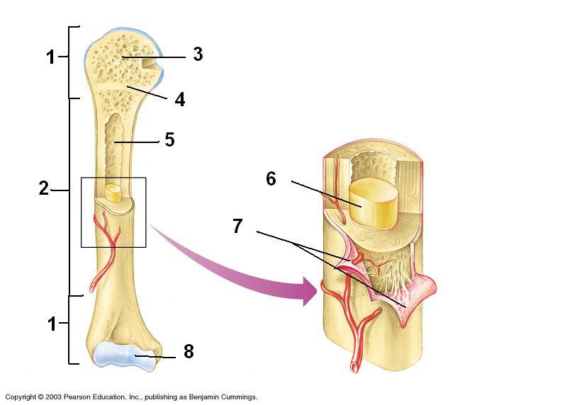
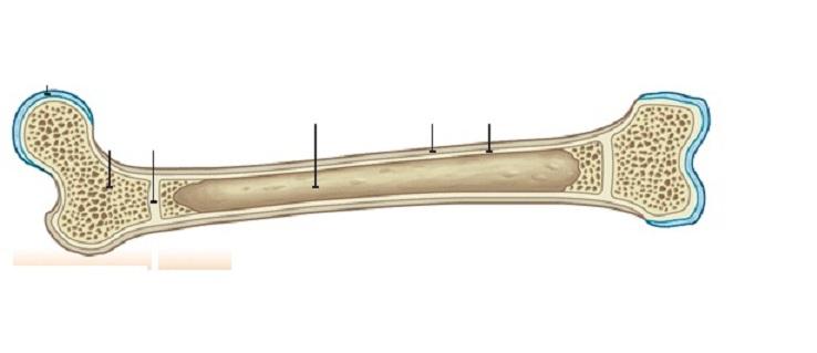
Where is spongy bone primarily found in long bones?
Diaphysis
Epiphysis
Periosteum
Endosteum
During Hematoma Formation, What happens at the site of the break?
Blood Clot or a Hematoma
Fibroblasts Produce Collagen Fibres
Osteoblasts Form Spongy Bone
Air gets to it and everything is fine
During this step of bone repair a strong, permanent "patch" is made based on mechanical stresses on the bone
hematoma forms
bony callus forms
fibrocartilage callus forms
bone remodeling
During fracture repair, the fibrocartilage callus is replaced with
compact bone
cartilage and collagen
spongy bone
compact bone and collagen
This structure acts as the body's natural splint for the broken bone
hematoma
fibrocartilage callus
bony callus
muscle tissue
This is the first event of bone repair
remodeling
hematoma forms
bony callus forms
fibrocartilage callus forms
This is the second step of fracture repair
hematoma forms
bony callus forms
fibrocartilage callus forms
bone remodeling

What do the bones make?
veins
cartilage
red blood cells
bone marrow

Identify #1
Frontal Bone
Parietal Bone
Sphenoid Bone
Ethmoid Bone
Lacrimal Bone
Identify #2
Frontal Bone
Parietal Bone
Sphenoid Bone
Ethmoid Bone
Lacrimal Bone

Identify #3
Frontal Bone
Parietal Bone
Sphenoid Bone
Ethmoid Bone
Lacrimal Bone
Identify #4
Frontal Bone
Parietal Bone
Sphenoid Bone
Ethmoid Bone
Lacrimal Bone

Identify #8
Nasal Turbinates
Vomer
Temporal Bone
Nasal Bone
Zygomatic Bone
Identify #9
Nasal Turbinates
Vomer
Temporal Bone
Nasal Bone
Zygomatic Bone
Identify #10
Nasal Turbinates
Vomer
Temporal Bone
Nasal Bone
Zygomatic Bone
Identify #11
Maxilla
Mandible
Teeth
Occipital Bone
Hyoid Bone
Identify #12
Maxilla
Mandible
Teeth
Occipital Bone
Hyoid Bone
Identify #14
Maxilla
Mandible
Teeth
Occipital Bone
Hyoid Bone
Which of these is NOT a function of the muscular system?
Allows for sensation
Creates body movement
Protects internal organs
Heat production
Maintain body posture and shape
What type of muscle makes of the walls of our internal organs?
smooth muscles
voluntary muscle
striated muscle
cardiac muscle
Point of origin refers to .......
the place where the muscles attach to the bone, but no movement occurs
the place where the muscles attach the and movement occurs
where a muscle attaches to another muscle
the membrane the covers and supports muscles
There are approximately how many individual muscles in the body?
(a)
Tendon
attaches a muscle to a bone
Ligament
attaches a bone to another bone
Fascia
creates a membrane-like covering for muscles
Named for it's size
gluteus minimus, gluteus medius
Named for their shapes
Deltoid, Rhomboid, Trapezius
Named for their locations
Vastus lateralis, vastus medialis, vastus intermedius
Named for the number of heads (origins)
Biceps brachii, triceps brachii,
Named for their functions
Adductors, flexors
Match the following
Origin
the point of a muscle which attaches to a stationary bone
Insertion
the point of a muscle which attaches to a bone that performs the movement
Belly
the largest part of the muscle containing the muscle cells
Think about the action of the triceps. Then label the origin, belly, and insertion.
Synergist
muscles working together in the same direction of motion
Prime Mover or agonist
a muscle that is most important in the creation of a specific action or movement
Antagonist
a muscle that opposes the action of another

smooth or visceral muscle

cardiac muscle

skeletal muscle
Which connective tissue surrounds fasicles?
epimysium
endomysium
perimysium
Which connective tissue surrounds the whole muscle?
epimysium
endomysium
perimysium
Which connective tissue surrounds individual muscle fibers?
epimysium
endomysium
perimysium
Actin myofilaments are
attached to the z disk
are found primarily in the h zone
are thicker than myosin myofilaments
all of these options
Which structure provides an attachment site for calcium?
actin
troponin
tropomyosin
m-line
Skeletal muscles...
Require energy in order to contract
Require energy in order to relax
Relax when calcium ions are transported into the sarcoplasmic reticulum
All of these

Which of the following puts the structures of the muscle in order from largest to smallest?
Whole muscle, muscle fiber, actin & myosin, myofibril, muscle fasicle
Whole muscle, muscle fasicle, muscle fiber, myofibril, actin & myosin
Whole muscle, muscle fiber, muscle fasicle, myofibril, actin & myosin
Whole muscle, muscle fasicle, muscle fiber, actin & myosin, myofibril

Identify the muscle labeled as #1
Latissimus Dorsi
Trapezius
Serratus Anterior
Vastus Medialis

Identify the muscle labeled as #2
Rectus Abdominis
Latissimus Dorsi
Trapezius
Semitendinosus

Identify the muscle labeled as #3
Biceps Brachii
Deltoid
Triceps Brachii
Brachioradialis

Identify the muscle labeled as #4
Biceps Femoris
Biceps Brachii
Brachioradialis
Brachialis

Identify the muscle labeled as #5
Triceps Brachii
Gastrocnemius
Semimembranosus
Biceps Femoris

Identify the muscle labeled #6
Vastus Lateralis
Biceps Femoris
Brachioradialis
Pectoralis Major

Identify the muscle labeled #7
Gluteus Maximus
Serratus Anterior
Pectoralis Major
Latissimus Dorsi

Identify the muscle labeled #9
Obliques
Serratus Anterior
Rectus Femoris
Rectus Abdominus

Identify the muscle labeled #10
Obliques
Trapezius
Semimembranosus
Rectus Femoris

Identify the muscle labeled as #11
Pectoralis Major
Gluteus Maximus
Latissimus Dorsi
Gastrocnemius

Identify the muscle labeled #12
Vastus Medialis
Rectus Femoris
Vastus Lateralis
Rectus Abdominus

Identify the muscle labeled #13
Rectus Femoris
Vastus Intermedius
Vastus Lateralis
Vastus Medialis

Identify the muscle labeled #14
Rectus Femoris
Vastus Intermedius
Vastus Lateralis
Vastus Medialis

Identify the muscle labeled #15
Biceps Femoris
Semimembranosus
Vastus Lateralis
Vastus Medius

Identify the muscle labeled #4
Soleus
Sartorius
Achilles Tendon
Gastrocnemius

Identify the muscle labeled #5
Soleus
Sartorius
Achilles Tendon
Gastrocnemius

Identify the muscle labeled as #6
Soleus
Sartorius
Achilles Tendon
Gastrocnemius
Identify the muscle labeled as #1
Latissimus Dorsi
Trapezius
Serratus Anterior
Vastus Medialis
Identify the muscle labeled as #2
Rectus Abdominis
Latissimus Dorsi
Trapezius
Semitendinosus
Identify the muscle labeled as #3
Biceps Brachii
Deltoid
Triceps Brachii
Brachioradialis
Identify the muscle labeled as #4
Biceps Femoris
Biceps Brachii
Brachioradialis
Brachialis
Identify the muscle labeled as #5
Triceps Brachii
Gastrocnemius
Semimembranosus
Biceps Femoris
Identify the muscle labeled #6
Vastus Lateralis
Biceps Femoris
Brachioradialis
Pectoralis Major
Identify the muscle labeled #7
Gluteus Maximus
Serratus Anterior
Pectoralis Major
Latissimus Dorsi
Identify the muscle labeled #9
Obliques
Serratus Anterior
Rectus Femoris
Rectus Abdominus
Identify the muscle labeled #10
Obliques
Trapezius
Semimembranosus
Rectus Femoris
Identify the muscle labeled as #11
Pectoralis Major
Gluteus Maximus
Latissimus Dorsi
Gastrocnemius
Identify the muscle labeled #12
Vastus Medialis
Rectus Femoris
Vastus Lateralis
Rectus Abdominus
Identify the muscle labeled #13
Rectus Femoris
Vastus Intermedius
Vastus Lateralis
Vastus Medialis
Identify the muscle labeled #14
Rectus Femoris
Vastus Intermedius
Vastus Lateralis
Vastus Medialis
Identify the muscle labeled #15
Biceps Femoris
Semimembranosus
Vastus Lateralis
Vastus Medius
Identify the muscle labeled #4
Soleus
Sartorius
Achilles Tendon
Gastrocnemius
Identify the muscle labeled #5
Soleus
Sartorius
Achilles Tendon
Gastrocnemius
Identify the muscle labeled as #6
Soleus
Sartorius
Achilles Tendon
Gastrocnemius

