
 Worksheets
HBS Semester 1 Review 2025
Total questions: 168
Worksheet time: 2hrs 19mins
What is the main function of bones in the human body?
Sensation, perception, and cognition.
Digestion, absorption, and elimination.
Support, protection, and structure
Movement, flexibility, and coordination.
Which type of bone cells are responsible for bone formation?
Osteocytes
Osteoblasts
Chondrocytes
Osteoclasts
What is the outer layer of a bone called?
periosteum
epiphysis
medullary cavity
endosteum
What is the inner, spongy part of a bone called?
compact bone
marrow
cortical bone
trabecular bone
What is the scientific term for the collarbone?
humerus
scapula
femur
clavicle
What is the purpose of bone marrow?
To produce and store blood cells
To produce and store fat cells.
To regulate body temperature.
To aid in digestion.
What are the two main types of bone tissue?
dense bone and porous bone
hard bone and soft bone
compact bone and spongy bone
solid bone and hollow bone
What are the three main types of bone fractures?
Closed fracture, Open fracture, Greenstick fracture
Avulsion fracture
Hairline fracture
Comminuted fracture
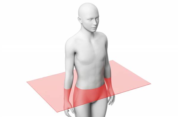
Identify the section or cut.
frontal section
lateral section
transverse section
sagittal section
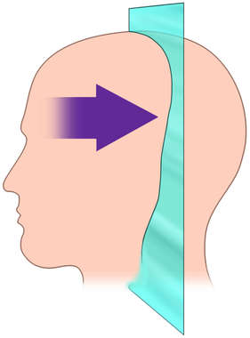
Identify the section or cut.
cross section
plane
midsagittal section
coronal section
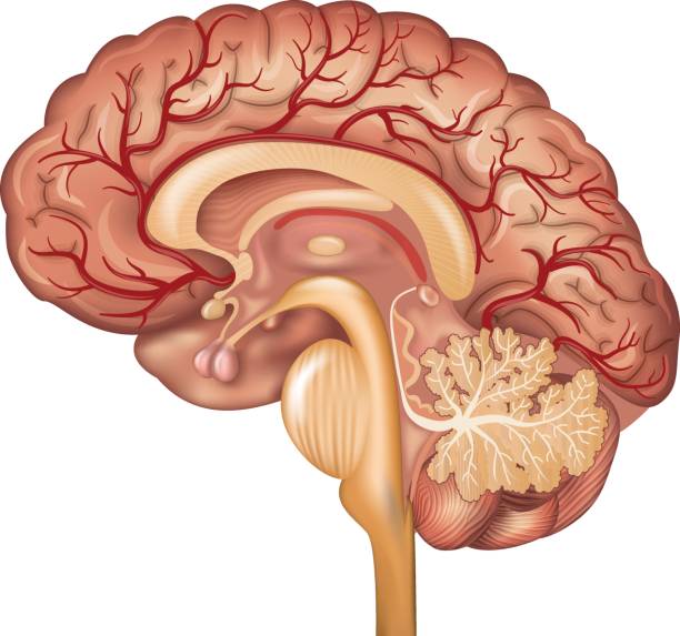
Identify the section or cut.
frontal section
cross section
transverse section
sagittal section
The shoulder is _____ to the elbow.
proximal
dorsal
distal
ventral
The cranial cavity is _____ to the thoracic cavity.
inferior
anterior
peripheral
superior
What is anatomical position?
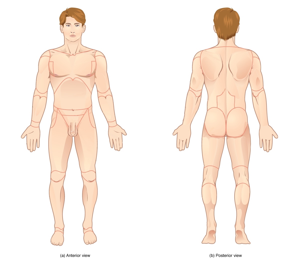
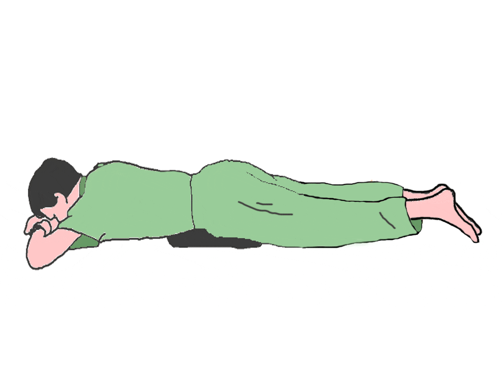
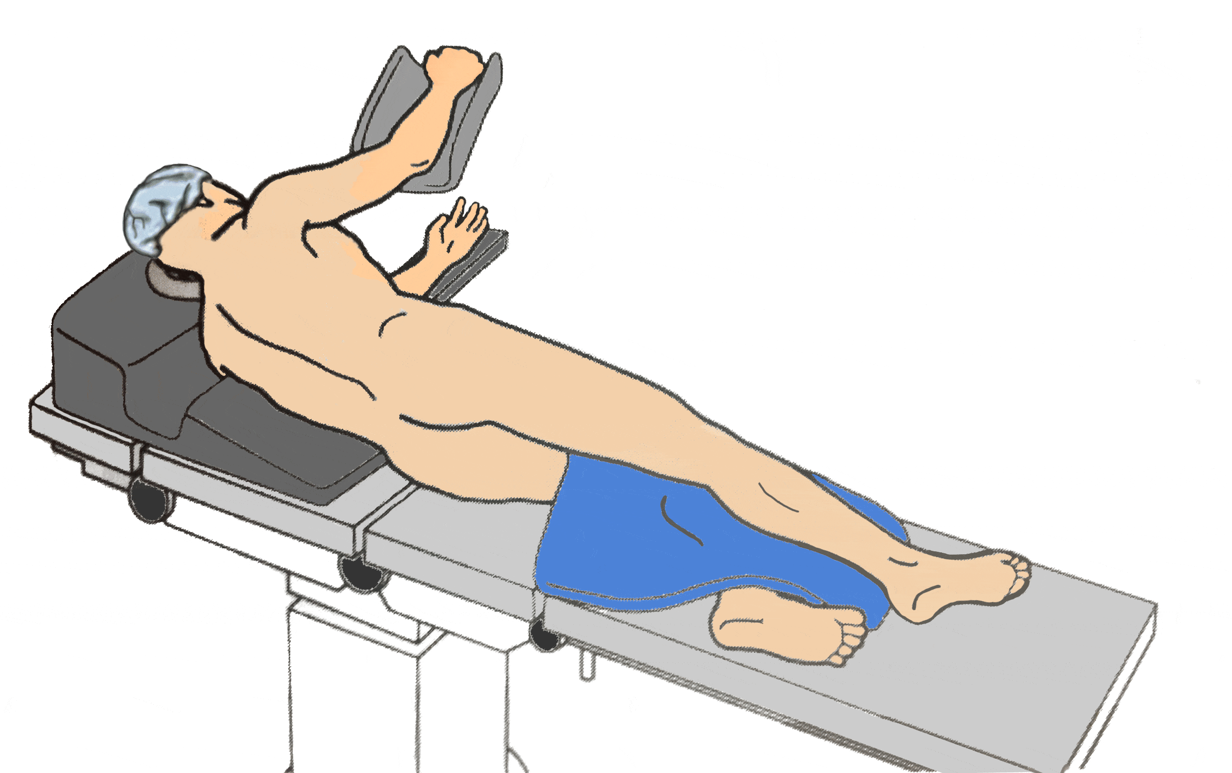
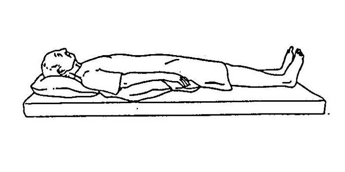
Which cavity includes the heart and lungs?
Thoracic cavity
Cranial cavity
Abdominal cavity
Retroperitoneal cavity
The pinky finger is ______ to the thumb in anatomical position.
posterior
superior
lateral
medial

[The eyes are _______ to the brain].

Match the following planes
Traverse
Separates top & bottom
Sagittal
Separates left & right
Coronal
Seperates front & back
Median
Even separation of left & right
Distal
Away from

The cavity surrounded by a box is?
Abdominal Cavity
Pelvic Cavity
Pleural Cavity
Cranial Cavity

Cranial Cavity
Pleural Cavity
Pericardial Cavity
Vertebral Canal

The cavity surrounded by a box is?
Abdominal Cavity
Pelvic Cavity
Thoracic Cavity
Pericardial Cavity

The cavity surrounded by a box is?
Pericardial Cavity
Pleural Cavity
Abdominal Cavity
Pelvic Cavity
Which of the following cavities holds the heart AND lungs?
pleural
pericardial
thoracic
abdominal
The head is _________________ to the navel.
anterior
posterior
superior
inferior
The nose is_________________ to the ears.
medial
lateral
superficial
deep
The thumbs are _________________ to the pinkies.
proximal
distal
medial
lateral
The skin is _________________ to the muscles.
ventral
dorsal
superficial
deep
The ankle is _________________ to the knee.
superior
inferior
proximal
distal

This muscle (B) allows you to close your eyes, squint, blink, and wink.
Orbicularis oris
Orbicularis oculi
Zygomaticus
Platysma
This muscle (E) closes the mouth and protrudes the lips. It is often called the "kissing" muscle.
Orbicularis oris
Buccinator
Zygomaticus
Orbicularis oculi
This muscle (H) lies over the temporal bone and helps the masseter close the jaw.
Trapezius
Platysma
Sternocleidomastoid
Temporalis

Right Lower Quadrant
Pelvic cavity
Ventral cavity
Thoracic cavity

Abdominal cavity
Pelvic cavity
Inguinal
Dorsal cavity

Axillary
Ventral cavity
Dorsal cavity
Pelvic cavity

Dorsal cavity
Cubital
Epigastric
Ventral

Pelvic cavity
Thoracic cavity
Abdominal cavity
Cephal

Hypogastric region
Abdominopelvic cavity
Dorsal cavity
Brachial

Identify Letter B
Brachial
Sagittal plane
Dorsal
Axillary

Identify letter A
Cubital
Cephal
inguinal
Thoracic cavity

Identify Letter C
Popliteal
Brachial
Cubital
Cephal

Identify the area behind the knee
Anterior
Cubital
Brachial
Popliteal
These type of bones are typically longer than they are wide.
Irregular
Flat
Short
Long
These type of bones have a variety of shapes.
Long
Short
Irregular
Flat
These bones are thin and flattened.
Flat
Long
Short
Irregular
These type of bones have roughly equal widths and lengths; cube shaped.
Long
Short
Flat
Irregular
Which of the following is NOT an example of a long bone?
forearm bone
wrist bone
femur
thigh bone
Either end of a long bone.
Diaphysis
Periosteum
Spongy Bone
Epiphyses
Shaft of a long bone.
Epiphysis
Compact Bone
Sternum
Diaphysis
Dense connective tissue covering the surface of a bone.
Trabeculae
Articuler cartilage
Periosteum
Spongy tissue


Radius
Humerus
Ulna
Clavicle


What bone is the arrow pointing to?
rib
spine
scapula
clavicle


Name this group of bones.
Articular cartilage found at the end of the long bones serves to
attach tendons
produce red blood cells
provide a smooth surface at the ends of synovial joints
form the synovial membrane
A sarcomere is
a nonfunctional unit of skeletal muscle
a repeating structural unit of skeletal muscle
the area between two intercalated discs
the wavy lines on the muscle seen in the microscope.
Which of the following are composed of myosin?
thick filaments
thin filaments
Z discs
all myofilaments
Which of the following surrounds the individual muscle cell?
perimysium
endomysium
epimysium
fascicle
What are the bundles of muscle fibers within the skeletal muscle called?
myofilaments
Muscle
myofibrils
fascicles
Which of the following muscle type is not striated?
smooth muscle
skeletal muscle
cardiac muscle
none of above
The bundle of collagen fibers at the end of a skeletal muscle that attaches the muscle to bone is called a(n)
fascicle
tendon
ligament
myofibril
Rigor mortis occurs because
the cells are dead
sodium ions leak out of the muscle
ATP is required to release the attached
actin and myosin molecules
proteins are beginning to break down, thus preventing the flow of calcium ions.
The hip joint, like the shoulder joint, is a ________ joint
pivot
saddle
hinge
ball and socket
Which of the following is not a way to classify muscles?
muscle location
the type of muscle action
the type of muscle fiber
muscle shape
The movement away from the midline of the body
abduction
adduction
rotation
flexion
Bending the foot in the direction of the plantar surface (sole)
extension
abduction
dorsiflexion
plantar flexion
Moving a bone around its own axis, with no other movement
rotation
abduction
adduction
flexion
An unbending movement around a joint in a limb (as the knee or elbow) that increases the angle between the bones of the limb at the joint
extension
flexion
circumduction
dorsiflexion

Identify the muscle type pictured above
Smooth muscle
Skeletal muscle
Cardiac muscle
What four bones make up the knee joint?
Tibia
Fibula
Patella
Femur
All of the above
What bone is the knee cap?
Tibia
Fibula
Patella
Femur
What does ACL stand for?
Atlanta Cruciate Ligament
Anterior Cruciate Ligament
Anterior Coolio Ligament
The ligament on the inside of the knee is the MCL.
True
False
A _________ force puts pressure from the lateral side and stresses the medial side of the knee
Big
Valgus
Varus
Superior

The cushion between the femur and tibia is the ________.
Ligament
Meniscus
LCL
Patellar Tendon
What type of joint is the knee joint?
Hinge
Ball in socket
Modified hinge
modified ball in socket
What two main movements occur at the knee?
Flexion and Extension
Rotation and Flexion
Abduction and Flexion
Adduction and Flexion
What action occurs with the quads?
Flexion
Extension
Rotation
Abduction

Select the correct range of motion shown in the video clip.
Hip extension
Hip flexion
Knee flexion

Select the correct range of motion shown in the video clip.
Hip extension
Hip flexion
Knee flexion

Select the correct range of motion shown in the video clip.
Knee extension
Hip flexion
Knee flexion

What is angle is being measured by this goniometer?
25
180
155
0
Thick filaments within a myofibril are:
ATPase
Sarcoplasmic reticulum
Actin
Myosin
Thin filaments in a myofibril are
Sarcomeres
Sarcoplasm
Actin
Myosin
The energy needed to perform a muscle contraction comes directly from
ATP
Oxygen
Glucose
Calcium

The neurotransmitter stored in the axon terminal identified as #3
Calcium Ion (Ca2+)
Protein Channel
Acetylcholine
Synaptic Cleft

sarcomere
myofilament
sarcolemma
sarcoplasm
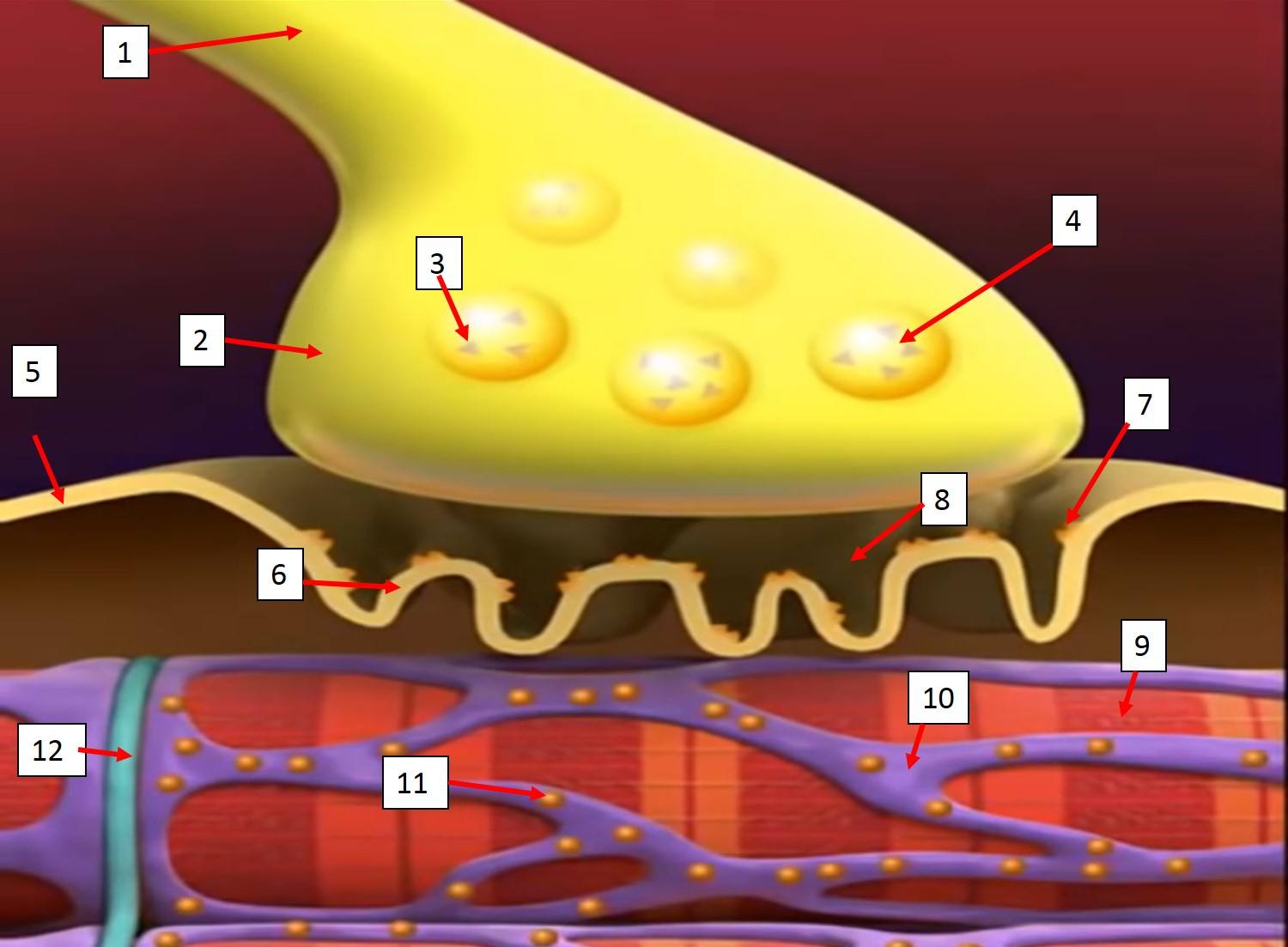
The organelle that stores Acetylcholine (ACh) in the axon is identified as #4
Motor Neuron
Axon Terminal of Motor Neuron
Sarcolemma
Synaptic Vesicle
Motor Neuron
Cardiac Muscle

What state is the sarcomere in B.
Relaxed
Contracting
Both
None of the above
Muscles are attached to bones by _____.
Ligaments
Tendons
Cartilage
Axons

Calcium is going to bind to the protein (a) This will cause (b) to change shape because the (c) moves and exposes the (d) .
Calcium binds to ___of the troponin-tropomyosin complex.
troponin
tropomyosin
actin
myosin
What is the structure that stores the Calcium in the muscle cell.
Sarcoplasm
Sarcolemma
Myoglobin
Sarcoplasmic Reticulum
Order the parts of a muscle from largest to smallest :
myofilaments
actin and myosin
muscle
muscle fascicle
myofibril
Muscle
Muscle Fascicle
Myofibril
Myofilaments
actin and myosin
What layer of connective tissue covers the muscle fiber?
endomysium
epimysium
perimysium
myomysium
What layer of tissue surrounds the entire muscle?
Endomysium
Epimysium
Perimysium
myomysium
Which of the following best describes the power stoke?
actin's troponin pulls on the myosin filament
myosin filament heads pull on binding sites of the actin filament
myosin filaments pull on the Z lines of the actin filament
The A bands shrink
Contractile unit of a muscle which is also the functional unit of a muscle is ...
sarcomere
motor unit
fascicle
myofibril
What element is important in directly triggering contraction?
Potassium
Calcium
Sodium
Chloride
Identify what occurs first during Phase 1 of an Action Potential of the muscular system?
Action Potential arrives at axon terminal of motor neuron
Calcium ion channels open and Calcium enters axon terminal
Calcium causes Acetylcholine (neurotransmitter) to be released
Action Potential travels across the entire sarcolemma


Which of these connects bone to bone?
tendon
ligament
Which type of connective tissue makes up the dermis of the skin?
Stratified squamous
Loose connective
Adipose
Bone
Where would you find hyaline cartilage?
ends of bones
nose
ear pinnae
vertebral discs
epiglottis
Where would you find elastic cartilage?
ends of bones
nose
ear pinnae
vertebral discs
epiglottis
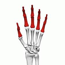
Which bones are highlighted
phalanges
carpals
metatarsals
metacarpals
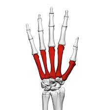
Which bones are highlighted
metacarpals
metatarsals
carpals
tarsals
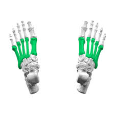
Which bones are highlighted
metatarsals
metacarpals
tarsals
carpals
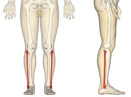
Which bone is highlighted
femur
radius
tibia
fibula
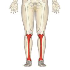
Which bone is highlighted
fibula
tibia
ulna
radius
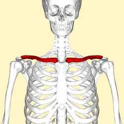

Which bones are highlighted
carpals
tarsals
metacarpals
metatarsals
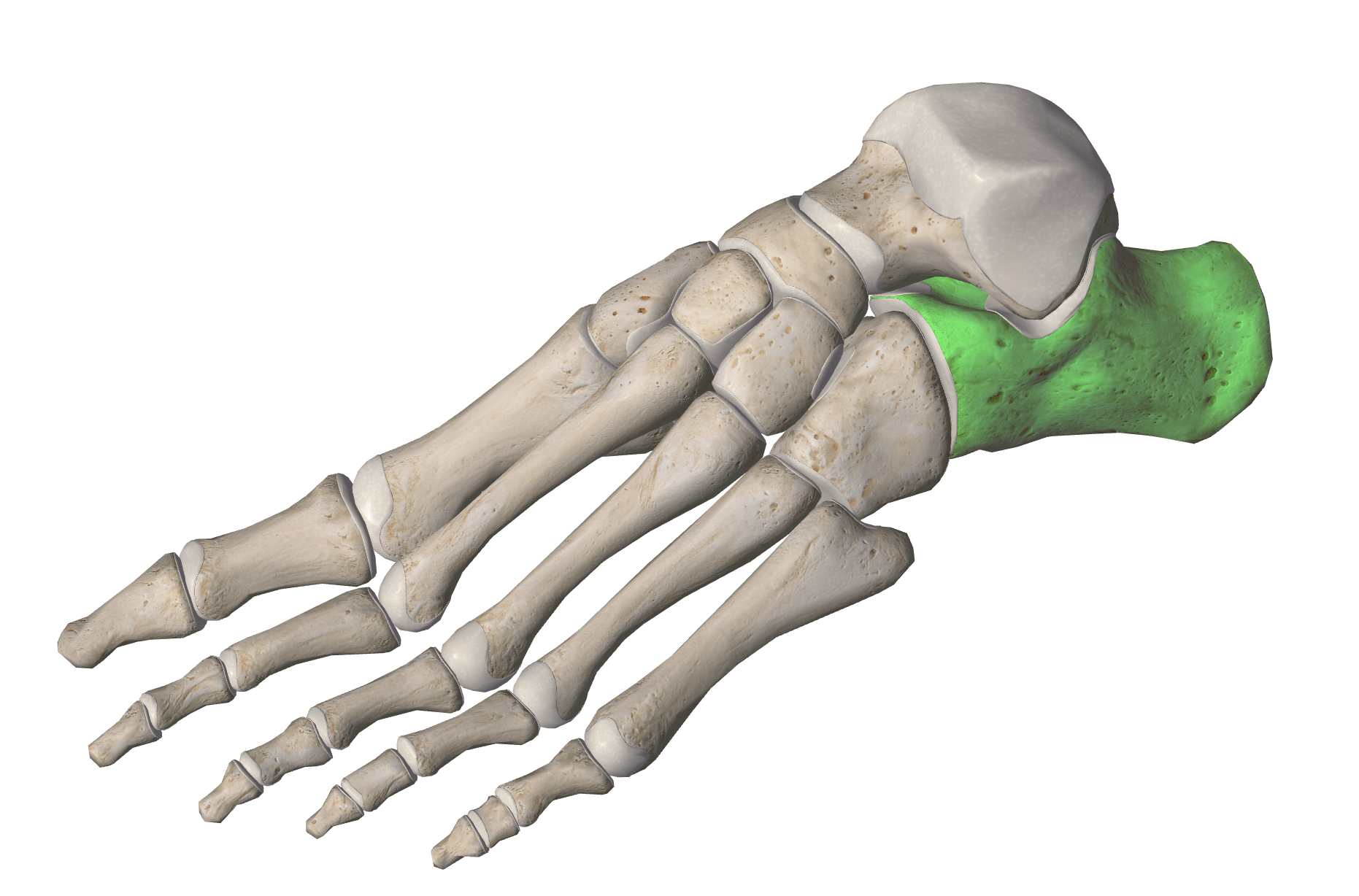


Identify this bone
Ulna
Radius
Humerus
Tibia

Identify this bone
Ulna
Scapula
Humerus
Clavicle
Match the following bones that articulate with each other
Femur
Tibia
Scapula
Humerus
Carpals
Radius
Clavicle
Sternum
Humerus
Ulna
Femur
Tibia
Fibula
Scapula
Clavicle
Coxal Bone
Radius
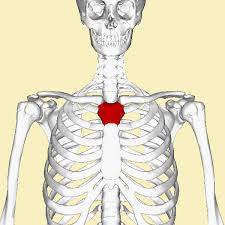
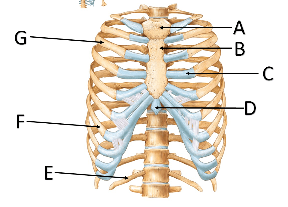

A female patient is brought to the emergency room with an injury to her lower leg from an ATV accident. You notice a white tip of a bone protruding through the skin as you cut her pant leg off. Her injury is described as what kind of fracture.
Comminuted
Pathologic
Compound
Stress


The xray shows a fracture that has bones broken into more than two pieces. What type of fracture is this?
Simple
Compound
Impacted
Comminuted
Closed Reduction
Treating a bone through realignment with one's hands
Treating a bone surgically with pins, rods, screws, and/or wires
Placing the broken bone within a cast, splint, or boot
Open Reduction
Treating a bone through realignment with one's hands
Treating a bone surgically with pins, rods, screws, and/or wires
Placing the broken bone within a cast, splint, or boot
Immobilization
Treating a bone through realignment with one's hands
Treating a bone surgically with pins, rods, screws, and/or wires
Placing the broken bone within a cast, splint, or boot
Which special test is used to determine the integrity of the MCL?
Lachman's Test
Posterior Drawer
Valgus Stress Test
Varus Stress Test
Which special test is used to determine the integrity of the LCL?
Apley's Compression Test
Posterior Drawer
Valgus Stress Test
Varus Stress Test
Which ligament prevents the tibia from moving backward on the femur?
MCL
LCL
PCL
ACL

