
 Worksheets
HBS Final Review
Total questions: 203
Worksheet time: 2hrs 52mins
What is the main function of the cerebellum?
Coordinating voluntary movements, balance, and posture
Producing hormones
Regulating body temperature
Processing visual information
Which part of the brain is responsible for decision making?
Cerebellum
Medulla oblongata
Prefrontal cortex
Occipital lobe
What is the primary function of the occipital lobe?
Processing visual information
Regulating body temperature
Controlling muscle movement
Processing auditory information
Which part of the brain is responsible for auditory processing?
Auditory cortex
Frontal lobe
Cerebellum
Visual cortex
What are the basic life functions controlled by the brain stem?
Vision, hearing, and balance
Digestion, muscle movement, and temperature regulation
Breathing, heart rate, and blood pressure
Memory, learning, and decision making
Which part of the brain is responsible for muscle coordination and maintaining posture?
Cerebellum
Hippocampus
Frontal lobe
Occipital lobe
What is the function of the frontal lobe in the brain?
Memory storage and retrieval
Digestion and metabolism
Emotional regulation and empathy
Decision making, problem solving, and voluntary movement
Which part of the brain is responsible for processing visual information?
Temporal lobe
Occipital lobe
Frontal lobe
Parietal lobe
What is the primary function of the temporal lobe?
Regulate body temperature
Process auditory information and formation of long-term memory
Control muscle movement
Produce digestive enzymes
What are the functions of the brain stem?
Controlling basic life functions such as breathing, heart rate, and blood pressure
Regulating body temperature
Controlling muscle movement
Processing visual information
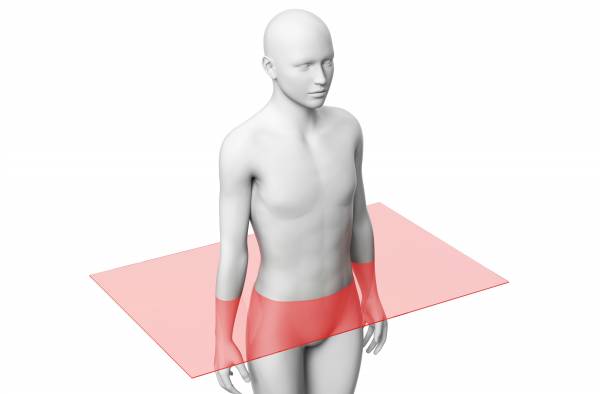
Identify the section or cut.
frontal section
lateral section
transverse section
sagittal section
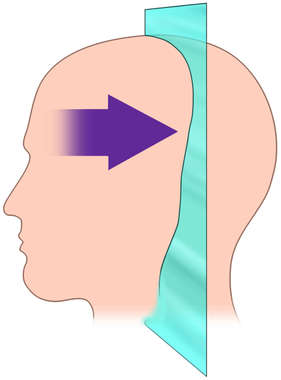
Identify the section or cut.
cross section
plane
midsagittal section
coronal section
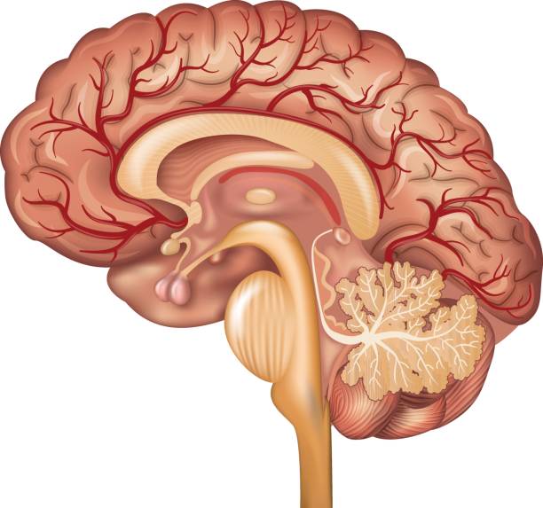
Identify the section or cut.
frontal section
cross section
transverse section
sagittal section
The shoulder is _____ to the elbow.
proximal
dorsal
distal
ventral
The cranial cavity is _____ to the thoracic cavity.
inferior
anterior
peripheral
superior
What is anatomical position?
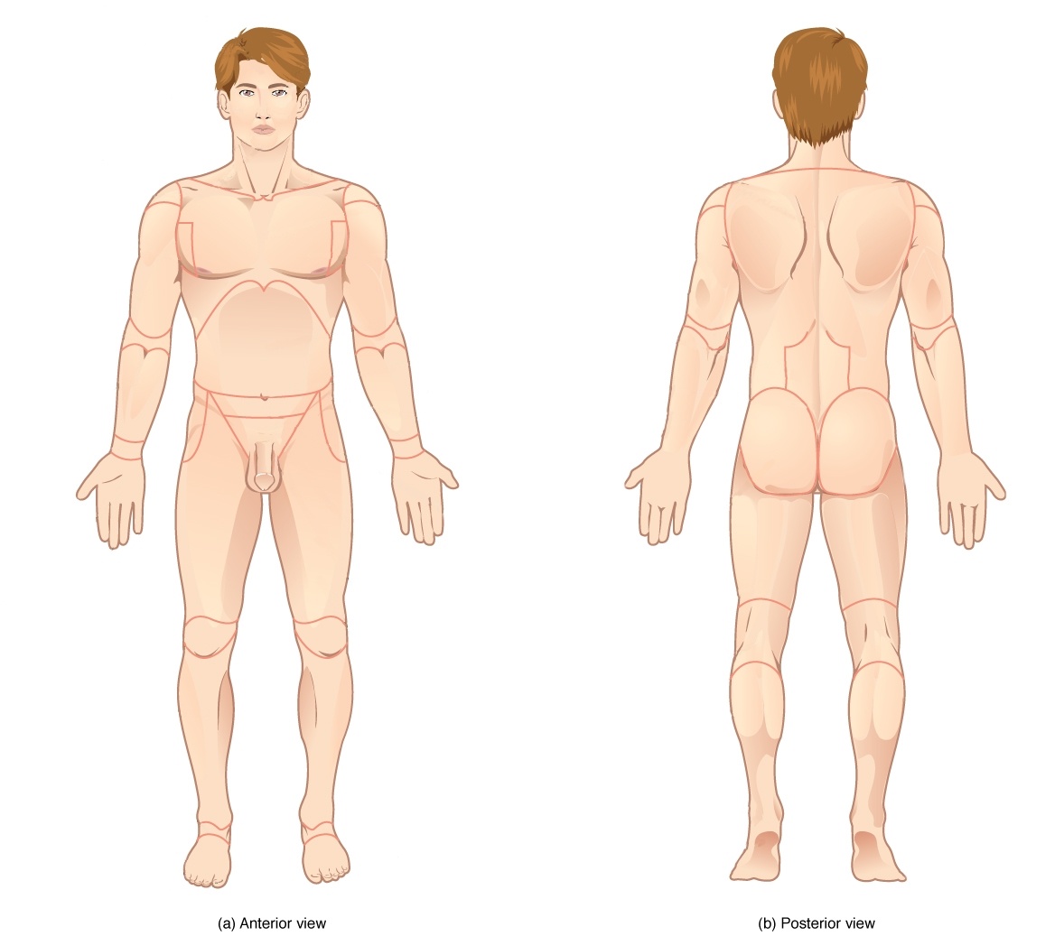
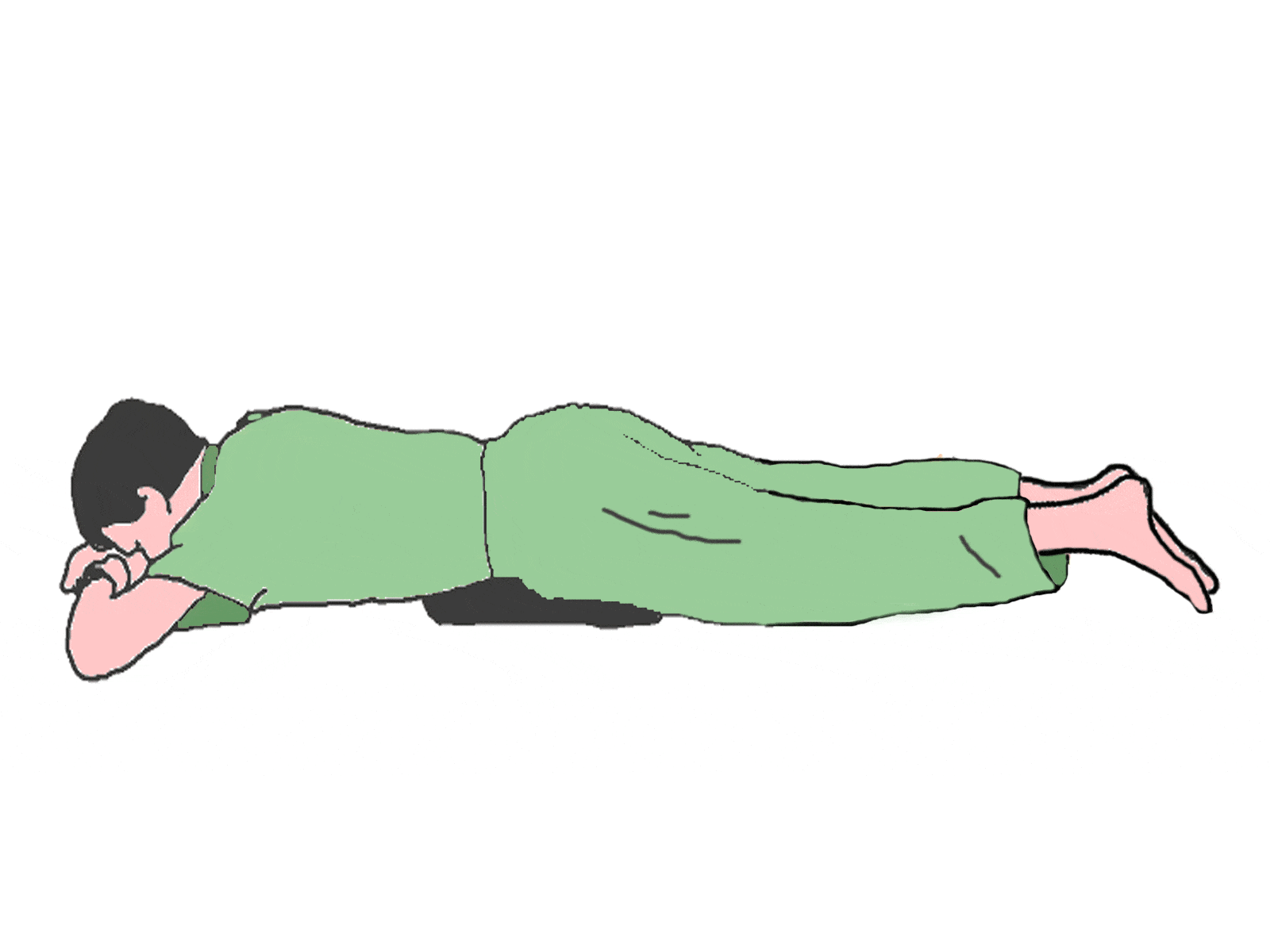
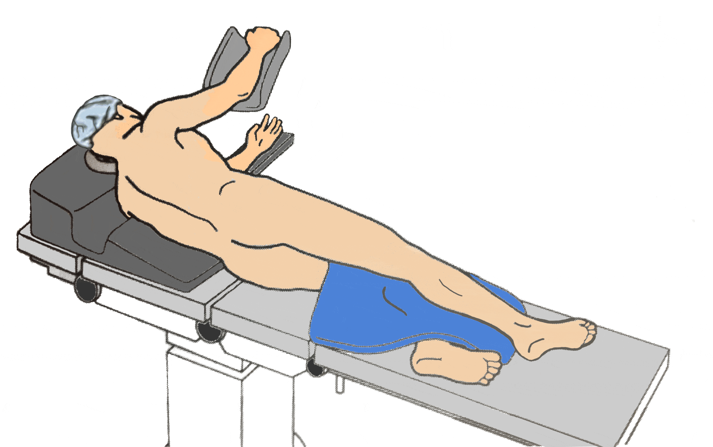
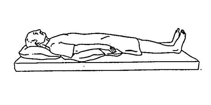
Which cavity includes the heart and lungs?
Thoracic cavity
Cranial cavity
Abdominal cavity
Retroperitoneal cavity
The pinky finger is ______ to the thumb in anatomical position.
posterior
superior
lateral
medial

What abdominal region is #1?
Right Hypochondriac Region
Left Hypochondriac Region
Right Lumbar Region
Right Lumbar Region

What abdominal region is #6?
Hypogastric
Right Inguinal
Left Lumbar
Umbilical

What is b?
Right Upper Quadrant
Left Upper Quadrant
Right Lower Quadrant
Left Lower Quadrant
What abdominal region is #5?
Right Lumbar
Left Hypochondriac Region
Epigastric
Umbilical
What abdominal region is #2?
Hypogastric
Right Inguinal
Epigastric
Umbilical

[The eyes are _______ to the brain].

Match the following planes
Traverse
Separates top & bottom
Sagittal
Separates left & right
Coronal
Seperates front & back
Median
Even separation of left & right
Distal
Away from

The cavity surrounded by a box is?
Abdominal Cavity
Pelvic Cavity
Pleural Cavity
Cranial Cavity

Cranial Cavity
Pleural Cavity
Pericardial Cavity
Vertebral Canal

The cavity surrounded by a box is?
Abdominal Cavity
Pelvic Cavity
Thoracic Cavity
Pericardial Cavity

The cavity surrounded by a box is?
Pericardial Cavity
Pleural Cavity
Abdominal Cavity
Pelvic Cavity
Which of the following cavities holds the heart AND lungs?
pleural
pericardial
thoracic
abdominal
The head is _________________ to the navel.
anterior
posterior
superior
inferior
The nose is_________________ to the ears.
medial
lateral
superficial
deep
The thumbs are _________________ to the pinkies.
proximal
distal
medial
lateral
The skin is _________________ to the muscles.
ventral
dorsal
superficial
deep
The ankle is _________________ to the knee.
superior
inferior
proximal
distal

What is the name of the yellow bone?
occipital
parietal
temporal
frontal
What is the name of the light red bone?
maxilla
palatine
mandible
zygomatic
What is the name of the purple bone?
maxilla
palatine
mandible
zygomatic

What is the name of the orange bone?
temporal
frontal
parietal
occipital

What is the name of the purple bone between the teeth?
maxilla
mandible
ethmoid
palatine
What is the name of the brown bone?
ethmoid
occipital
sphenoid
zygomatic
What is the name of the pink bone?
ethmoid
occipital
sphenoid
zygomatic
What is the name of the dark blue bone?
ethmoid
maxilla
nasal
zygomatic

This muscle (B) allows you to close your eyes, squint, blink, and wink.
Orbicularis oris
Orbicularis oculi
Zygomaticus
Platysma
This muscle (E) closes the mouth and protrudes the lips. It is often called the "kissing" muscle.
Orbicularis oris
Buccinator
Zygomaticus
Orbicularis oculi
This muscle (H) lies over the temporal bone and helps the masseter close the jaw.
Trapezius
Platysma
Sternocleidomastoid
Temporalis

Right Lower Quadrant
Pelvic cavity
Ventral cavity
Thoracic cavity

Abdominal cavity
Pelvic cavity
Inguinal
Dorsal cavity

Axillary
Ventral cavity
Dorsal cavity
Pelvic cavity

Dorsal cavity
Cubital
Epigastric
Ventral

Pelvic cavity
Thoracic cavity
Abdominal cavity
Cephal

Hypogastric region
Abdominopelvic cavity
Dorsal cavity
Brachial

Identify Letter B
Brachial
Sagittal plane
Dorsal
Axillary

Identify letter A
Cubital
Cephal
inguinal
Thoracic cavity

Identify Letter C
Popliteal
Brachial
Cubital
Cephal

Identify the area behind the knee
Anterior
Cubital
Brachial
Popliteal
These type of bones are typically longer than they are wide.
Irregular
Flat
Short
Long
These type of bones have a variety of shapes.
Long
Short
Irregular
Flat
These bones are thin and flattened.
Flat
Long
Short
Irregular
These type of bones have roughly equal widths and lengths; cube shaped.
Long
Short
Flat
Irregular
Which of the following is NOT an example of a long bone?
forearm bone
wrist bone
femur
thigh bone
Either end of a long bone.
Diaphysis
Periosteum
Spongy Bone
Epiphyses
Shaft of a long bone.
Epiphysis
Compact Bone
Sternum
Diaphysis
Dense connective tissue covering the surface of a bone.
Trabeculae
Articuler cartilage
Periosteum
Spongy tissue


Radius
Humerus
Ulna
Clavicle


What bone is the arrow pointing to?
rib
spine
scapula
clavicle


Identify the section or cut.
frontal section
lateral section
transverse section
sagittal section
Identify the section or cut.
cross section
plane
midsagittal section
coronal section
The shoulder is _____ to the elbow.
proximal
dorsal
distal
ventral
Which cavity includes the heart and lungs?
Thoracic cavity
Cranial cavity
Abdominal cavity
Retroperitoneal cavity
The pinky finger is ______ to the thumb in anatomical position.
posterior
superior
lateral
medial
The head is _________________ to the navel.
anterior
posterior
superior
inferior
The nose is_________________ to the ears.
medial
lateral
superficial
deep
What is the name of the yellow bone?
occipital
parietal
temporal
frontal
What is the name of the purple bone?
maxilla
palatine
mandible
zygomatic
This muscle (E) closes the mouth and protrudes the lips. It is often called the "kissing" muscle.
Orbicularis oris
Buccinator
Zygomaticus
Orbicularis oculi
Hypogastric region
Abdominopelvic cavity
Dorsal cavity
Brachial
Identify the area behind the knee
Anterior
Cubital
Brachial
Popliteal
These type of bones have roughly equal widths and lengths; cube shaped.
Long
Short
Flat
Irregular
Shaft of a long bone.
Epiphysis
Compact Bone
Sternum
Diaphysis
Dense connective tissue covering the surface of a bone.
Trabeculae
Articuler cartilage
Periosteum
Spongy tissue
Radius
Humerus
Ulna
Clavicle




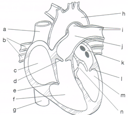


Motor neurons carry impulses __________ the brain
towards
away from
within
Which of the following describes the function of the Na/K Pumps in the neuron membrane?
depolarizing the cell by allowing Na into the cell
repolarizing the cell by allowing K out of the cell
rearranging Na and K back to return cell to resting state
Which of the following correctly outlines a reflex arc?
Motor neuron - interneuron - sensory neuron
Sensory neuron - interneuron - motor neuron
Sensory neuron - interneuron - brain - motor neuron
The dendrites of a neuron carry a stimulus
Towards the cell body
Away from the cell body
Through the cell body
During resting membrane potential, the inside of a neuron has a _______________ charge
negative
positive
neutral
Neurotransmitters are stored in which structure of the neuron?
Cell body
Dendrite
Axon terminals
Myelin is located on the ____________ and functions to ____________ nerve transmission.
dendrite - speed up
dendrite - slow down
axon - speed up
axon - slow down
Neurotransmitters bind to complementary sites on a cell called
receptors
axon terminals
cell bodies
Reaction time is ___________________ than reflex time.
faster
slower
same
Depolarization occurs when the inside of a cell becomes _________________ due to _________ gates opening.
positive - potassium
negative - potassium
positive - sodium
positive - sodiium
The spaces between myelin on an axon are called
axon gaps
nodes of ranvier
dendrite dents
Which of the following nervous system disorders occurs as a result of tangled plaques forming in brain tissue?
Huntington's disease
Alzheimer's disease
Parkinson's disease
Which of the following disorders is associated with a decrease in the neurotransmitter called dopamine?
parkinson's disease
epilepsy
ALS
Which of the following diagnoses best describes a disorder that involves seizure activity?
MS
ALS
epilepsy
Which of the following diseases does NOT affect memory or mental cognition?
ALS
Huntington's
Alzheimers
A sarcomere is
a nonfunctional unit of skeletal muscle
a repeating structural unit of skeletal muscle
the area between two intercalated discs
the wavy lines on the muscle seen in the microscope.
Which of the following are composed of myosin?
thick filaments
thin filaments
Z discs
all myofilaments
Which of the following surrounds the individual muscle cell?
perimysium
endomysium
epimysium
fascicle
Which of the following muscle type is not striated?
smooth muscle
skeletal muscle
cardiac muscle
none of above
The bundle of collagen fibers at the end of a skeletal muscle that attaches the muscle to bone is called a(n)
fascicle
tendon
ligament
myofibril
Rigor mortis occurs because
the cells are dead
sodium ions leak out of the muscle
ATP is required to release the attached
actin and myosin molecules
proteins are beginning to break down, thus preventing the flow of calcium ions.
Where is cardiac muscle found?
In the walls of hollow organs
In the walls of the heart
Around the skeleton
In between ribs
What is the function of skeletal muscle?
Movement of the body
Moving food toward the stomach
Pumping blood through the heart
Moving the lungs during breathing
What influence does the structural makeup of the muscle have on its function?
Structure does not relate to function
There are big fibers that flex at different times to contract
Tiny fibers all contract together in order to perform a stronger contraction all together
Tiny fibers contract all together but make a weaker contraction
The place where neurons meet muscles cells is called the
point of joint origin.
site of insertion.
neuromuscular junction.
fiber that covers the muscle.
What are the three types of muscle?
Open, closed and striated
Smooth, rough and striated
Cardiac, skeletal and closed
Smooth, cardiac and skeletal
A syndrome that affects the median nerve in the wrist is called:
ATP
rigor mortis
carpal tunnel
deletion

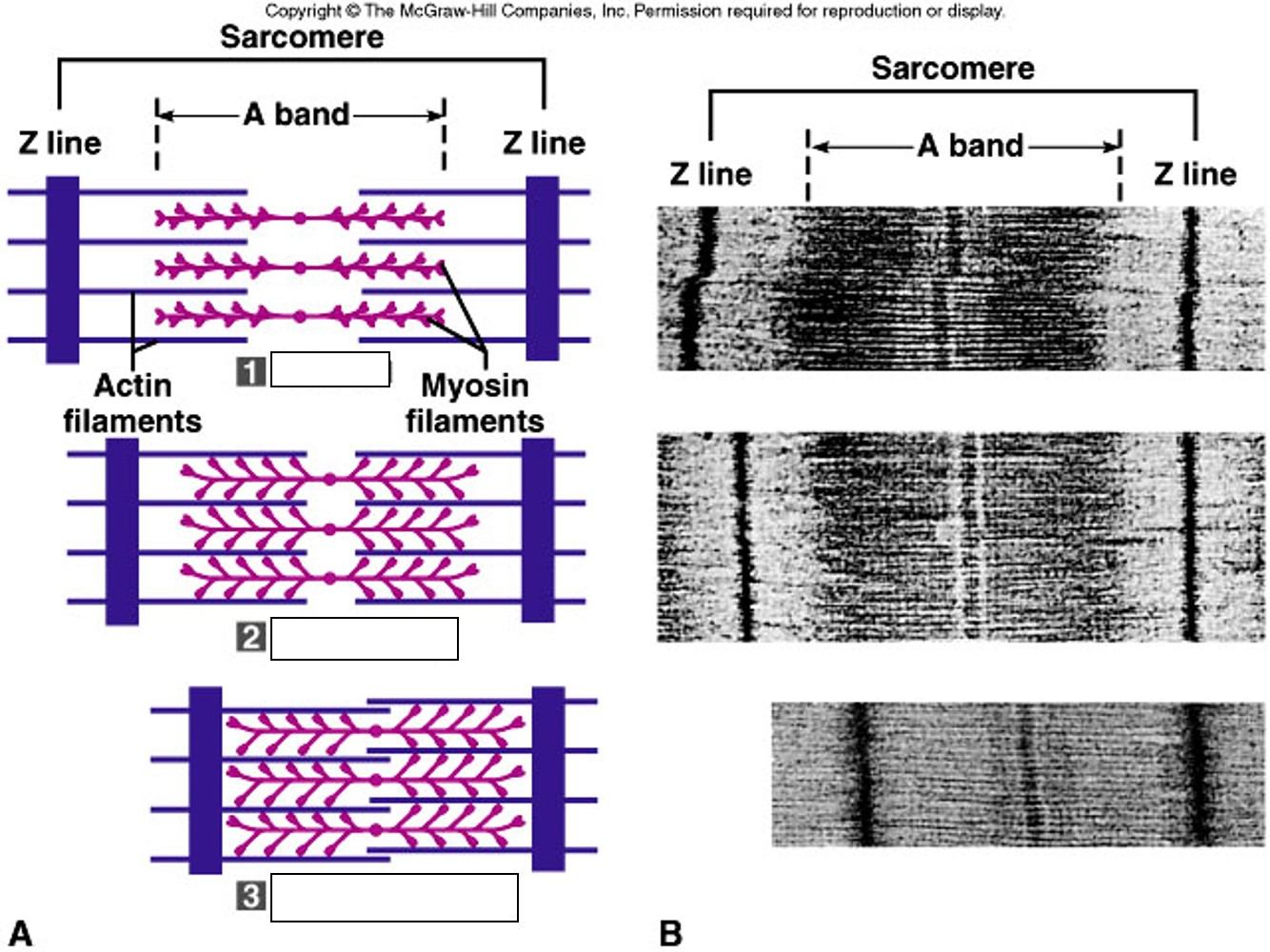
Found attached to bones throughout the body
smooth
cardiac
skeletal
all
Found in the heart
smooth
cardiac
skeletal
all
Found in the walls of hollow organs; also called visceral
smooth
cardiac
skeletal
all
Use to move materials through contractions
smooth
cardiac
skeletal
all


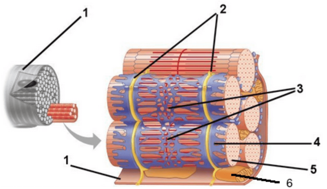
What is shown in the colored part of this diagram?
a whole muscle
five muscle fibers
five muscle fascicles
five myofibrils
What is labeled "4" (the blue/purple part) in the diagram?
sarcolemma
sarcoplasmic reticulum
transverse tubules
myofibril
Name the following in order from largest to smallest:
muscle, muscle fascicle, muscle cell, myofibril, filaments
muscle, muscle fascicle, muscle cell, filaments, myofibril
muscle, muscle cell, muscle fascicle, myofibril, filaments
muscle, muscle cell, muscle fascicle, filaments, myofibril
What is the length between Z-lines of an actin molecule called?
fascicle
sarcomere
myosin binding site
sarcoplasmic length
____________ attaches to actin to form the cross bridge.
Troponin
Tropomyosin
Myosin
Calcium

