
 Worksheets
Wrist/forearm RP
Total questions: 20
Worksheet time: 13mins

What is the projection of this image?
PA wrist
PA scaphoid
PA Oblique wrist
PA Oblique Scaphoid

What carpal is demonstrated in this image?
(a)

How was this image obtained?
Wrist in ulnar deviation
Stretcher method
Select the most medial carpal

What deviation is demonstrated in this image?
Ulnar deviation
Radial deviation
How is the CR directed for a Lateral wrist projection?
Perp to mid-carpal area
Perp to 2nd MCP
Perp to wrist joint
Perp to radius
For a PA oblique wrist projection with lateral/external rotation, what carpals are demonstrated?
Medial
Lateral
Central
None
How is the CR directed for the stretcher method (standard) of the scaphoid?
CR angled 20 degrees toward fingers
CR angled 20 degrees toward elbow
Perp to scaphoid
Perp to mid carpal area
Of the following, which are variations of the stretcher method?
Hand flat, IR flat- CR 20 degrees toward elbow
Hand and IR angled 20 degrees- CR angled 20 degrees toward elbow
Hand and IR flat- CR perp
Hand and IR angled 20 degrees- CR perp
Tightly clenched fist
Select the structures that are demonstrated on a PA oblique wrist projection.
Radius
Ulna
Trapezium
Scaphoid
Lateral carpals
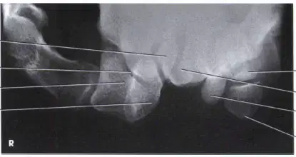
What is the method name of this image?
(a)
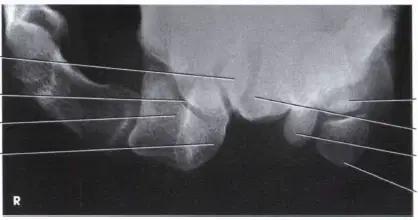
How is the CR directed for this projection?
Inferiosuperior
superoinferior
proximal distal
distal proximal
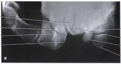
What projection is this image?
PA
Lateral
AP
PA Oblique
Tangential
When the ulnar styloid is projected in profile medially, it indicates that the humeral condyles are ______ to the IR?
Perpedicular
Parallel
Obliqued
How are the humeral epicondyles positioned to the IR in an AP projection of the forearm?
Obliqued
Perpendicular
Parallel
Select the following that will increase recorded detail.
decreased OID
Collimation
Patient position
Small focal spot
Technique
How is the wrist positioned for the lateral projection of the forearm?
Pronated
supinated
oblique 45 degrees
true lateral
For the AP forearm projection, when the coronoid process is visible on end, this indicates....
rotation
anode heel effect was used
Elbow was extended
Hand was supinated
On a a PA projection of the wrist the CMC joints are demonstrated as closed, and there is no superimposition of the posterior margin of the radius and the lunate carpal. what does this indicate in regard to the position of the patient?
Rotation
Elbow bent at 90 degrees
Hand flexed
Hand not flexed
Elbow not bend to 90 degrees
An image of a lateral projection of the wrist demonstrates the pisiform anterior to the scaphoid, how was the wrist positioned?
True lateral
Under rotated
Over rotated

