
 Worksheets
Anatomy Final Review SM2
Total questions: 168
Worksheet time: 3hrs 48mins

Accurate facts about the bone(s) are
Zygomatic, 2 irregular
Lacrimal, 2, flat
Nasal bone, 1 irregular bone
Nasal bones, 2 flat bones

Zygomatic
Temporal
Parietal
Maxilla

Mandible
Zygomatic
Temporal
Maxilla

Maxilla
lacrimal
Mandible
zygomatic

Two bones that make up the lateral parts of the nasal cavity; lined with membranes that help to filter warm air. It is represented by #2 in the image.
inferior nasal concha
nasal bones
zygomatic bones
sphenoid bone

How is this bone connected to your head?
frontal bone
zygomatic bones
temporomandibular joint
maxilla bones

This bone along with the nasal bones forms the ____________ canal which collects __________.
nasolacrimal, nasal discharge
nasolacrimal, tears
ear, ear wax
nasal, boogers
Maxilla
Frontal
Mandible
Zygomatic

Zygomatic
Vomer
Ethmoid
Lacrimal

The thin, flat bone that forms the back and inferior portion of the nasal septum. It is #3 on the image.
ethmoid
lacrimal
inferior nasal conchae
vomer

The purple bone(s) is (are) called the
palatine bone(s)
lacrimal bone(s)
septum bone(s)
vomer bone(s)

This bone(s) forms
part of the nasal cavity & part of the roof of the mouth or hard palate
part of the orbit or eye socket
part of the orbit or eye socket, part of the nasal cavity, part of the roof of the mouth or hard palate
part of the orbit or eye socket & part of the roof of the mouth or hard palate

This bone is the ________ bone and forms part of the ________.
palatine, nasal septum
vomer, nasolacrimal canal
vomer, tear duct canal
vomer, nasal septum

Identify the bone highlighted in the image.
occipital
parietal
temporal
frontal
maxilla

Identify the bone highlighted in the image.
occipital
parietal
temporal
frontal
maxilla

Identify the bone highlighted in the image.
occipital
parietal
temporal
frontal
maxilla

Identify the bone highlighted in the image.
occipital
parietal
temporal
frontal
maxilla

suture highlighted in red
coronal
lamboidal
sagittal
metopic

suture highlighted in red
lamboidal
frontal
sagittal
squamosal

occipital bone
foramen magnum
coronal suture
fontanelle
Identify the lambdoid suture below.


Identify the squamous suture below.




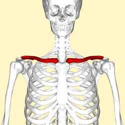
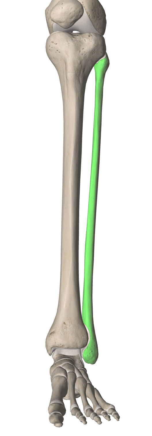
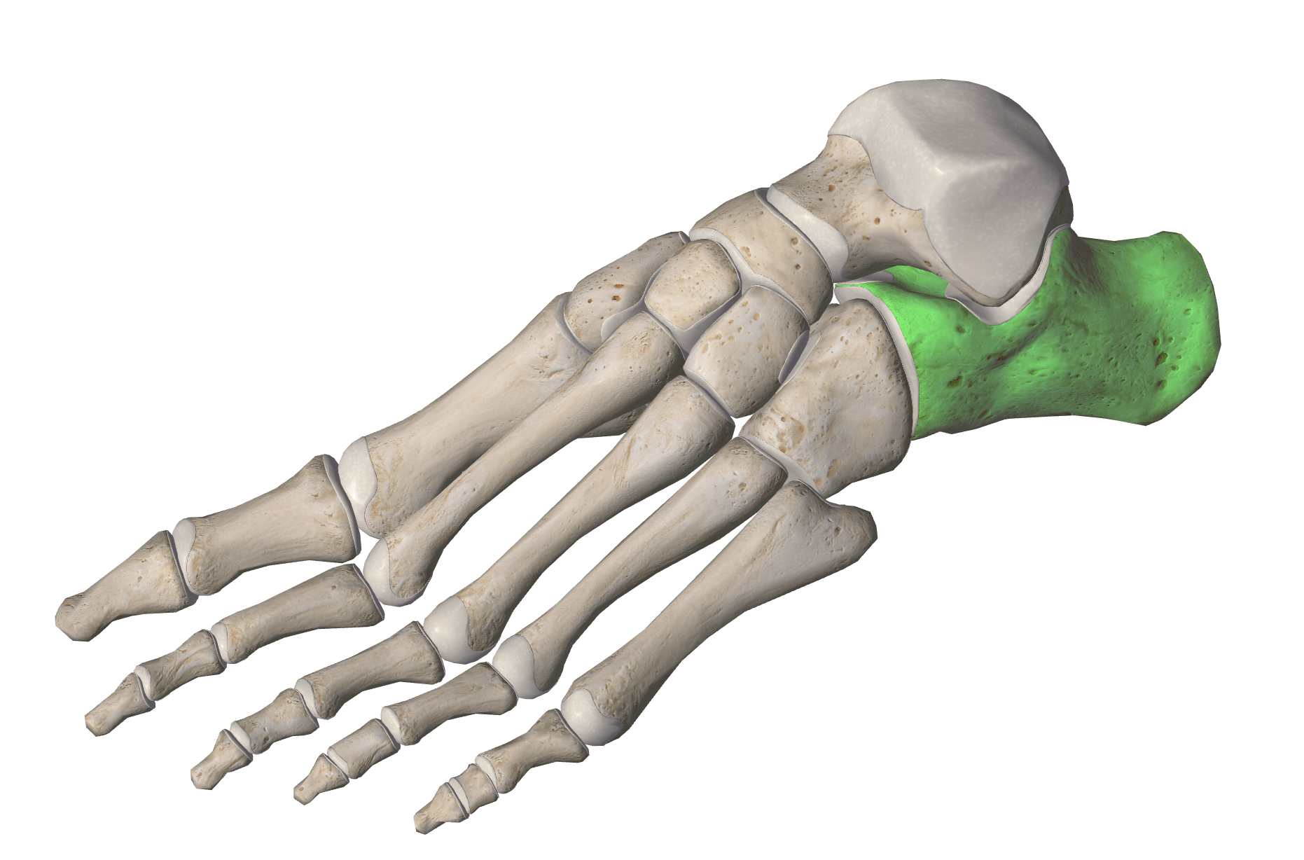
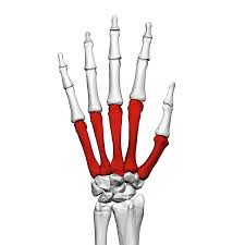
Which bones are highlighted
metacarpals
metatarsals
carpals
tarsals

Which bones are highlighted
carpals
tarsals
metacarpals
metatarsals
The anatomical name for the big toe is the
Popliteal
Pollex
Hallux
Patella

clavicle
scapula
humerus
radius
ulna

clavicle
scapula
humerus
radius
ulna

clavicle
scapula
humerus
radius
ulna

clavicle
scapula
humerus
radius
ulna

carpals
metacarpals
phalanges
ilium
ischium

carpals
metacarpals
phalanges
ilium
ischium

carpals
metacarpals
phalanges
ilium
ischium

carpals
metacarpals
phalanges
ilium
ischium

coxal
femur
patella
fibula
tibia

coxal
femur
patella
fibula
tibia

coxal
femur
patella
fibula
tibia

coxal
femur
patella
fibula
tibia

coxal
femur
patella
fibula
tibia

Tarsals
Metatarsals
phalanges of feet
iliac crest
calcaneus

Tarsals
Metatarsals
phalanges of feet
iliac crest
calcaneus
Which muscle lies across the forehead and raises the eyebrows?
mentalis
Frontalis
sterno cleido mastoid
masseter
The name given to the two small triangular muscles that sit between the eyebrows and draw them down and together?
zygomaticus
platysma
mentalis
corrugator
The muscle that covers the sides of the neck & chin, it draws the mouth's corners downwards.
platysma
sterno cleido mastoid
masseter
corrugator
The muscle that covers the front of the chin and raises the lower lip causing the chin to wrinkle.
corrugator
zygomaticus
mentalis
platysma
The circular muscle that surrounds the eyes and closes the lid.
orbicularis oculi
orbicularis oris
orbicularis norris
orbicularis coris
The muscle that extends diagonally from the cheek to the corners of the mouth and causes grinning.
mentalis
risorius
zygomaticus
nisorius
The muscle that sits inside the cheeks between the upper and lower jaw.
mentalis
corrugator
buccinator
orbicularis oris
The muscle that extends diagonally across the cheek, causes smiling and laughing.
zygomaticus
buccinator
corrugator
mentalis
The muscle that covers the front of nose and surrounds the nostrils.
mentalis
zygomaticus
nasalis
corrugator
The muscle that sits between the eyebrows at the top of the nose.
platysma
fontalis
mentalis
procerus
The circular muscle that surrounds the mouth causing pouting & kissing action.
orbicularis oris
mentalis
orbicularis oculi
procerus
The muscle that sits at the temple area.
procerus
frontalis
mentalis
temporalis
The muscle that sits on the lower cheek area and clenches the teeth and raises the lower jaw.
frontalis
masseter
temporalis
trapezius
The large triangular muscle covering the back and neck.
corrugator
trapezius
masseter
mentalis
The muscle that runs down the sides of the front of the neck.
occipitalis
trapezius
sterno cleido mastoid
platysma
The muscle that covers the back of the base of the skull.
occipitalis
temporalis
orbicularis oris
orbicularis oculi
The muscle that sits on the top of shoulder.
masseter
deltoid
trapezius
occipitalis
What muscle lies across the forehead? (a)
Which muscles lies across the forehead?
(a)
Which muscle is highlighted?
orbicularis oris
orbicularis oculi
occipitalis
What muscle is highlighted?
corrugator
platysma
frontalis
Which muscle is highlighted?
mentalis
corrugator
platysma
What muscle is highlighted?
corrugator
temporalis
nasalis
What muscle is highlighted?
orbicularis oris
orbicularis oculi
mentalis
What muscle is highlighted?
buccinator
mentalis
frontalis
What muscle is highlighted?
temporalis
zygomatic
mentalis
What muscle is highlighted?
orbicularis oris
temporalis
metalis
What muscle is highlighted?
zygomaticus
sterno cleido mastoid
platysma

?
deltoid
pectoralis major
subclavius
diaphragm

?
pectoralis minor
diaphragm
pectoralis major
subclavius

?
external intercostals
subclavius
serratus anterior
pectoralis minor

?
external oblique
serratus anterior
external intercostals
subclavius

?
omohyoid
sternocleidomastoid
deltoid
subclavius

?
diaphragm
rectus abdominis
external oblique
transversus abdominis

?
pectoralis minor
internal intercostals
external intercostals
thyrohyoid

?
diaphragm
internal intercostal
external intercostals
sternohyoid

?
external oblique
internal oblique
serratus anterior
rectus abdominis

yellow
subclavius
transverse abdominis
internal oblique
infraspinous

red
rectus abdominis
transverse abdominis
internal oblique
diaphragm

blue
internal oblique
transversus abdominis
rectus abdominis
external intercostals

?
latissimus dorsi
teres major
supraspinatus
infraspinatus

?
erector spinae
latissimus dorsi
scalenes
trapezius

?
sartorius
levator scapulae
supraspinatous
rhomboids

?
rhomboid major
rhomboid minor
latissimus dorsi
teres major

green
supraspinatus
infraspinatus
teres major
subscapularis

?
teres minor
glenoid cavity
teres major
infraspinatus

?
subscapularis
teres minor
infraspinatus
teres major

?
infraspinatus
levator scapulae
latissimus dorsi
trapezius

?
subscapularis
infraspinatus
teres major
supraspinatus
rotator cuff muscles
supraspinatus
infraspinatus
teres minor
subscapularis


These vessels carry blood away from the heart.
arteries
veins
capillaries
superior vena cava
The exchange of oxygen and carbon dioxide takes place in these vessels.
arteries
veins
inferior vena cava
lung capillaries
Brings blood to the heart from the upper portion of the body.
pulmonary artery
superior vena cava
pulmonary vein
inferior vena cava
Valve between the left atrium and the left ventricle.
tricuspid valve
mitral valve
pulmonary valve
aortic valve
Thickest walled chamber of the heart.
left atrium
right ventricle
left ventricle
right atrium
Valve between the left ventricle and the aorta.
aortic valve
pulmonary valve
tricuspid valve
mitral valve
These vessels carry blood to each lung from the right ventricle.
superior vena cava
pulmonary arteries
pulmonary veins
aorta
Valve between the right atrium and the right ventricle.
mitral valve
pulmonary valve
aortic valve
tricuspid valve
Blood in the right atrium:
Oxygenated
Deoxygenated
Blood in the left ventricle:
Oxygenated
Deoxygenated
Blood in the pulmonary artery:
Oxygenated
Deoxygenated
Blood in the pulmonary veins:
Oxygenated
Deoxygenated
Blood in the aorta:
Oxygenated
Deoxygenated
Blood in the left atrium:
Oxygenated
Deoxygenated
Blood in the Inferior Vena Cava:
Oxygenated
Deoxygenated

