
Font size
 Worksheets
Facial Bones/Mandible/Sinuses
Total questions: 40
Worksheet time: 20mins
What are the two smallest bones in the skull called?
Nasal bones
Lacrimal bones
Zygomatic bones
Palatine bones
Which of the following is NOT a pair of facial bones?
R and L nasal
R and L lacrimal
Vomer
R and L zygomatic
What passes through the lacrimal foramen in each lacrimal bone?
Blood vessels
Nerves
Tear duct
Air
How many facial bones are there in total?
12
14
16
18
What is the only movable bone of the face?
Vomer
R and L palatine
Mandible
R and L maxillary
Where are the lacrimal bones located?
In the lower jaw
In the medial wall of orbits between labyrinth of ethmoid and maxilla
In the posterior part of the skull
In the upper part of the nasal cavity
With which of the following do the maxillary bones not articulate?
Frontal cranial bones
Ethmoid cranial bones
Mandible
Zygomatic bones
The maxillary bones also form part of the floor of which structure?
The cranium
The orbit
The mouth
The cheek
What is the name of the large, pyramidal cavity contained in the body of the maxillary bones?
Maxillary sinus
Mandibular sinus
Frontal sinus
Sphenoidal sinus
What does the temporal process extend posteriorly to join?
mandibular process of temporal bone
zygomatic process of frontal bone
zygomatic process of temporal bone
maxillary process of zygoma
What is formed by the union of the temporal process and the zygomatic process?
Temporal arch
Zygomatic arch
Maxillary arch
Mandibular arch
With which bone does the zygomatic bone articulate superiorly?
Maxilla
Temporal bone
Frontal bone
Sphenoid
Which bone forms the anterior articulation of the zygomatic bone?
Frontal bone
Temporal bone
Sphenoid
Maxilla
What is the largest and densest bone of the face?
Maxilla
Zygomatic bone
Mandible
Nasal bone
The anterior nasal spine is an aspect of the ______ bone.
maxillary
zygomatic
mandibular
inferior nasal conchae
What is another name for the angle of the mandible?
Alveolar process
Gonion
Condyle
Symphysis
What is the mandibular notch?
Anterior process on top of ramus
Posterior process on top of ramus
Concave area at top of ramus between coronoid and condylar processes
Articulates with the zygomatic arch
Which of the following facial bones is unpaired?
Maxillary
Palatine
Lacrimal
Vomer
For a lateral projection of the paranasal sinuses, the central ray is directed
2 in anterior ro EAM
2 in above EAM
1 inch posterior to the outer canthus
Where will the Petrous Ridges be located on a Parietoacanthial Projection?
within the maxillary sinus
below the maxillary sinus
filling the orbits
in the lower third of the orbits
What plane is parallel to the IR for a lateral facial bone projection?
MCP
oblique plane
MSP
which sinuses are best demonstrated for the following projections of the Paranasal Sinuses: Parietoacanthial Projection (Waters)
ethmoid
maxillary
sphenoid
frontal
To place the mandibular symphysis closer to the IR, how much should the head be rotated towards the IR?
0
45
30
15
For the Waters method of the facial bones, the orbitomeatal line is placed at what angle to the IR?
30
55
37
63
Several methods area available to perform the axiolateral oblique projection of the mandible to demonstrate the symphysis, body, or ramus. What is the central-ray angulation for all of these projections?
15 cephalad
25 cephalad
25 caudad
15 caudad
All of the following should be clearly demonstrated on a lateral projection of the paranasal sinus, except
all four paranasal sinus groups.
petrous ridge in lower third of orbits.
sella turcica in profile.
superimposed orbital roofs.
Which of the sinuses is developed at birth and visible radiographically?
frontal
ethmoid
maxillary
sphenoid
Which two of the following are functions of the sinuses? (Select all that apply.)
Decrease the weight of the skull.
Deepens the voice.
Improve respiration.
Warm and moisten inhaled air.
The largest sinus is the
maxillary
frontal
ethmoid
sphenoid
Which of the following is placed perpendicular to the front edge of the IR for a lateral projection of the facial bones?
Infraorbitomeatal line
Acanthiomeatal line
Orbitomeatal line
MML
All of the following are clearly demonstrated on the parietoacanthial projection (Waters method), except
foramen magnum
zygomatic bone
orbits
maxilla
Which of the following is true regarding the lateral projection of the nasal bones?
1.MSP is parallel with the tabletop.
2. Both sides are done for comparison.
3. The interpupillary line is perpendicular to the tabletop.
1 only
1 and 2 only
1,2 and 3
2 and 3 only
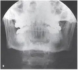
What projection and anatomy is demonstrated in the image below?
AP axial of the TMJs
PA axial of the mandibular rami
PA of the mandibular body
Axiolateral oblique of the mandibular body
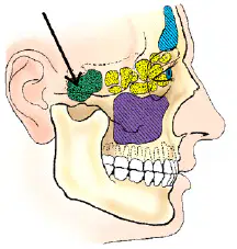
The sinus identified in the figure below is the
maxillary
frontal
sphenoid
ethmoid
At what age are all of the sinuses completely developed?
5
10
18
25
Which sinus is located immediately below the sella turcica?
frontal
sphenoid
ethmoid
maxillary
When using the angled grid technique for the PA axial (Caldwell method) projection of the sinuses, the vertical grid device must be angled _____ degrees.
25
10
15
30
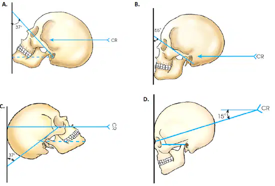
Which drawing in the figure below demonstrates the correct head position for the Waters method?
A
B
C
D
At which level should the central ray enter the base of the skull for the SMV projection of the sinuses?
½ inch posterior to the level of the EAM
½ inch anterior to the level of the EAM Correct
½ inch below the mental protuberance
1 inch below the mental protuberance
What is the radiographic appearance if the neck is extended too little for the Waters method of the sinuses?
Petrosae demonstrated immediately below the maxillary sinuses.
Petrosae are projected over the inferior portions of the maxillary sinuses. Correct
Maxillary sinuses are elongated.

