
NEW
Font size
 Worksheets
CXR Speed Round
Total questions: 18
Worksheet time: 9mins

Interpret the following radiograph:
Pneumothorax
RML consolidation
normal radiograph
tracheal deviation to the right

Interpret the following radiograph:
Pulmonary Edema
RML consolidation
bilateral pleural effusions
pulmonary fibrosis

Interpret the following radiograph:
normal radiograph
RML pneumonia
pneumothorax
Right sided pleural effusion

Interpret the following radiograph:
tension pneumothorax
air bronchogram on left side
improper ETT placement
CHF

Interpret the following radiograph:
RML consolidation
RLL consolidation
CHF
Right sided pleural effusion
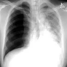
Interpret the following radiograph:
pulmonary edema
left sided pleural effusion
pneumothorax
tension pneumo

Interpret the following radiograph:
RML consolidation
sail sign
right sided pleural effusion
lung mass RLL

Interpret the following radiograph:
ARDS
RML,RLL,LLL pneumonia
increased CT ratio
bilateral pleural effusions

Interpret the following radiograph:
air bronchograms
atelectasis
pneumothorax
normal radiograph

Interpret the following radiograph:
TB
lung mass
atelectasis
cardiomegaly

Interpret the following radiograph:
ARDS
RUL and LUL consolidation
reticulogranular opacities
pulmonary edema

Interpret the following radiograph:
lung mass
TB
right sided pneumo
lobectomy

Interpret the following radiograph:
CHF
left sided pneumo
pleural effusion
RLL pneumonia
Interpret the following radiograph:
normal radiograph
pneumothorax
cardiomegaly
lung nodules

CHF
bilateral pleural effusions
improper radiograph, have tech retake image
ARDS

Interpret the following radiograph:
pulmonary edema
reticulogranular opacities
pleural effusions
hemothorax

Interpret the following radiograph:
tension pneumo
pneumothorax
meniscus
sail sign

Interpret the following radiograph:
pneumothorax
normal radiograph
RML consolidation
LLL pneumonia

