
 Worksheets
Cardiology
Total questions: 52
Worksheet time: 28mins
Fxn; of the circulatory system
regulates blood supply
Generates blood pressure
routes for blood
Ensures 1 way blood flow
Heart characteristics
-Size; Fist and less than 1 pound
True
False

Where is the heart located?
- center
- Between lungs and thoracic cavity
The heart is located in the thoracic cavity/ Mediastinum
Orientation of heart;
Apex ( Bottom ) towards the left side
Deep ( inside ) towards the left side
Superficial ( outside )
towards the left side

Heart coverings
; double layered sac that anchors and protects the heart
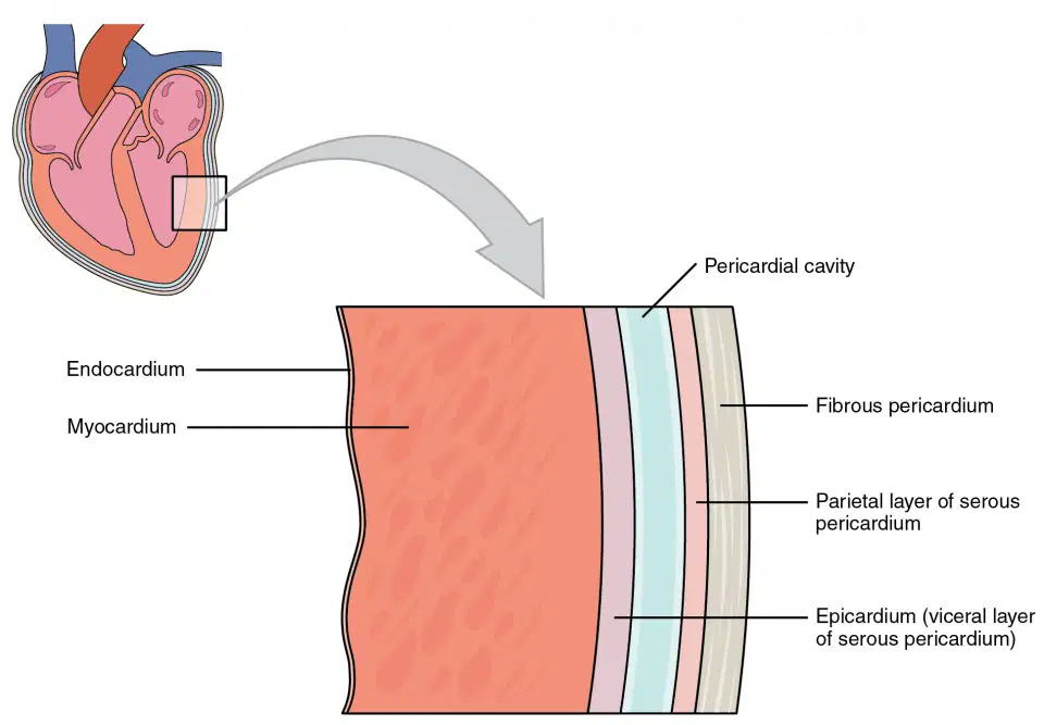
Heart coverings
; membrane around heart cavity
Parietal
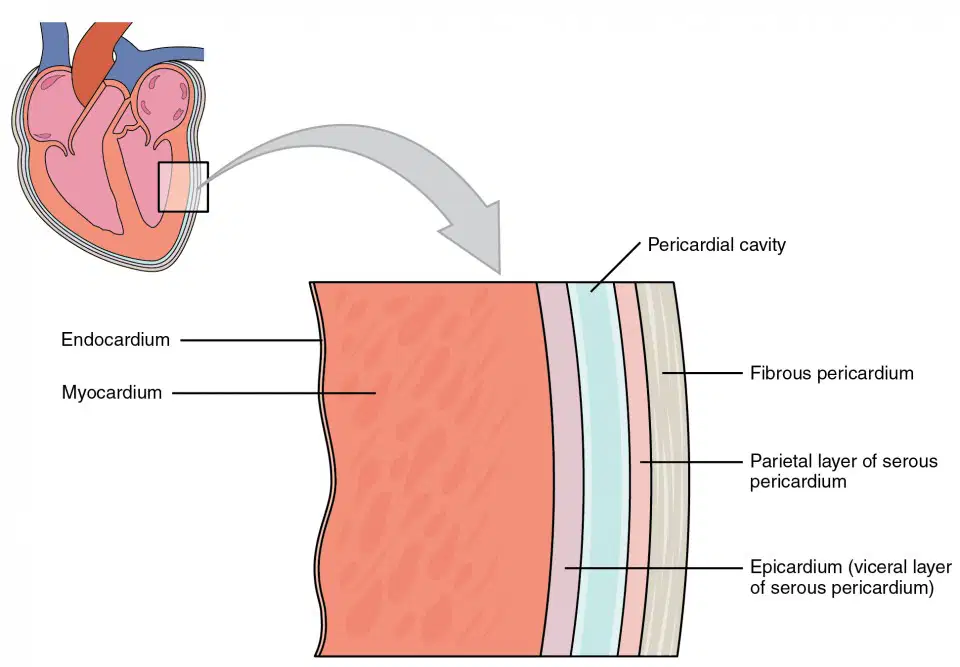
; surface of the heart
Visceral Pericardium
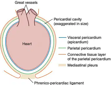
Heart coverings
; space around the heart inbetween of parietal and visceral pericardium

Heart layers
surface of the heart
Heart layers
- 2nd layer
- thickest, middle layer

Heart layers
-pinakaloob
- smooth inner surface
- 1 centrally located nucleus
- BRANCHING CELLS
- rich in mitochondria
- striated ( actin and myosin )
- Calcium and ATP
- Intercalated discs connect cells
Smooth muscle
Skeletal muscle
Cardiac muscle
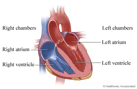
Chambers of the heart
Right Atrium (RA)
Right ventricle
( RV )
Left Atrium ( LA )
Right Atrium ( LV )
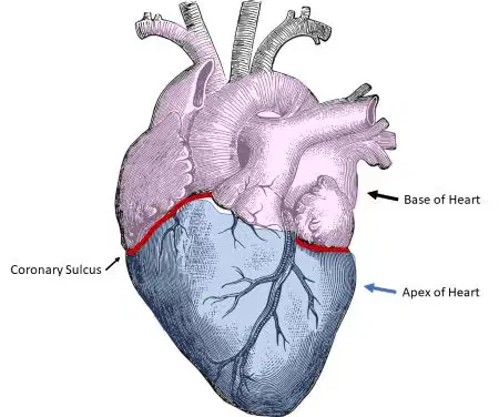
separates atria from ventricles
coronary sulcus

- a thin wall of tissue that separates the two upper chambers of the heart, called the right and left atria.
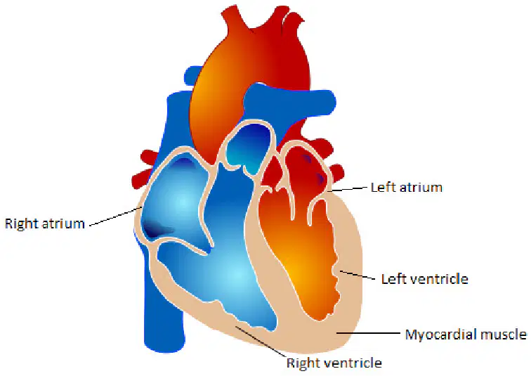
- upper portion
holding chambers
small, thin wall
contracts to push blood into the ventricles
Right atrium
Left atrium
Atrium
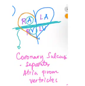
lower portion
“pumping” chambers
thick, strong wall
contract “forcefully” to propel blood into the different parts of the body
Right ventricle
Left ventricle
Ventricle

Parts of the heart
- structures that ensure one way blood flow
Superior Vena Cava
Valves
Aorta
Tricuspid valves
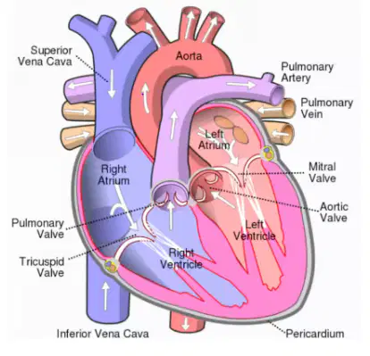
- located between atria and ventricles

3 cusps/leaflets
AV valve between right atrium and right ventricle
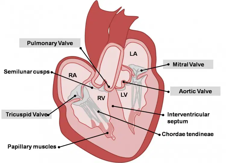
- 2 cusps/leaflets
Av valve between left atrium and left ventricle
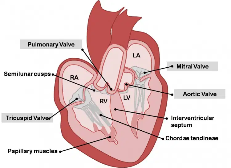
attached to AV valve flap
it supports valves
- STRING LIKE
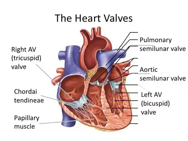
SEMILUNAR VALVES
- located on the base of the pulmonary trunk
Biscupid valve
Aortic semilunar valve
Pulmonary semilunar valve
Papillary muscle
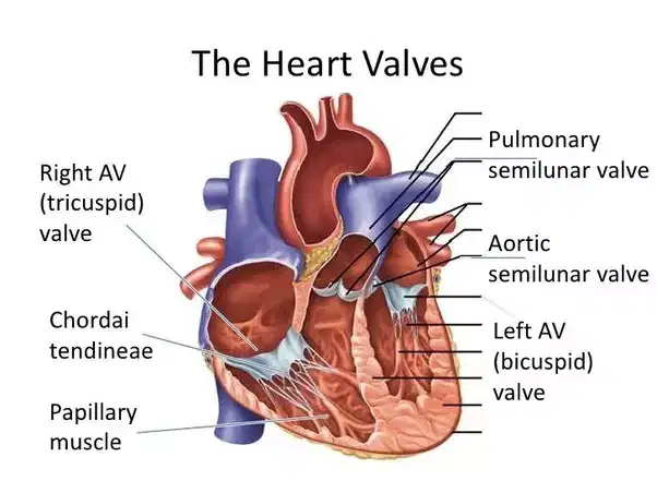
SEMILUNAR VALVES
- located on the base of aorta
Right AV valve
Aortic semilunar valve
Pulmonary semilunar valve
Papillary muscle
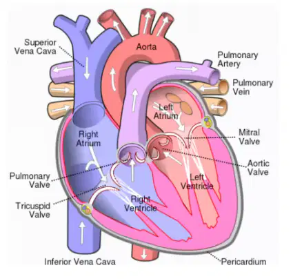
- drains blood: upper
head, neck, thorax, upper limbs
higher than diaphragm
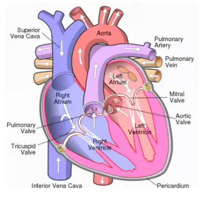
- drains blood: lower
below diaphragm
abdominopelvic cavity
lower limbs
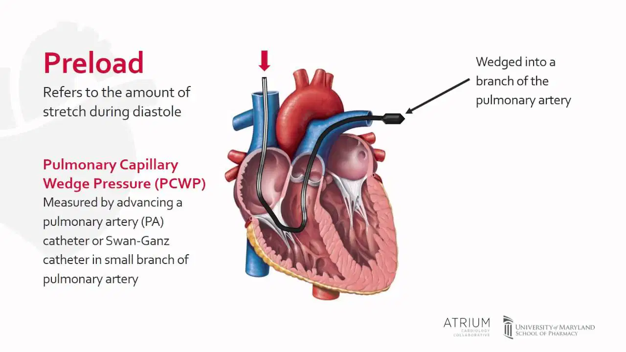
- amount of blood returning to the heart
blue
oxygen deficient
PRE-LOAD
AFTER-LOAD

- amount of blood going out of the heart
red
oxygenated
AFTER LOAD
PRE-LOAD
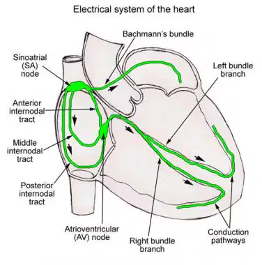
contraction of atria & ventricles by cardiac muscle cells
- same as electrical system
SA NODE (SINOATRIAL NODE)
CONDUCTION SYSTEM
CORONARY ARTERY
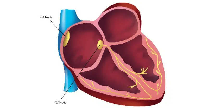
- located in the right atrium
where action potential originates
“pacemaker”
more/large calcium channels
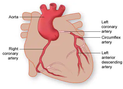
- supply blood to heart wall
originates from base of aorta (aortic valve)
- has two parts
- Left coronary artery
-Right coronary artery

- 3 branches
supplies blood to anterior heart wall & left ventricle
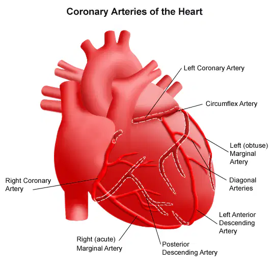
- located in right side of aorta
supplies blood to right ventricle
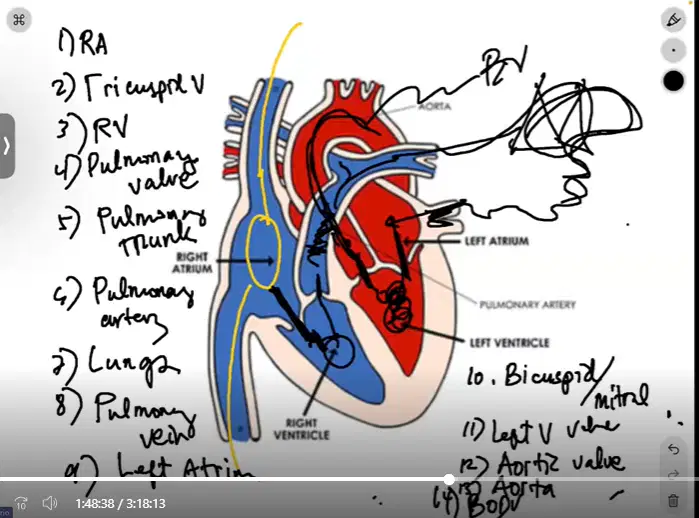
what chamber of the heart firstly gathers deoxygenated blood
- Superior vena cava = upper body
- inferior vena cava = lowerbody
- FIRST STEP
LEFT ATRIUM
RIGHT ATRIUM (RA)
RIGHT VENTRICLE
LUNGS
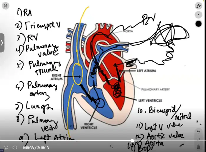
next step, the deoxygenated blood goes thru the what?
Pulmonary artery
bicuspid valve
Tricuspid valve
Pulmonary vein
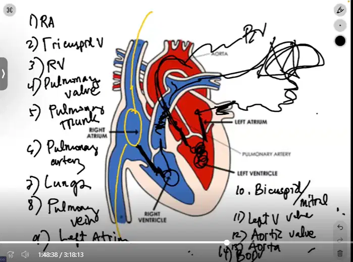
next, where does the dexoygenated blood go?
3rd step
LUNGS
RA
LA
RV
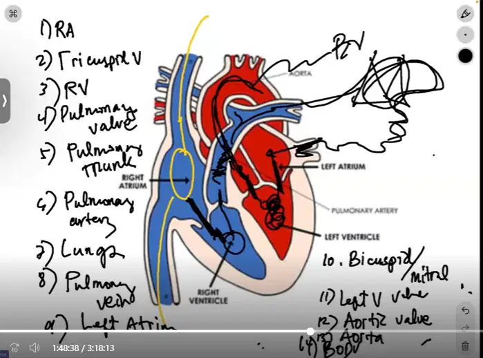
after going thru the RV, where would be the deoxygenated blood go?
4th step
Pulmonary valve
Pulmonary artery
Pulmonary trunk
Lungs
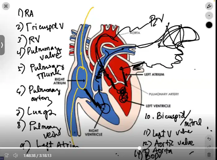
next, where does the deoxygenated blood go after entering the pulmonary valve?
- step 5
Pulmonary atrium
Pulmonary artery
Pulmonary trunk
Lungs
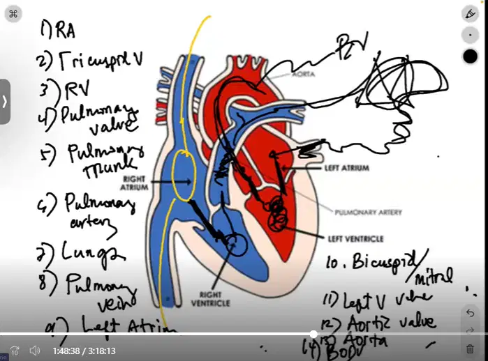
Next, the only BLUE ARTERY in the heart
- typically arteries are RED
- step 6
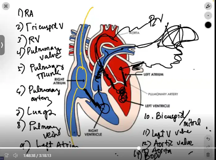
where oxygenation occurs
- step 7
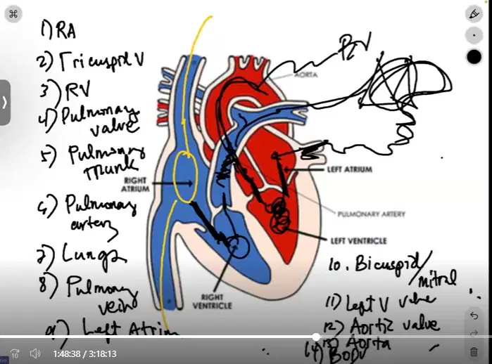
after oxygenation, where does the oxygen rich blood enter the heart?
- beside the left atrium
- step 8
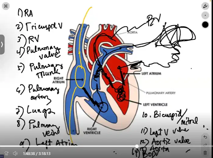
next, what chamber does and collects the blood from the pulmonary vein
- step 9
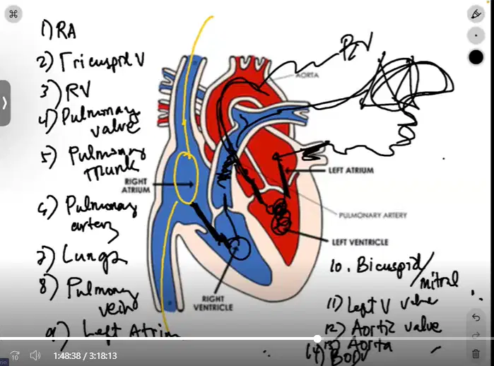
next, where does the blood go thru in order to go to the left ventricle
- step 10
Left atrium through the mitral/bicuspid valve to the left ventricle.
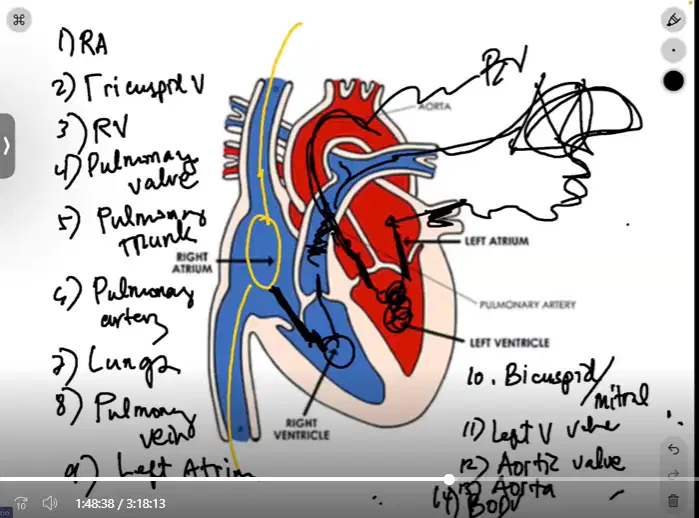
- where does the blood go after passing the bicuspid valve/mitral valve
step 11
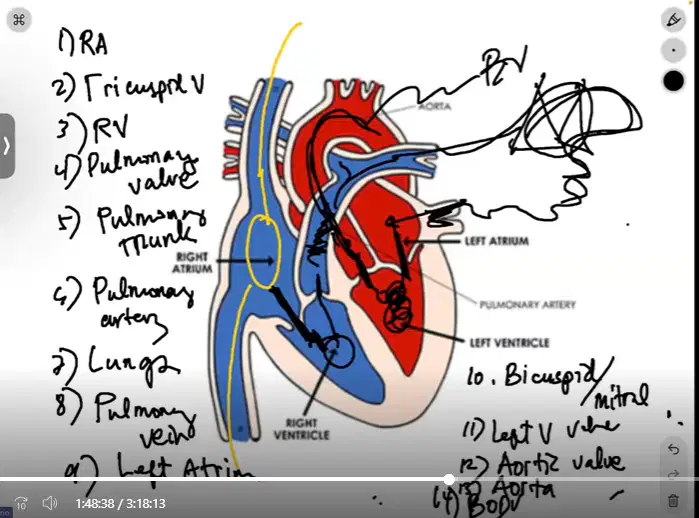
next, where is the blood going thru after the left ventricle?
Aortic valve
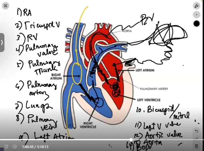
After the aortic valve, where does the oxygenated blood go before being pumped into the body?
-step 13
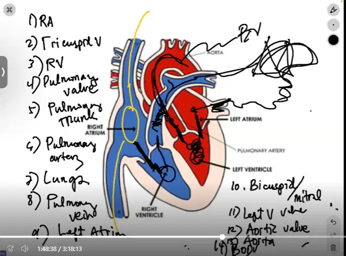
where does the oxygenated blood go after everything?
step 14

Conduction system of heart
- what are the parts of the left coronary artery?
Diagonal branches
Left circumflex
Left anterior decensding
Right circumflex
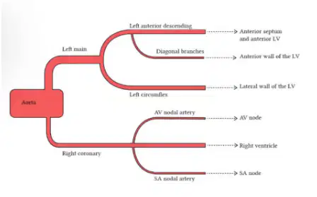
What are the parts of the Right coronary arteries?
AV Nodal Artery
SA Nodel Artery
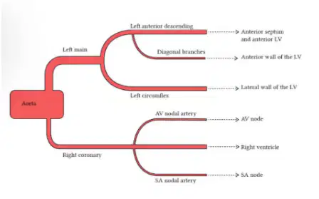
What does the left coronary artery provide blood to?
Anterior septum and anterior LV
Anterior wall
Left ventricle
Lateral wall of LV
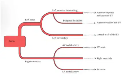
What does the right coronary artery provide blood to?
Right atrium, right ventricle, SA node, AV node.

Pathway of action potential thru the heart
- answers from left to right
SA NODE
AV NODE
AV BUNDLE
R AND L VENTRICLE

