
 Worksheets
Ophtha 250 Quizlet
Total questions: 25
Worksheet time: 17mins
Which of the following should be asked FIRST when assessing a patient who presents with a red eye?
How long have you had the redness?
Do you wear contact lenses
Is the redness associated with any discharge
Have you experienced any trauma to the eye
A 50-year-old diabetic patient presents with blurry vision and reports no pain. What question would be most helpful to assess if diabetic retinopathy is the cause?
Do you have high blood pressure?
Are you on any maintenance medications?
How well is your blood sugar control?
Do you have a family history of eye disease?
A 60-year-old man reports seeing halos around lights and difficulty seeing at night. Which additional question would help differentiate between cataracts and glaucoma?
Do you have a family history of eye problems?
Have you had any recent changes in your prescription glasses?
Do you feel any eye pain or pressure?
Do you experience double vision?
A patient's visual acuity is recorded as 20/40. What does this notation mean?
The patient can see at 40 feet what a person with normal vision can see at 20 feet.
The patient can see at 20 feet what a person with normal vision can see at 40 feet.
The patient can see at 40 feet what a person with normal vision can see at 40 feet.
The patient has vision twice as good as normal vision.
If a patient is unable to read the largest letter on the Snellen chart at 20 feet, what is the next appropriate step in assessing their vision?
Move the patient closer to the chart.
Perform a pinhole test.
Use the Jaeger chart for near vision instead.
Test for color vision using the Ishihara plates.
What is the correct procedure for testing visual acuity in a patient with glasses or contact lenses?
Test each eye individually without correction, then with correction.
Test both eyes together without correction.
Test each eye individually with correction, then without correction.
Test only with correction.
When performing a visual acuity test in a child, what is an appropriate action if the child is uncooperative or unable to focus on the chart?
Use a lower-resolution chart.
Perform a visual assessment using light reflex testing.
Use the Snellen chart at a shorter distance.
Stop the test and assume the child has poor vision.
Which of the following is the correct method for recording near visual acuity using a Jaeger chart?
Record the distance at which the patient reads the smallest line of text.
Record the fraction, similar to a Snellen chart.
Use the J scale and record the smallest line number the patient can read.
Use the pinhole to record near visual acuity.
Which of the following is assessed during the external inspection of the eye in a gross examination?
Visual acuity
Pupil reaction to light
Eyelid position and symmetry
Red reflex
Which of the following would be considered a normal finding in a gross eye examination?
Mild ptosis
Conjunctival pallor
Clear cornea with no opacities
Constricted pupils in dim light
When one eye is exposed to bright light, a ____________ occurs (constriction of that pupil) as well as a _____________(simultaneous constriction of the other pupil) occurs in normal patients.
Consensual light reflex, Primary light reflex
Primary light reflex, Secondary light reflex
Direct light reflex, Consensual light reflex
Consensual light reflex, direct light reflex
What would be the expected finding in the pupillary light reflex of a patient with complete oculomotor nerve palsy in the right eye?
Both pupils constrict equally when light is shined in either eye
The right pupil remains dilated when light is shined in either eye
The left pupil constricts when light is shined in the right eye, but the right pupil does not
Both pupils remain dilated when light is shined in the left eye
Which of the following is/are NOT a component/s of the near triad (accommodation reflex)?
Pupil dilation
Lens thickening
Convergence of the eyes
Pupil constriction
Thinning of the lens
In which of the following situations would intraocular pressure measurement be contraindicated?
A patient with a corneal infection
A patient using topical steroid eye drops
A patient with a history of cataract surgery
A patient with dry eye syndrome
A patient's intraocular pressures are reported as follows:
Right Eye = 16 mm Left Eye = 12 mm
How should these findings be interpreted?
The patient has normal IOPs; both measurements fall within the normal IOP range.
The patient has abnormal IOPs; both IOP measurements fall outside of normal IOP range.
A pathology should be ruled out; while both IOP readings are within normal IOP range, they exceed the allowable difference of 2 mm.
Which tonometry method is most appropriate for use in children and non-cooperative patients?
Goldmann applanation tonometry
Schiotz tonometry
Rebound tonometry
Dynamic contour tonometry
Which of the following factors can lead to falsely elevated intraocular pressure measurements
using Goldmann applanation tonometry?
Thin corneas
Thick corneas
Use of anesthetic eye drops
Dilated pupils
Which of the following symptoms is commonly associated with extraocular muscle palsy?
Photophobia
Blurred vision
Diplopia
Eye pain
When performing motility testing, a patient is asked to look at a specific direction to assess the function of an extraocular muscle. Which of the pairings between direction and EOM is/are correct?
Inferior rectus:
down and out
Inferior oblique: down and in
Superior rectus:
up and out
Superior oblique:
down and in
Superior oblique: down and out
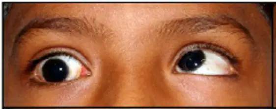
Based on this photo of a patient on right upgaze, which EOM exhibits limitation in movement?
Right superior rectus
Right inferior oblique
Right inferior rectus
Right superior oblique
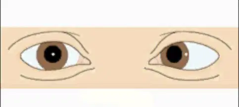
If the corneal light reflection is displaced outward as shown in the photo, what does the patient have?
hypertropia
hypotropia
esotropia
exotropia
In direct ophthalmoscopy, what is the purpose of adjusting the diopter settings?
To focus the ophthalmoscope on different depths of the eye
To illuminate the retina more clearly
To adjust the field of view
To magnify the optic disc
Which of the following is the normal color of a healthy optic disc observed
during a fundus examination?
Pale white
Pinkish-yellow
Bright red
Dark brown
Select disc/s with normal cup:disc ratio.


Select the normal fundus.



