
Font size
 Worksheets
SPI Review Part 3
Total questions: 84
Worksheet time: 42mins
What is a characteristic of read magnification in image processing?
Rescans and acquires new data
Reads old data
Increases line density
Improves spatial resolution
Which type of magnification can improve temporal resolution if the region of interest is shallower?
Read magnification
Write magnification
Both read and write magnification
Neither read nor write magnification
In write magnification, what happens if the pixel size is unchanged?
Number of pixels decreases
Number of pixels increases
Spatial resolution decreases
Temporal resolution remains unchanged
What is the appearance of speckle in tissues?
Smooth and uniform
Grainy or granular
Bright and clear
Dark and shadowy
What causes speckle in imaging artifacts?
Reflection of light
Refraction of sound
Interference effects of scattered sound
Absorption of heat
What is the effect of speckle on image quality?
Enhances image contrast
Reduces image contrast and spatial resolution
Improves diagnostic accuracy
Increases image brightness
What is clutter in Doppler artifact?
Strong reflections from blood cells
Weak reflections from anatomic structures
Strong reflections from anatomic structures
Weak reflections from blood cells
What is the purpose of fill-in interpolation in imaging?
To remove noise
To fill in gaps between lines
To enhance color
To increase brightness
What does interpolation improve in images?
Color accuracy
Image detail (spatial resolution)
Image brightness
Image contrast
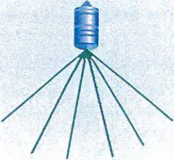
Diagram showing fill-in interpolation with sector-shaped image and circular structure
Fill-in interpolation with sector-shaped image
Circular structure with fill-in interpolation
Sector-shaped image with circular interpolation
Diagram showing fill-in interpolation with sector-shaped image and circular structure
What is the main benefit of using spatial compounding in ultrasound imaging?
Increases artifacts
Reduces spatial resolution
Improves signal-to-noise ratio
Decreases frame rate
Which type of transducers are used for spatial compounding?
Linear array transducers
Phased array transducers
Convex array transducers
Mechanical transducers
What is another name for temporal compounding?
Spatial averaging
Frequency compounding
Temporal averaging
Dynamic compounding
What effect does temporal compounding have on temporal resolution?
Increases temporal resolution
Decreases temporal resolution
No effect on temporal resolution
Eliminates temporal resolution
What is the primary benefit of frequency compounding in image processing?
Increases the speed of image creation
Reduces the need for manual adjustments
Improves signal-to-noise ratio
Enhances color accuracy
Which artifact is reduced by frequency compounding?
Motion artifact
Speckle artifact
Color artifact
Shadow artifact
In frequency compounding, what remains unchanged?
Spatial resolution
Temporal resolution
Frequency resolution
Color resolution
What is dynamic aperture primarily used for in image processing?
Enhancing color depth
Electronic receive focusing
Increasing image brightness
Reducing image size
How does dynamic aperture optimize lateral resolution?
By increasing the number of sound pulses
By varying the number of elements used to receive the signal
By reducing the frequency range
By enhancing color contrast
What is the primary purpose of edge enhancement in imaging?
To reduce noise in the image
To increase the contrast at a boundary
To add color to grayscale images
To decrease the resolution
What does coded excitation improve in imaging?
Color accuracy
Image brightness
Axial resolution
Image size
What is the main function of elastography?
To measure temperature changes
To identify tissues of different mechanical properties
To enhance color contrast
To reduce image size
What is shear wave elastography used to measure?
Blood flow speed
Tissue temperature
Speed of sideways created sound waves
Image brightness
What is the purpose of rendering in image processing?
To create 2-D images
To enhance audio quality
To add realism to 3-D or 4-D images
To reduce file size
What is the unit of measurement for dynamic range?
Hertz
Decibels (dB)
Lumens
Pixels
Which of the following is a function of PACS in recording and archiving?
Enhancing image resolution
Storing and forwarding teleradiology
Compressing video files
Editing audio tracks
What does DICOM stand for?
Digital Imaging and Communications in Medicine
Data Integration and Communication in Media
Dynamic Imaging and Computation in Medicine
Digital Information and Communication in Media
What does "hyperechoic" refer to in image characteristics?
Portions of an image that are brighter than surrounding tissues
Portions of an image that are not as bright as surrounding tissues
Describes structures with equal echo brightness
Displaying a variety of different echo characteristics
What is the meaning of "anechoic" in the context of image characteristics?
With echoes; echo-rich
Without echoes; echo-free
Equal echo brightness
Varied echo characteristics
Which term describes structures with equal echo brightness?
Hyperechoic
Hypoechoic
Isoechoic
Heterogeneous
What does "heterogeneous" mean in image characteristics?
Equal echo brightness throughout
Displaying a variety of different echo characteristics
Brighter than surrounding tissues
Less bright than surrounding tissues
Which of the following is NOT a cause of artifacts in imaging?
Violation of assumptions
Equipment malfunction
Proper brightness
Operator error
Which of the following is one of the basic assumptions of imaging systems?
Sound travels in a straight line.
Sound travels in a circular path.
Sound travels at varying speeds.
Sound does not reflect back.
What happens to artifacts when corrective measures are taken?
Artifacts vanish.
Artifacts become more prominent.
Artifacts change color.
Artifacts multiply.
Reverberations in imaging systems are created when:
US "ping-pongs" between two reflectors.
Sound travels in a circular path.
The imaging plane is extremely thick.
Reflections arise from random structures.
What is a characteristic of a comet tail artifact in sonography?
Single, solid hyperechoic line
Multiple, scattered hypoechoic lines
Circular, anechoic region
Irregular, hypoechoic pattern
What can create comet tail artifacts in ultrasound imaging?
Large fluid-filled structures
Small metal objects
Dense bone structures
Air pockets
What is a common cause of shadowing in ultrasound imaging?
Excessive reflection from the surface
Too much attenuation in the structure above the shadow
Increased transmission through the structure
Enhanced echo from the region beneath
What does shadowing indicate in an ultrasound scan?
Presence of true anatomy
Absence of true anatomy in the region of the shadow
Increased echo intensity
Uniform tissue density
What is the cause of edge shadowing by refraction in ultrasound imaging?
Hyperechoic foreground color
Refraction and beam spreading
Increased attenuation
High-frequency sound waves
How can edge shadowing by refraction be eliminated in ultrasound imaging?
By increasing the frequency
By using spatial compounding
By reducing the gain
By adjusting the focus
What does hyperechoic enhancement indicate in ultrasound imaging?
Too few reflections on the scan
Brighter echoes on the scan
Absence of true anatomy
Increased attenuation
What results from too little attenuation in the structure above the artifact in ultrasound imaging?
Edge shadowing
Hypoechoic background
Hyperechoic enhancement
Increased refraction
What is the role of a strong reflector, called a mirror, in ultrasound imaging?
It absorbs sound waves completely.
It redirects sound waves along a different path.
It amplifies the sound waves.
It changes the frequency of the sound waves.
In ultrasound systems, what assumption is made about the travel of sound?
Sound travels in a circular path.
Sound travels directly to a reflector and back to the transducer.
Sound travels in a zigzag pattern.
Sound travels in a spiral path.
What is an artifact in the context of ultrasound imaging?
A real anatomical structure.
A duplicate of real anatomy appearing on a scan.
A sound wave distortion.
A color change in the image.
What happens if the media through which ultrasound travels does not propagate at 1.54 km/s?
The image will be clearer.
The assumed relationship between time and distance is invalid.
The scan will have fewer reflections.
The depth will be accurate.
What are the two main results of propagation speed errors in ultrasound imaging?
Increased image brightness and reduced noise.
Correct number of reflections and proper depths.
Correct number of reflections and improper depths.
Fewer reflections and accurate depths.
How do speed errors appear on an ultrasound display?
As a blur.
As a step-off, split, or cut.
As a bright spot.
As a shadow.
What happens when the speed of sound is slower than in soft tissue?
Reflector is placed too shallow on the display.
Reflector is placed too deep on the display.
Reflector is not visible on the display.
Reflector is placed at the correct depth.
What is a refraction artifact in sonography?
An artifact that appears as a duplicate image at a different depth
An artifact that appears side-by-side with the true anatomic structure
An artifact that appears as a shadow behind a structure
An artifact that enhances the brightness of a structure
What effect does a refraction artifact have on lateral resolution?
It improves lateral resolution
It has no effect on lateral resolution
It degrades lateral resolution
It enhances lateral resolution
Under what conditions does refraction occur in sonography?
When sound travels through a uniform medium
When sound reflects off a smooth surface
When sound occurs with oblique incidence and different speeds
When sound is absorbed by a dense structure
What is the main cause of side lobes in ultrasound imaging?
Mechanical transducers
Array transducers
Subdicing
Apodization
How can grating lobes be reduced in ultrasound imaging?
By increasing the beam's main axis
By using mechanical transducers
By subdicing
By ignoring the lobe artifact
What is the process called that involves exciting subelements with different voltages to reduce grating lobes?
Subdicing
Apodization
Reflection
Diffraction
What effect do lobe artifacts have on ultrasound imaging?
Improve lateral resolution
Degrade lateral resolution
Enhance image clarity
Increase image size
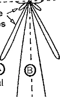
Diagram showing side lobes and grating lobes in ultrasound imaging
Diagram showing main lobe and side lobes in ultrasound imaging
Diagram showing only main lobe in ultrasound imaging
Diagram showing only grating lobes in ultrasound imaging
Diagram showing side lobes and grating lobes in ultrasound imaging
What is the term used to describe the artifact that occurs when an ultrasound beam has a greater width than the reflector?
Partial volume artifact
Elevational resolution
Speckle
Linear resolution
Which type of transducer is used to cure the slice thickness artifact?
Linear array transducer
Convex array transducer
Annular array transducer
Phased array transducer
What is the appearance of speckle in ultrasound images?
Clear and sharp
Grainy
Blurry
Colorful
What causes the speckle artifact in ultrasound images?
Reflection from a single surface
Refraction of sound waves
Interference effects of scattered sound
Absorption of sound waves
What is a Range Ambiguity Artifact?
Created by reflection from a structure located deeper than the maximum imaging depth
Caused by a shallow reflector in the image
Occurs when the sound beam is smaller than the reflector
Happens when the imaging depth is less than 5 cm
How can a Range Ambiguity Artifact be cured?
By increasing the PRF
By imaging deeper
By using a wider beam
By decreasing the frequency
In the given example, if the maximum imaging depth is 8 cm and the boundary is located 11 cm deep, where will the boundary appear on the image?
At a depth of 11 cm
At a depth of 8 cm
At a depth of 3 cm
At a depth of 5 cm
What happens when the sound beam is larger than the reflector?
Artifacts are minimized
Artifacts are created
Resolution is improved
No effect on the image
Which type of pulse is better to avoid artifacts?
Long pulses
Short pulses
Medium pulses
Pulses with high frequency
What is the purpose of routine periodic evaluation of the US system in a clinical environment?
To increase patient visits
To guarantee optimal image quality
To reduce staff workload
To enhance software features
Which of the following is a requirement for quality assurance in the clinical environment?
Hiring more staff
Multiple evaluations of the system's components
Increasing patient appointments
Reducing equipment costs
What is one of the objectives of quality assurance in the clinical environment?
Increase patient wait time
Detect gradual changes
Decrease equipment usage
Limit staff training
According to the document, who is ultimately responsible for quality assurance in a clinical environment?
The manufacturer’s service engineer
The hospital administrator
The sonographer
The patient
What is a phantom used for in quality assurance?
To provide a personal opinion
To serve as an objective standard
To replace the sonographer
To increase image size
What is the speed of sound in the AIUM 100mm Test Object - Water-Filled Phantom?
1,540 m/s
1,500 m/s
1,600 m/s
1,450 m/s
What does the pin set labeled 'D' evaluate in the AIUM 100mm Test Object?
Dead Zone
Lateral Resolution
Horizontal Registration
Vertical Registration
What is the minimum distance between two pins that are still individually identified in the test object?
1 mm
2 mm
3 mm
4 mm
What is the purpose of a Tissue Equivalent Phantom?
To mimic soft tissue with pins, mock cysts, and masses
To simulate bone density
To replicate muscle tissue
To imitate brain tissue
What is the speed of sound in a Tissue Equivalent Phantom?
1,540 m/s
1,200 m/s
1,800 m/s
1,000 m/s
What is a Doppler Phantom used to assess?
The accuracy of pulsed, continuous wave, and color flow systems
The density of bone structures
The elasticity of muscle tissue
The temperature of body fluids
What type of phantoms may be used to evaluate Doppler systems?
Vibrating string and moving belt phantoms
Static and dynamic phantoms
Gel and liquid phantoms
Solid and hollow phantoms
What determines the minimum sensitivity in a test object?
The gain setting at maximum value
The TGC set to maximum
Increasing the gain from its minimum value
The use of a gel pad
What is the normal sensitivity setting in the AIUM test object?
Lower than minimum sensitivity
The same as minimum sensitivity
Higher than minimum sensitivity
Not related to gain settings
What is the purpose of using an acoustic standoff or gel pad?
To increase the gain
To eliminate the dead zone
To adjust the TGC
To enhance the CRT display
What is the machine's ability to display echoes in the proper depth called?
Horizontal Calibration
Registration Accuracy
Range Accuracy
Minimum Sensitivity
What is the speed of sound in the phantom that may cause an error in range accuracy?
1.54 km/sec
1.54 m/sec
1.54 cm/sec
1.54 mm/sec

