
 Worksheets
EKG Module 5 Basic Cardiac Anatomy
Total questions: 20
Worksheet time: 10mins

8
right ventricle
left ventricle
aorta
pulmonary artery
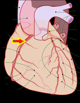
Identify the structure
right coronary artery
left coronary artery
pulmonary artery
aorta
Artifact:
Patient becomes dislodged from the electrode or broken wires.
Improper calibration of the device.
Interference from nearby electronic devices.
Incorrect placement of the electrodes.

14
superior vena cava
inferior vena cava
pulmonary artery
aorta

5
internodal pathways
sinoatrial node
atrioventricular node
bundle of His
SA node artery
Branches from the RCA (Right circumflex artery)
Branches from the LCA (Left circumflex artery)
Branches from the LAD (Left anterior descending artery)
Branches from the Aorta

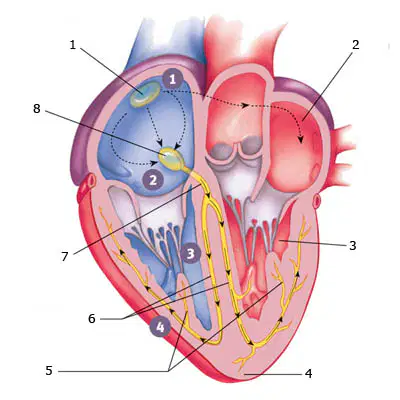
What is #2
pulmonary artery
aorta
coronary artery
jugular vein

15
superior vena cava
pulmonary artery
inferior vena cava
aorta
6
Right and left bundle branches
Aorta and pulmonary artery
Superior and inferior vena cava
Left atrium and right atrium
Artifact: Wandering baseline
Improper skin prep, leaving traces of oil or gel on the skin.
Excessive movement during the procedure.
Using outdated equipment.
Incorrect electrode placement.

11
left ventricle
right atrium
right ventricle
pulmonary artery
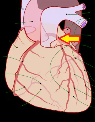
identify the structure
left coronary artery
right coronary artery
pulmonary artery
aorta

9
apex
base
vertex
summit

5
left atrium
right atrium
left ventricle
right ventricle
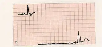
Identify the artifact.
Interrupted baseline.
Continuous waveform.
Flat line.
Irregular rhythm.
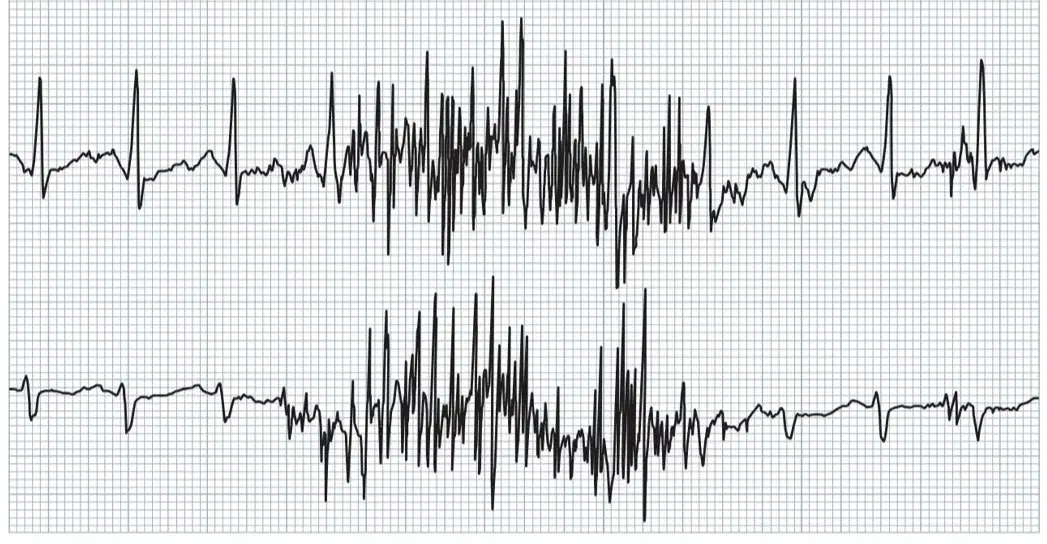
Identify the artifact.
Somatic Tremor
Cerebral Wave
Neural Oscillation
Electromagnetic Interference
occlusion of SA node artery=
extreme sinus bradycardia
atrial fibrillation
ventricular tachycardia
sinus tachycardia

7
tricuspid valve
pulmonary valve
mitral (bicuspid valve)
aortic valve
SA node artery
Perfuses the SA node
Supplies blood to the AV node
Provides oxygen to the left atrium
Nourishes the right ventricle

What is #1?
Aorta
Pulmonary Artery
Superior Vena Cava
Coronary Artery

