
 Worksheets
Intro: Nervous System, Special Senses & Endocrine
Total questions: 66
Worksheet time: 37mins
SensoryNeuron
Integration
Thermogensis
Motor Neuron
Motor Neuron
Integration
Sensory Neuron
Thermogensis
None of the options. They are all part of the CNS
Cerebellum
Cerebrum
Spinal Cord
Brain Stem
Cerebellum
Thalmus
Brainstem
Pituitary Gland
Occiptal
Parietal
Temporal
Frontal
Temporal
Frontal
Parietal
Occipital
Brain Stem
Diencephalon
Cerebrum
Cerebellum
Exchange of nutrients (nourishes) and waste products between the blood and nervous tissue
connect the brain and spinal cord
Detect changes and carry impulses to the brain and spinal cord
Is the main control center of the body
Olfactory Receptors
Motor Neurons
Optic Nerve
Mechanoreceptors
Middle Ear
Inner Ear
Outer Ear
Cochlea

Match the following definitions to their vocabulary words.
a sensory organ responsible for vision. It contains specialized structures like the cornea, lens, and retina that work together to capture light, focus it, and convert it into electrical signals that can be interpreted by the brain.
Eye
the body's largest sensory organ, responsible for the sense of touch. It contains various types of nerve endings that detect pressure, temperature, pain, and other sensations, sending signals to the brain for interpretation.
Skin
an intricate network of nerve cells, or neurons, that transmits signals between different parts of the body. It is divided into the central nervous system (CNS), which includes the brain and spinal cord, and the peripheral nervous system (PNS), which connects the CNS to the rest of the body.
Nervous System
specialized structures in the body that detect and respond to external stimuli, such as light, sound, touch, taste, and smell. They convert these stimuli into electrical signals that are transmitted to the brain for interpretation.
Sensory Organs
a sensory organ responsible for the sense of smell, or olfaction. It contains specialized cells called olfactory receptors that detect and identify odor molecules, sending signals to the brain for interpretation.
Nose


What body system controls other body systems?
Skeletal System
Nervous System
Digestive System
Circulatory System
Respiratory System

The major function of your nervous system demonstrated by #3 is:
sensory input
integration
motor output

The major function of your nervous system shown by #2 is
Accommodation
Integration
Regulation
Motor input
Adaptation


Which 2 are parts of the central nervous system?
brain
nose
spinal cord
left big toe

What is number 3 pointing at on the neuron?
Dendrite
Axon
Myelin Sheath
Soma

What part of the neuron is number 4 pointing at?
Dendrite
Axon Terminal
Axon
Myelin Sheath

Which part of the Neuron is number 1 pointing at?
Dendrite
Cell body
Axon
Nucleus
The Central Nervous System is made up of which TWO parts?
Brain
Arms
Legs
Spine

This division consists of sensory and motor neurons that transmit information to and from the central nervous system
Central nervous system
Cerebrum
Peripheral nervous system
Medulla oblongata
What hormone increase our heart rate
(a)
How many pairs of spinal nerves are there?
(a)
Excess hormone production that causes moon face and buffalo hump
Huntington's disease
GBS
Cushing's syndrome
TBI
FIGHT OR FLIGHT:
decreases heart rate
increase heart rate
reverses symptoms
makes you fly
Neurological disorder marked by reoccurrence of episodes or a sensory disturbance.
Epilepsy
Concussion
Traumatic Brain Injury
ALS

Which organ is represented by letter B?
Brain
Spinal Cord
Nerves
Which body system coordinates all of the systems by controlling every activity that takes place?
The Nervous System
The Digestive System
The Endocrine System
The Circulatory System
The Immune System
Which organ is represented by letter A?
Brain
Spinal Cord
Nerves
Which organ is represented by letter C?
Brain
Spinal Cord
Nerves
Digestion, Heartbeat, and Reflexes are examples of
Voluntary Processes
Involuntary Processes
Speaking, Running, and Karate Chopping are examples of
Voluntary Processes
Involuntary Processes
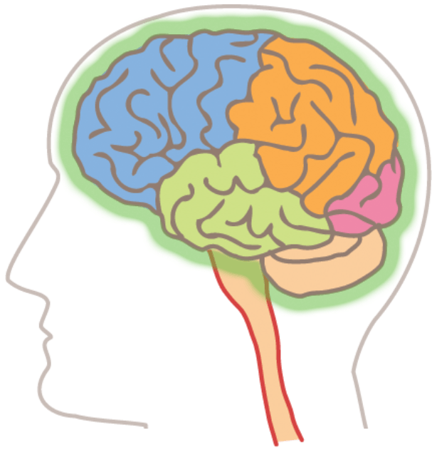
Identify all the parts of the brain by their color (only the parts highlighted green).
Frontal lobe
Blue
Temporal Lobe
Green
Parietal Lobe
Orange
Occipital Lobe
Red
Cerebellum
Beige

What part is the highlighted box pointing to. It is number 4 on the image.
Tympanic Cavity
Malleus
Auditory Tube
Ear Drum

What part of the ear is the highlighted box pointing to. it is number 1 on the image.
Tympanic Cavity
Ear Drum
Auricle
Stapes

What part is the highlighted box pointing to? It is number 5 on the image.
Malleus
Stapes
Auditory Canal
Tympanic Cavity

What part is the highlighted box pointing to? It is number 2 on the image
Stapes
Incus
Semicircular Ducts
Ear Drum

What part is the highlighted box pointing to? It is number 3 on the image.
Auricle
Ear Drum
Malleus
Incus

What part is the highlighted box pointing to? It is number 12 on the image.
Auditory Tube
Auditory Canal
Tympanic Cavity
Cochlear Nerve


What part is the highlighted box pointing to? It is number 7 on the image.
Cochlea
Cochlear Nerve
Tympanic Cavity
Auditory Canal


Which number identifies the external auditory meatus?
1
2
3
8
Which number identifies the auricle?
1
2
3
none of these
Which number identifies the malleus?
3
4
5
6
Which number identifies the tympanic membrane?
1
2
3
9
Which number identifies the stapes?
3
4
5
6
Which number identifies the semicircular canals?
(a)
Which number identifies the incus?
3
4
5
6
Which number identifies the cochlea?
(a)
Which number identifies the eustachian tube?
(a)

