
 Worksheets
Jatin Shah Campur
Total questions: 50
Worksheet time: 50mins
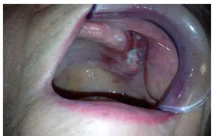
A 65-year-old woman presents with an ulcerated lesion of the left upper gum measuring 3 × 2 cm. A staging CT scan shows erosion of the bone of the upper alveolus with extension into the maxillary sinus. No enlarged cervical lymph nodes were present. The stage of the cancer in this patient is
I
II
III
IVa
IVb
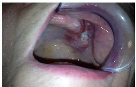
A 65-year-old woman (Figure 5-8) presents with an ulcerated lesion of the left upper gum measuring 3 × 2 cm. A staging CT scan shows erosion of the bone of the upper alveolus with extension into the maxillary sinus. No enlarged cervical lymph nodes were present. Management of the primary tumor is by
Chemoradiation therapy
Upper alveolectomy with a split-thickness skin
graft
Upper alveolectomy and an obturator
prosthesis
Subtotal maxillectomy with an obturator
prosthesis
Total maxillectomy with a rectus flap
Which oral cancer subsite is associated with the
highest rate of occult neck metastases?
Buccal mucosa
Hard palate
Oral tongue
Lower gum
Upper gum
Which is not an indication for segmental mandibulectomy?
Osteosarcoma of the mandible
Metastases to the mandible
Gross invasion of the mandible by SCC
Invasion of the mandibular canal and inferior
alveolar nerve
Odontogenic keratocyst
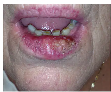
The patient shown in picture an SCC of the middle third of the lower lip. What are the most common lymph nodes at risk?
Submandibular lymph nodes
Submental nodes
Level II nodes
Level III nodes
Level IV nodes
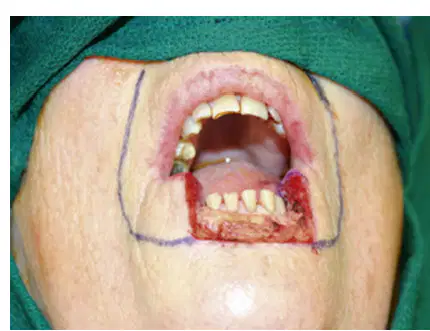
The defect after resection is shown in Figure. This is best repaired by a
Skin graft.
Free radial forearm flap
V-Y advancement flap.
Abbe-Estlander flap.
Bilateral Karapandzic flaps.
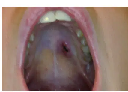
The patient in Figure has an adenoid cystic cancer of the hard palate. Which of the following is false regarding adenoid cystic cancer?
High incidence of local recurrence
Characteristic perineural invasion
Metastasizes to regional lymph nodes
Metastasizes to the lung
Classified by histology into cribriform, solid,
and tubular subtypes
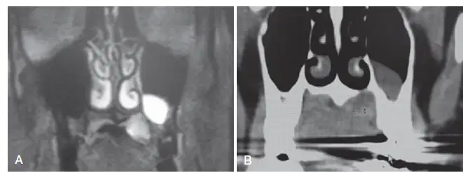
Figure shows a CT scan of the patient in Question 54. The patient should be managed by
Local resection and secondary intention.
Composite resection and an obturator prosthesis.
Composite resection and a local flap.
Soft tissue resection and a skin graft.
Local resection and a rotation flap.
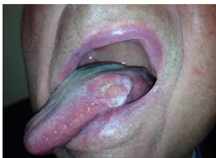
The patient in Figure has an SCC of the lateral border of the oral tongue. The tumor measures 3 cm × 2 cm. A CT scan shows no enlarged lymph nodes. The stage of the patient is
I
II
III
IVa
IVb

The patient in Figure has an SCC of the lateral border of the oral tongue. The tumor measures 3 cm × 2 cm. A CT scan shows no enlarged lymph nodes.
Elective treatment of the neck in this case involves dissection of levels
I, II, III
I, II, III, IV
II, III, IV
I, II
I, II, III, IV, V
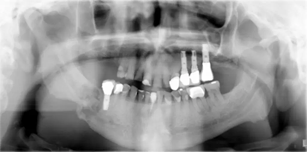
Optimal management of the osteoradionecrosis
shown in Figure is
Curettage and antibiotics
Hyperbaric oxygen.
Hemimandibulectomy and free fibula flap
reconstruction.
Hemimandibulectomy and primary closure
Hemimandibulectomy and a free rectus flap

The patient in this Figure SCC of the left lower gum. Assessment of the mandible is best done by
CT scan
Magnetic resonance imaging
Clinical examination
Positron emission tomography
Orthopantomogram

The primary tumor in this Figure measures 5 cm × 3 cm. A CT scan shows erosion of the cortex of the mandible. Two enlarged lymph nodes with central necrosis are shown in level 1b. One lymph node measures 4 × 2 cm and the other 1.5 × 2 cm. Under the TNM staging system, this patient’s tumor is classified as
T3N2a.
T3N2b.
T2N2b.
T4N2b.
T4N2a.
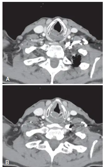
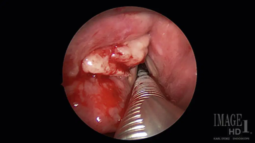
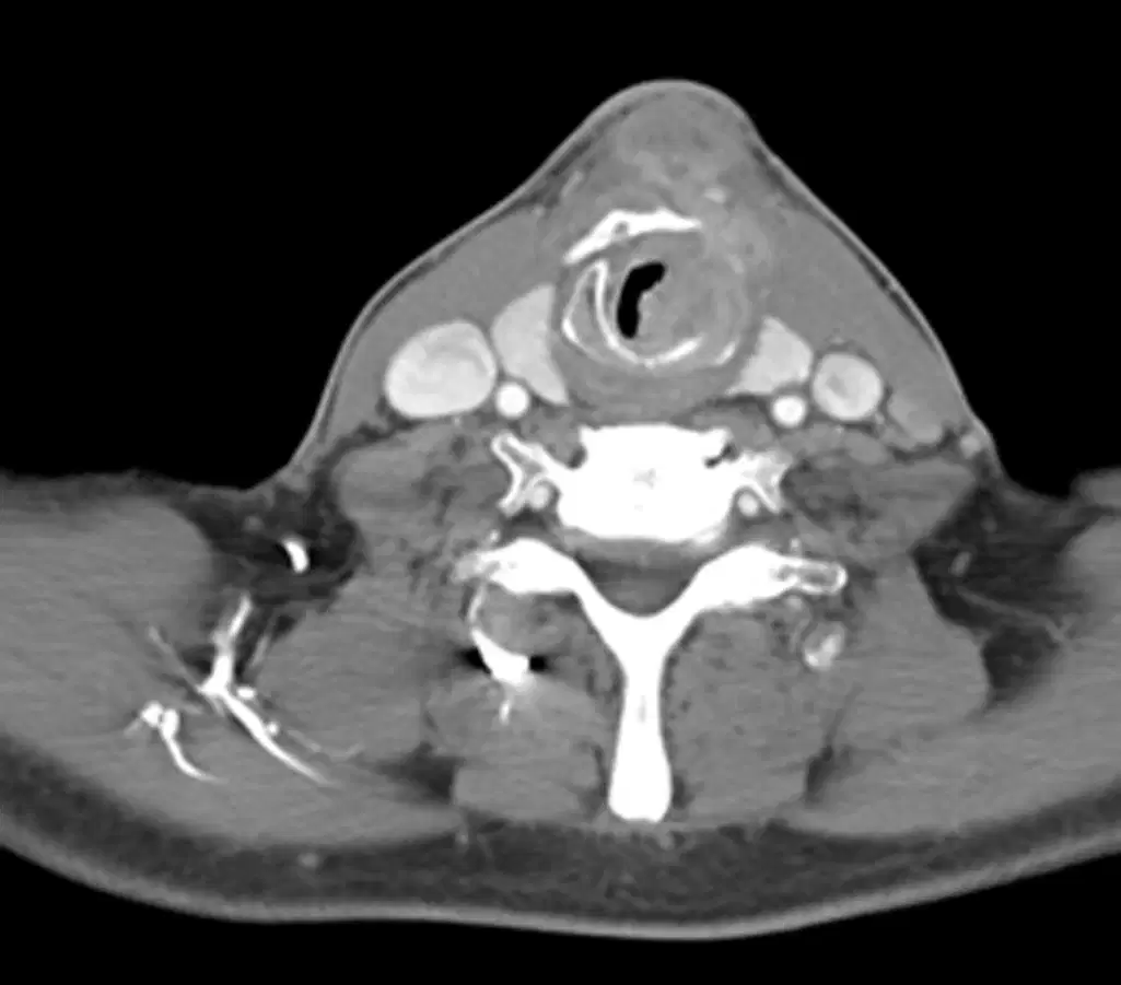
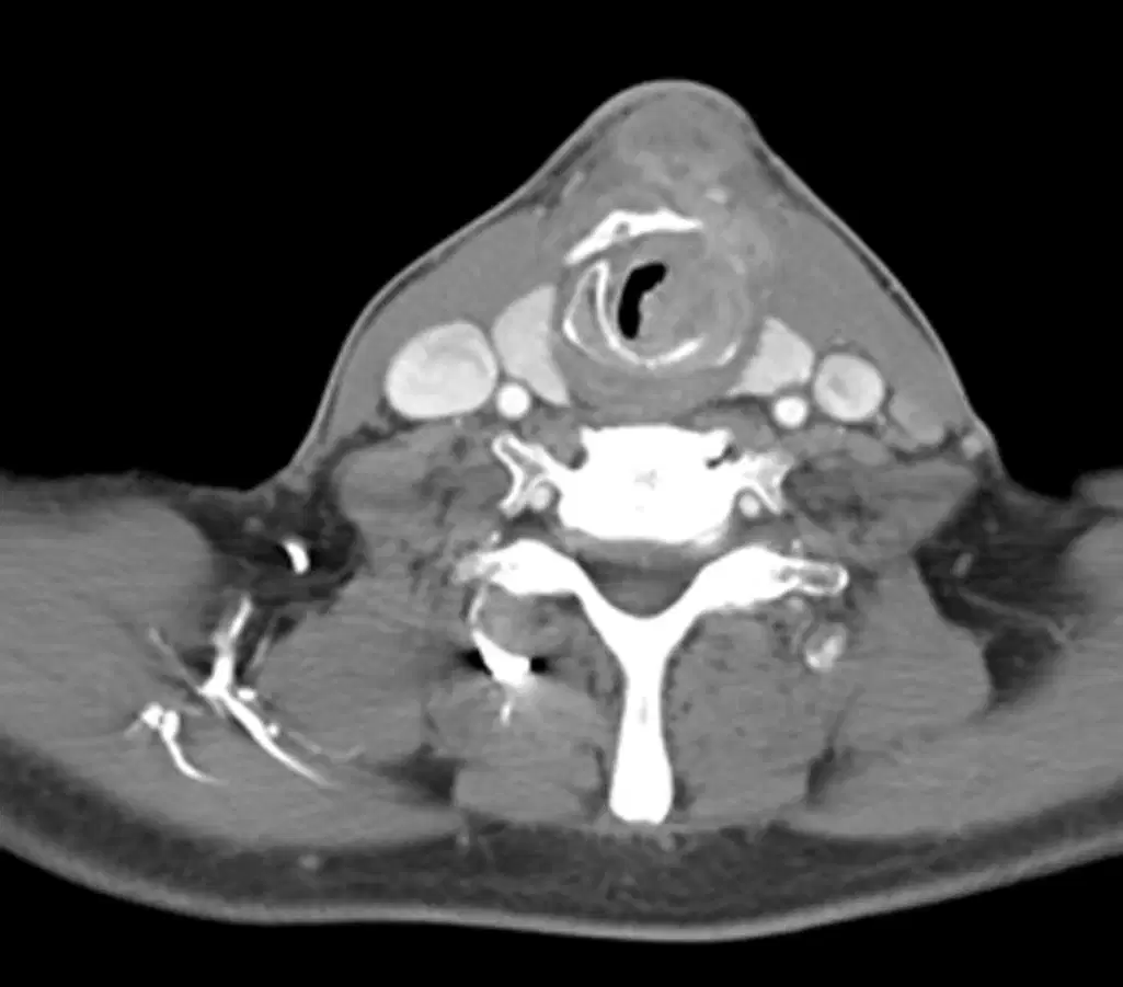
A 65-year-old man presents with 6 months of occasional hoarseness. A complete head and neck examination is performed and shows no lymphadenopathy. Flexible laryngoscopy shows a fullness of the subglottis that is incompletely visualized. Normal vocal cord movement is noted. A computed tomographic (CT) scan is displayed. The next best step in the management of should be
Positron emission tomography (PET)/CT
Magnetic resonance imaging (MRI).
Direct laryngoscopy with biopsy.
Videostroboscopy
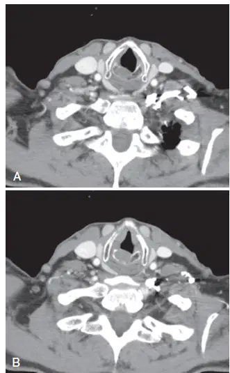
A 65-year-old man presents with 6 months of occasional hoarseness. A complete head and neck examination is performed and shows no lymphadenopathy. Flexible laryngoscopy shows a fullness of the subglottis that is incompletely visualized. Normal vocal cord movement is noted. A computed tomographic (CT) scan is displayed Which of the following is the least likely diagnosis of the patient in this Case?
Chondrosarcoma
Chondroma
SCC
Benign cricoid cyst
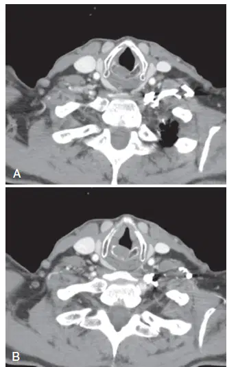
Biopsy of this lesion in this picture demonstrates
cartilage elements, but the pathologist is unable
to confirm chondroma versus chondrosarcoma.
Further metastatic evaluation results are negative.
The next step in management should be
Endoscopic cricoidectomy with or without
tracheostomy.
Total laryngectomy.
Open partial cricoidectomy with appropriate
reconstruction.
Observation with serial CT scans.
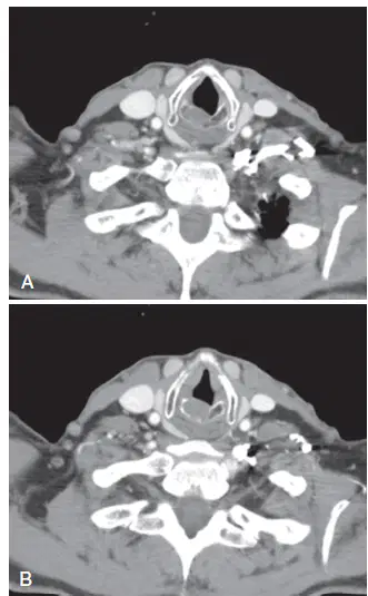
Biopsy of the lesion in this picture confirms chondrosarcoma.
Which of the following surgical options is
best for this patient?
Endoscopic cricoidectomy with or without
tracheostomy
Total laryngectomy
Open partial cricoidectomy with appropriate
reconstruction
Chemoterapy
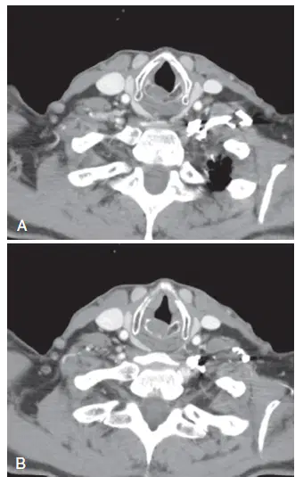
Biopsy of this lesion in this picture shows SCC in
the subglottis, with no vocal cord impairment.
Regional and distant metastatic workup results are
negative. The next appropriate treatment is
Total laryngectomy with adjuvant radiation
therapy.
Partial or endoscopic cricoidectomy with adjuvant
radiation therapy.
Primary radiation therapy.
Concomitant chemoradiation therapy with
cisplatin.
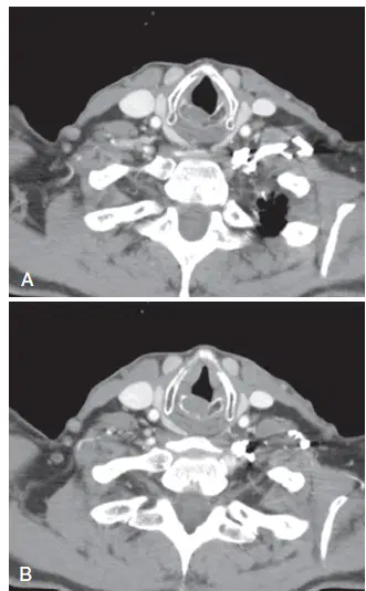
Biopsy of this lesion in this picture shows adenoid
cystic carcinoma. Regional and distant metastatic
workup results are negative. The next appropriate
treatment is
Total laryngectomy with adjuvant radiation
therapy.
Partial or endoscopic cricoidectomy with adjuvant
radiation therapy.
Primary radiation therapy.
Concomitant chemoradiation therapy with
cisplatin.
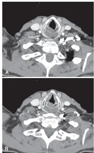
Biopsy of this lesion in this picture shows small cell neuroendocrine carcinoma. Regional and distant metastatic workup results are negative. The next appropriate treatment is
Total laryngectomy with adjuvant radiation
therapy.
Partial or endoscopic cricoidectomy with adjuvant
radiation therapy.
Primary radiation therapy.
Concomitant chemoradiation therapy with
possible additional chemotherapy.
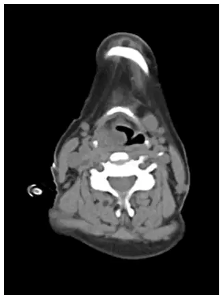
A 70-year-old man with a history of long-term smoking, as well as chronic obstructive pulmonary disease, presents with 1 year of hoarseness. Neck examination shows right level II lymphadenopathy with a 1.5-cm lymph node. Flexible fiberoptic laryngoscopy shows a right transglottic lesion with right vocal cord fixation. The disease extends to the left aryepiglottic fold. There is no impairment or involvement of the left vocal cord. PET/CT shows no distant metastasis, fluorodeoxyglucose (FDG)-avid lymphadenopathy as previously described, and an FDG-avid primary lesion of the larynx. The CT scan
What is the next appropriate step in management?
Direct laryngoscopy with biopsy
Referral to medical oncology
Needle biopsy of an enlarged FDG-avid lymph
node
Tracheostomy
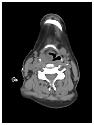
Tissue biopsy of the lesion in this case confirms
SCC. The patient desires surgery. What is the best
surgical treatment for this patient?
Total laryngectomy with bilateral neck dissection
Total laryngectomy with dissection of the left
side of the neck
Transoral endoscopic laser partial laryngectomy
with bilateral neck dissection
Transoral endoscopic laser partial laryngectomy
with dissection of the left side of the neck
Vertical partial laryngectomy, reconstruction with
strap musculature, and bilateral neck dissection
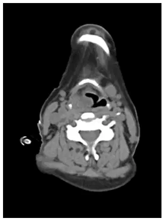
What adjuvant therapy will likely be indicated for
the tumor described in this case? with pathologi SCC
Radiation therapy to the laryngeal bed and left
side of the neck
Radiation therapy to the laryngeal bed and
both sides of the neck
Chemoradiation therapy to the laryngeal bed
and left side of the neck
Chemoradiation therapy to the laryngeal bed
and both sides of the neck
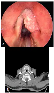
A 69-year-old man presents with the lesion pictured in this Figure. The left TVC is mobile, but the right TVC shows impaired movement without fixation. Regional and distant workup results are negative. Direct laryngoscopy shows an exophytic lesion confined to the vibratory surface of the TVC only; the lesion does not involve either arytenoid. The neuroradiology department states that no obvious erosion of the thyroid cartilage is present on the CT scan this picture, only age-related changes. Which of the following is the most appropriate T
stage for the glottis tumor described in the Case?
T1a
T1b
T2
T3
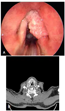
Which of the following is the most appropriate
nonsurgical treatment for the patient in the Case?
Primary radiation therapy alone to the larynx
Primary radiation therapy to the larynx and
bilateral neck
Concomitant chemoradiation therapy
Induction chemotherapy with cisplatin, followed
by radiation therapy alone

A 55-year-old man with good pulmonary function presents with the lesion in Figure. The lesion is confined to the laryngeal surface, and no disease is seen on the lingual surface of the epiglottis. The true vocal folds are mobile and are free of disease. The lesion extends to the false vocal fold only. On a CT scan, no lymphadenopathy is seen and the preepiglottic space is not involved. Which of the following is the most appropriate T stage for the supraglottic tumor described in Case?
T1
T2
T3
T4a
T4b

Which of the following is the most appropriate
nonsurgical treatment for the patient in this Case?
Primary radiation therapy alone to the larynx
Primary radiation therapy to the larynx and
bilateral neck
Concomitant chemoradiation therapy
Induction chemotherapy with cisplatin, followed
by radiation therapy alone
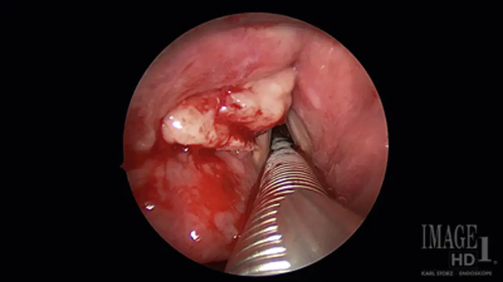
A 55-year-old man with good pulmonary function presents with the lesion in Figure 7-4. The lesion is confined to the laryngeal surface, and no disease is seen on the lingual surface of the epiglottis. The true vocal folds are mobile and are free of disease. The lesion extends to the false vocal fold only. On a CT scan, no lymphadenopathy is seen and the preepiglottic space is not involved.
Which of the following is the most appropriate surgical management of the disease described in
this Case ?
Transoral laser endoscopic resection
Open supraglottic partial laryngectomy
Open supracricoid laryngectomy
Total laryngectomy
Which of the following is appropriate management
of the neck after surgical management of the
primary lesion described in this Case?
No neck dissection; observation only
No neck dissection; radiation therapy only
Unilateral neck dissection
Bilateral neck dissection
A 65-year-old man presents with the lesion pictured in this Figure. He has bilateral bulky 3-cm cervical lymphadenopathy but no distant metastasis. Which of the following is the most appropriate T stage for this tumor based on data presented in this Case?
T1
T2
T3
T4a
T4b
What is the preferred method of treatment for the
patient in this Case?
Radiation therapy alone
Concomitant chemoradiation therapy with
cisplatin
Total laryngectomy with bilateral neck dissection
Induction chemotherapy with cisplatin, followed
by concomitant chemoradiation therapy
Which of the following would not be recommended as part of the treatment for a T1 piriform sinus hypopharynx tumor?
Transoral laser resection of the primary tumor
with negative margins
Radiation therapy alone
Transcervical lateral pharyngectomy
Bilateral neck dissection after surgery for the
primary tumor
Concomitant chemoradiation therapy
Why is the hypopharynx SCC survival rate consistently
worse than that of other head and neck
cancers?
These cancers are routinely more aggressive
than other head and neck cancers.
These cancers are HPV-related.
These cancers tend to be seen at more advanced
stages.
These cancers are difficult to resect with clear
margins.
These cancers tend to be chemotherapy and
radiation therapy resistant.
A 75-year-old woman presents with an incidentally found T1 hypopharynx tumor of the medial piriform sinus. CT and PET findings are negative for cervical disease. She undergoes surgical resection of the primary lesion. Which of the following is the best next step in treatment of the neck?
No neck dissection necessary
Ipsilateral neck dissection only
Bilateral neck dissection
Ipsilateral radiation therapy to the neck
Bilateral radiation therapy to the neck

The woman in Question 54 is taken to the operating room, an antrostomy is done endoscopically, and tissue is removed and sent for pathological analysis. The histological specimen is shown in this Figure. What is your diagnosis
SCC
Adenocarcinoma
Mucoepidermoid carcinoma
Olfactory neuroblastoma
Adenoid cystic carcinoma
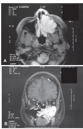
A 48-year-old woman has left-sided facial pressure, pain, and hypoesthesia of her left cheek. She has no visual problems. She has a tumor filling her maxillary sinus and biopsy by another ear, nose, and throat physician shows adenoid cystic carcinoma. MRI of the sinuses is shown in this Figure. What are the appropriate next interventions?
Consult an ophthalmologist.
Consult a plastic surgeon.
Consult a prosthodontist.
Obtain a PET/CT scan
Do all of the above
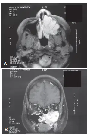
What maxillectomy approach do you recommend
for the patient in the case?
Lateral rhinotomy
Endoscopic approach combined with an inferolateral
orbitotomy
Weber-Ferguson incision with a Lynch extension
Lateral rhinotomy with a Lynch extension
Weber-Ferguson incision with a subciliary extension
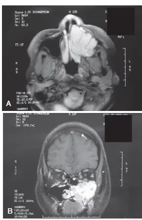
You perform a total maxillectomy with removal of the orbital floor. The periorbita was sampled, and results were negative on frozen-section analysis. You spare the eye. What are acceptable reconstructive options?
Affix a palatal prosthetic and close the facial
incisions.
Plate Medpor to reconstruct the orbital floor,
affix the palatal prosthetic, and close all
incisions.
Plate across the orbital floor defect, affix the
palatal prosthetic, and close the incisions.
Use a temporalis flap to support the eye soft tissues,
affix the palatal prosthetic, and then close
all incisions
Use a composite microvascular free flap with or
without a palatal prosthetic to reconstruct the
orbital floor and palatal defect.
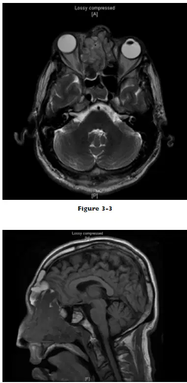
A 48-year-old man presented with right-sided proptosis and diplopia. His monocular vision was intact bilaterally. A biopsy of the mass showed small blue cell tumor cells in rosette formation in a neurofibrillary background with no necrosis this Figures. The cells had indistinct borders with scant cytoplasm and “salt and pepper” chromatin. What is the most likely diagnosis?
Intestinal-type adenocarcinoma
Adenoid cystic carcinoma
B-cell lymphoma
Olfactory neuroblastoma
SCC
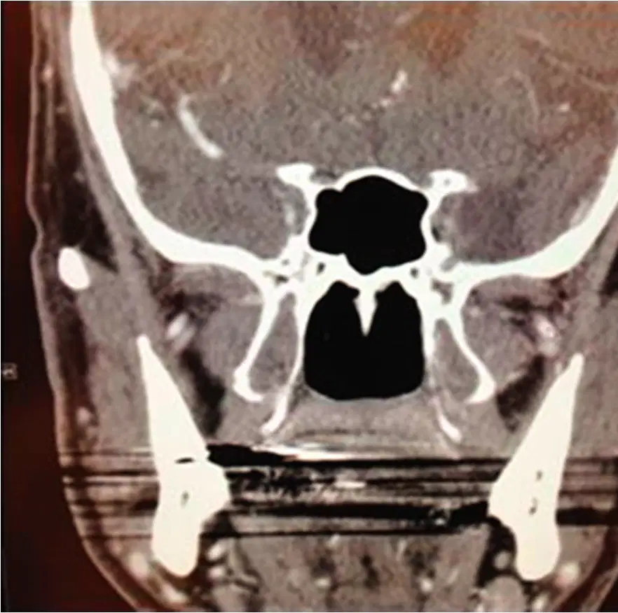
Which nerve is transmitted through the skull baseforamen shown in this Figure?
Vidian nerve
Mandibular nerve
Maxillary nerve
Optic nerve
Alveolar nerve
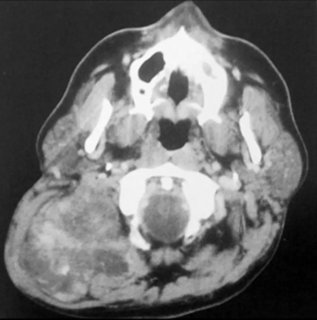
Which of the following statements is correct regarding malignant peripheral nerve sheath tumors (MPNST) as shown in Figure?
MPNSTs account for approximately 40% of all
soft tissue sarcomas.
MPNSTs are commonly found in the head and
neck.
MPNSTs are more commonly seen in pediatric
age groups.
MPNST is a relatively new term for tumors
formerly called malignant schwannoma, neurofibrosarcoma,
or neurogenic sarcoma.
Neoadjuvant cisplatin/5-fluorouracil therapy is
broadly recommended for offsetting the risk of
lung metastases.

The buccal branch of the facial nerve, as shown in this Figure?
Extends along the path of the Stensen duct.
Extends between the tragus and the lateral
canthus of the eye.
Extends along the path of the marginal
mandibular nerve.
Extends superior to the zygomatic branch.

Which of the following statements about the deep lobe of the parotid, as shown in Figure, is correct?
Most tumors present in the retromandibular
location.
Most tumors present in the parapharyngeal space
Most tumors present in the retromandibular
and parapharyngeal space.
Most tumors present in the retromandibular
and retropharyngeal space.
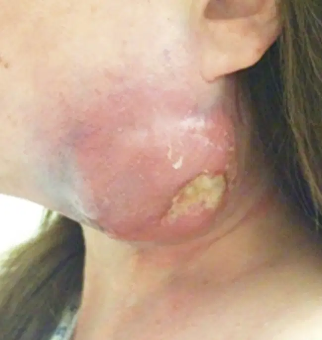
Which of the following descriptions is true about
lymphoma (Figure 10)?
Parotid involvement most commonly seen with
diffuse large A-cell lymphoma
Parotid involvement most commonly seen with
diffuse large B-cell lymphoma
Parotid involvement most commonly seen with
diffuse large C-cell lymphoma
Parotid involvement most commonly seen with
diffuse large D-cell lymphoma

From the options shown, choose the single most likely diagnosis of the tumor on the ventral tongue shown in Figure
Fibrosarcoma
Squamous cell carcinoma
Adenoid cystic carcinoma
Melanoma
Pleomorphic adenoma
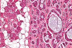
From the options shown, choose the single most likely diagnosis of the tumor on the ventral tongue shown in Figure?
Fibrosarcoma
Squamous cell carcinoma
Adenoid cystic carcinoma
Melanoma
Pleomorphic adenoma

From the options shown below, choose the single most likely diagnosis of the cutaneous tumor in Figure
Squamous cell carcinoma
Basal cell carcinoma
Melanoma
Merkel cell carcinoma
Adenoid cystic carcinoma

From the options shown below, choose the single most likely diagnosis of the thyroid tumor in Figure
Medullary carcinoma
Hurthle cell carcinoma
Papillary thyroid carcinoma
Follicular carcinoma
Anaplastic thyroid carcinoma
From the options below choose the single most likely diagnosis of the thyroid tumor shown in Figures
Medullary carcinoma
Papillary thyroid carcinoma
Follicular carcinoma
Anaplastic thyroid carcinoma
Follicular adenoma

From the options below, choose the single most likely diagnosis of the parotid tumor shown in Figure
Adenoid cystic carcinoma
Mucoepidermoid carcinoma
Epithelial myoepithelial carcinoma
Myoepithelial carcinoma
Acinic cell carcinoma

