
 Worksheets
Liver Anatomy and Structures HW
Total questions: 122
Worksheet time: 2hrs 57mins
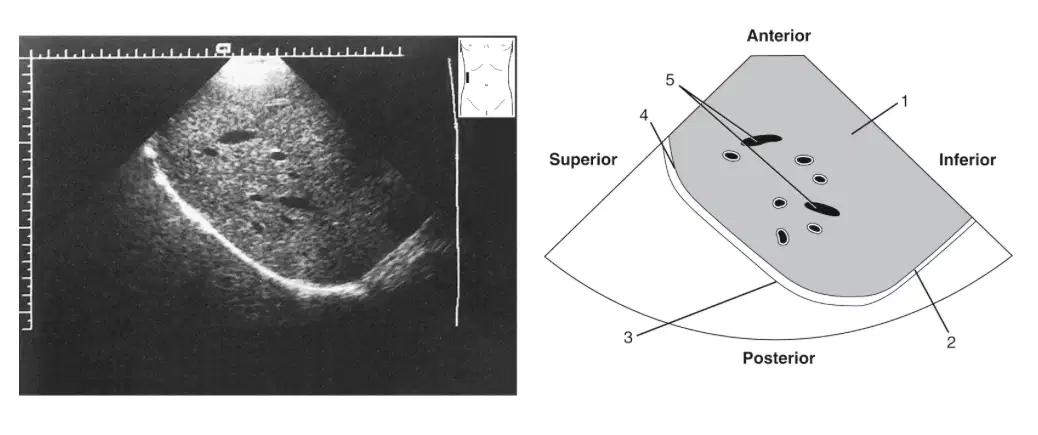
In this image, what is labeled 1?
(a)
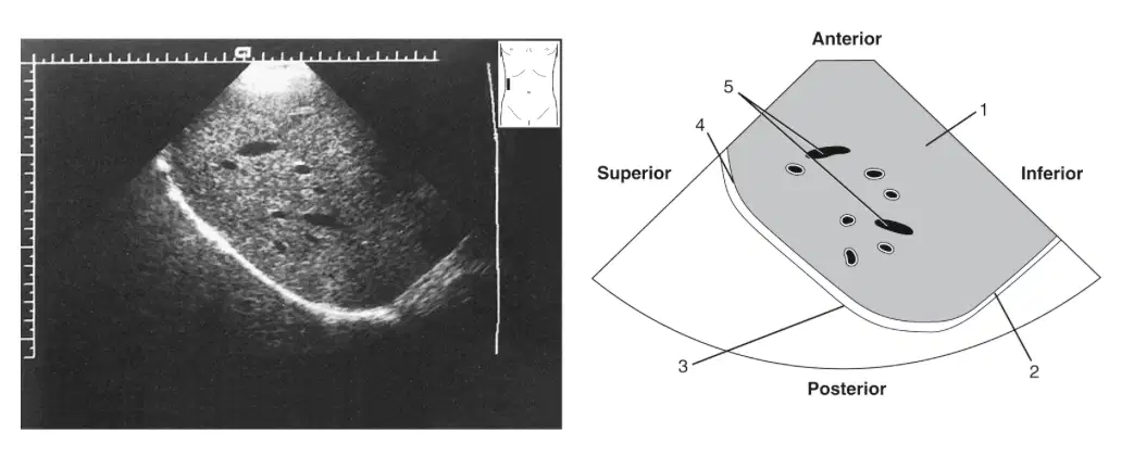
In this image, what is labeled 2
(a)
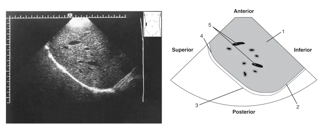
In this image, what is labeled 3?
(a)
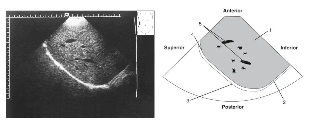
In this image, what is labeled 4?
(a)
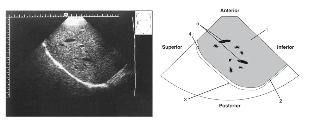
In this image, what is labeled 5?
(a)
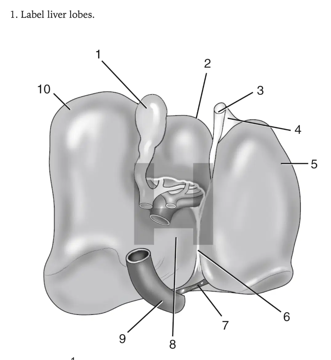
In this image, what is labeled 1?
(a)
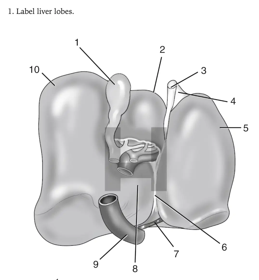
In this image, what is labeled 2?
(a)

In this image, what is labeled 3?
(a)

In this image, what is labeled 4?
(a)
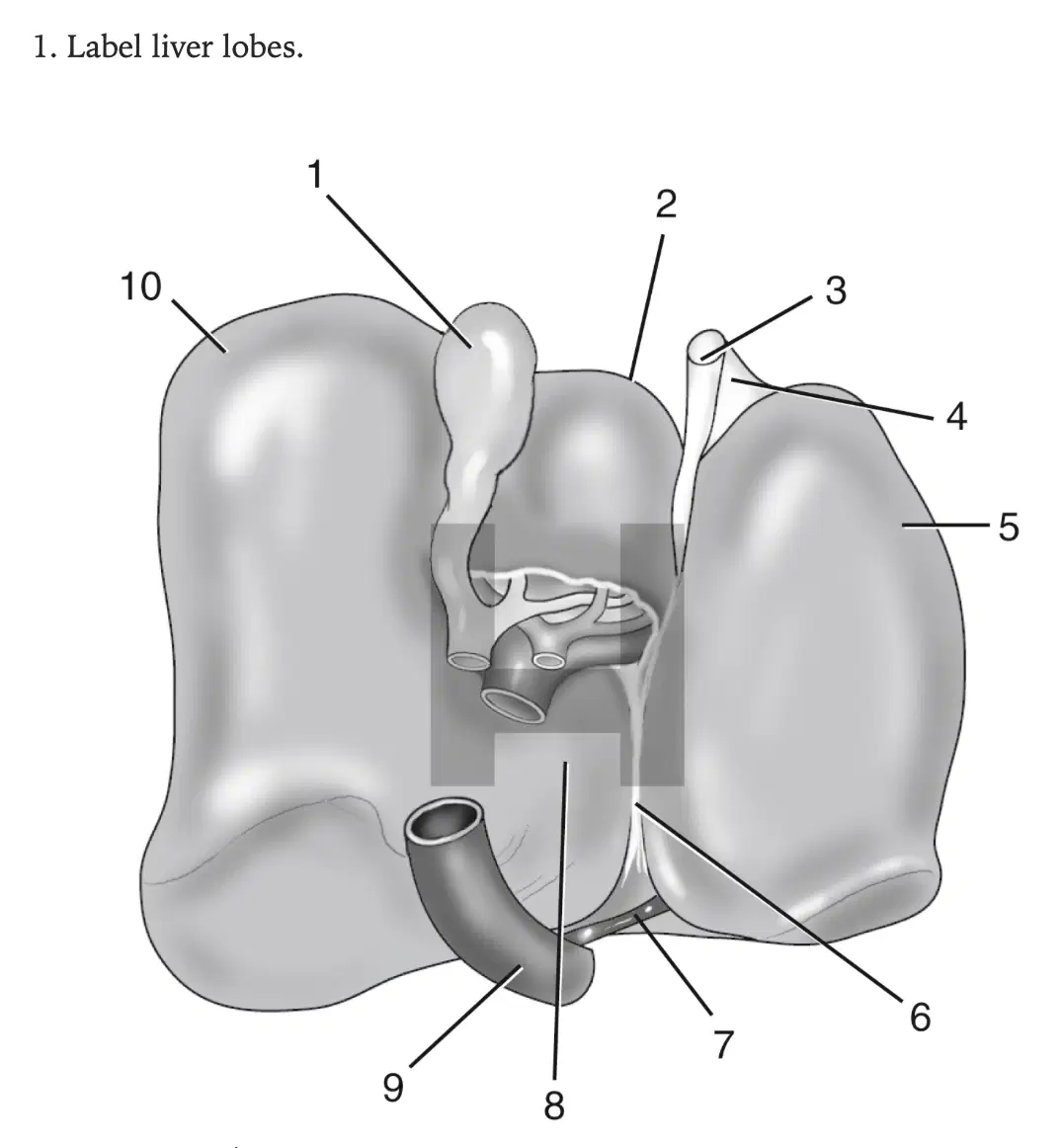
In this image, what is labeled 5?
(a)

In this image, what is labeled 6
(a)

In this image, what is labeled 7?
(a)
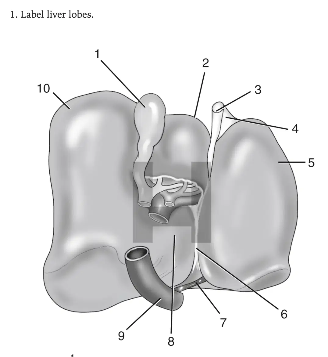
In this image, what is labeled 8?
(a)

In this image, what is labeled 9
(a)

In this image, what is labeled 10?
(a)
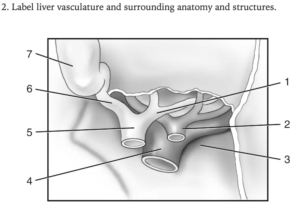
In this image, what is labeled 1?
(a)
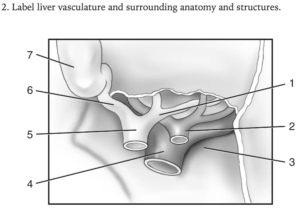
In this image, what is labeled 2
(a)
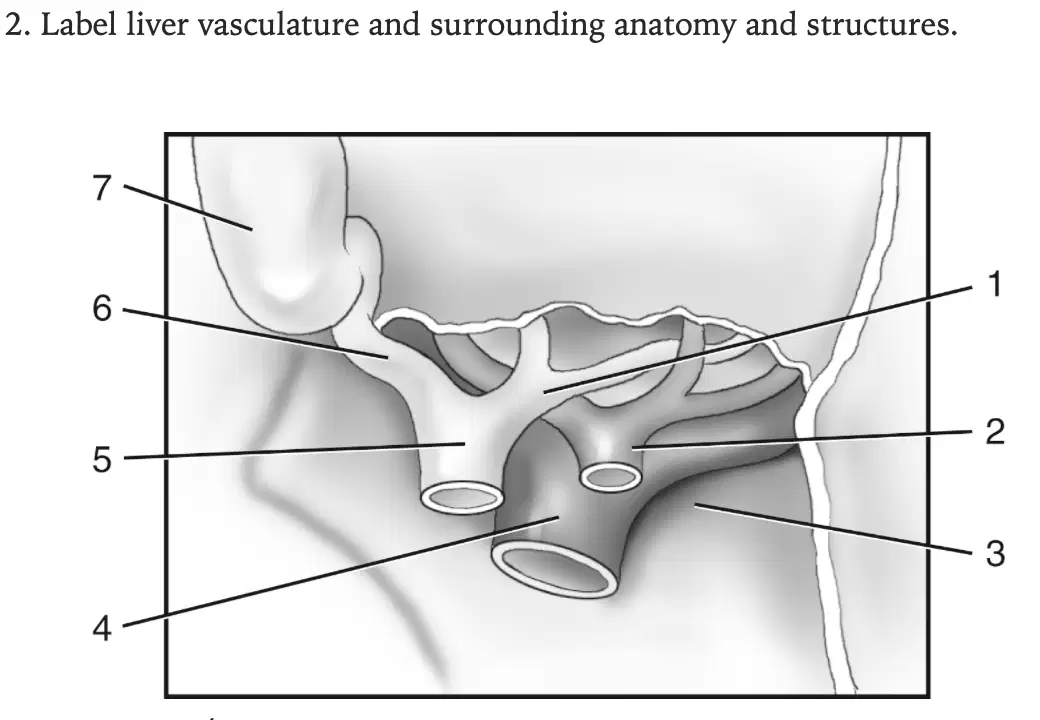
In this image, what is labeled 3?
(a)
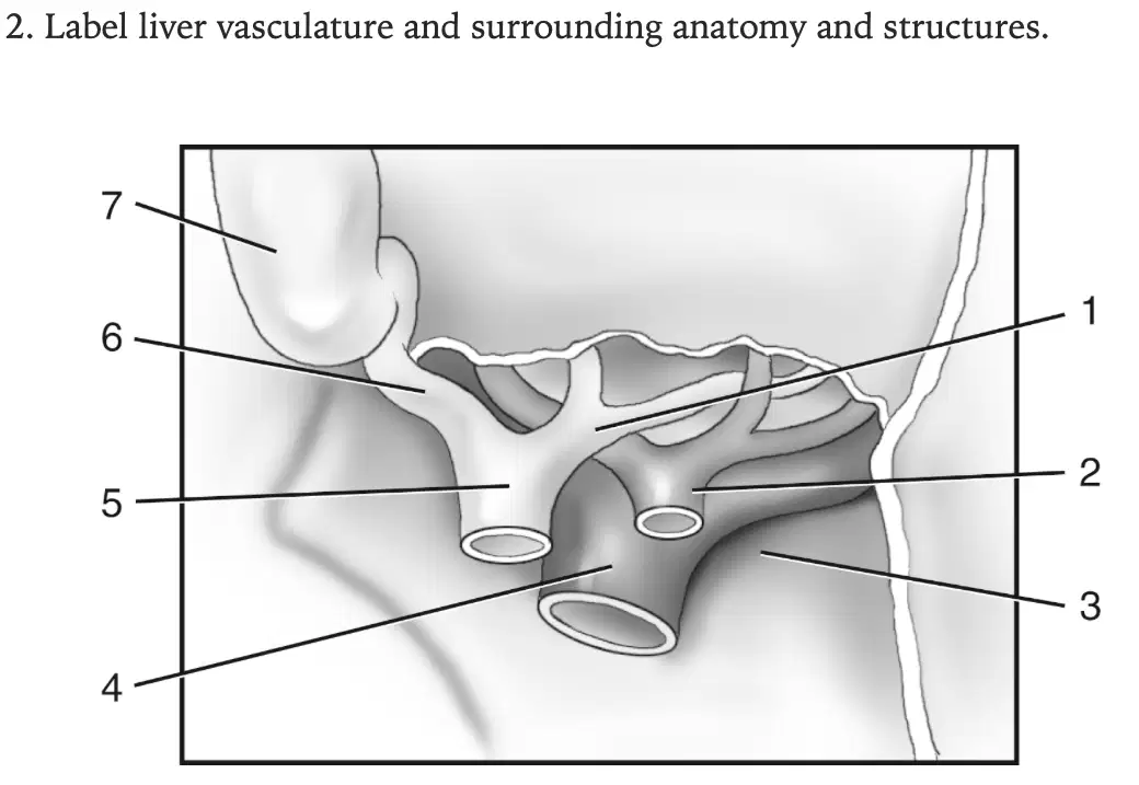
In this image, what is labeled 4?
(a)
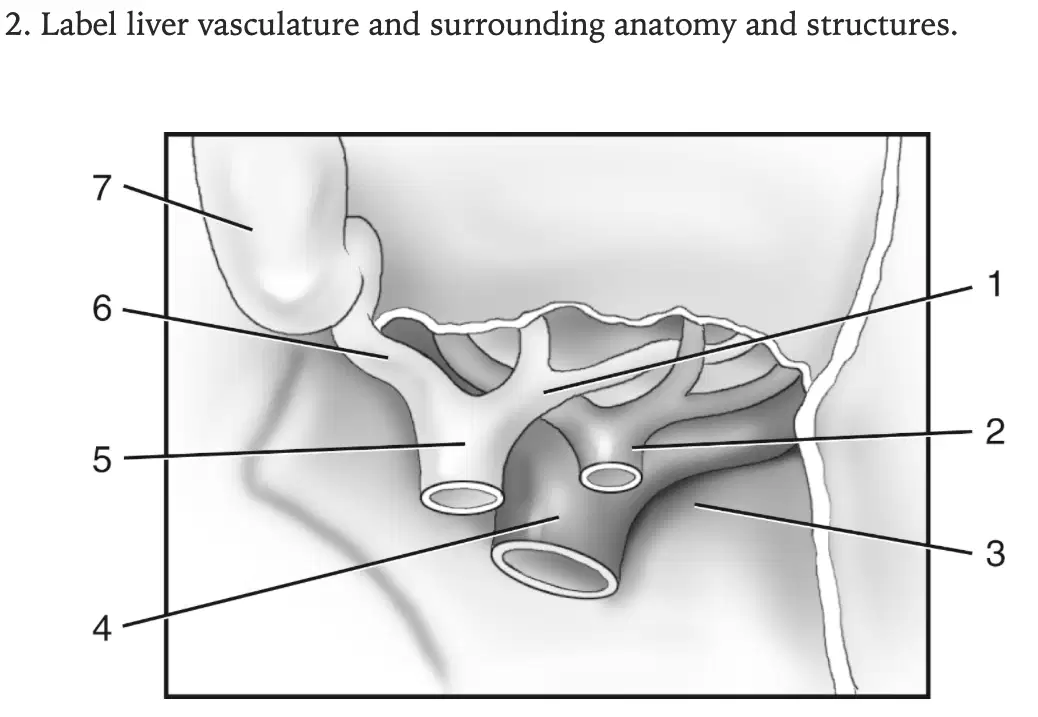
In this image, what is labeled 5?
(a)
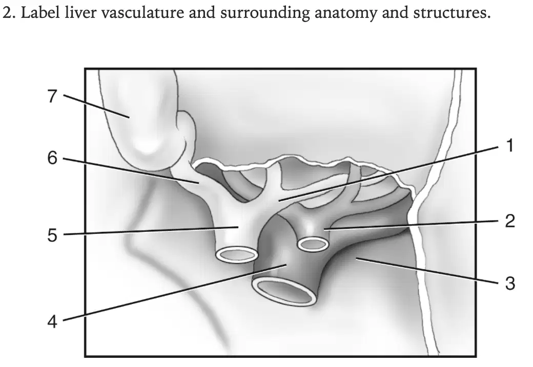
In this image, what is labeled 6?
(a)
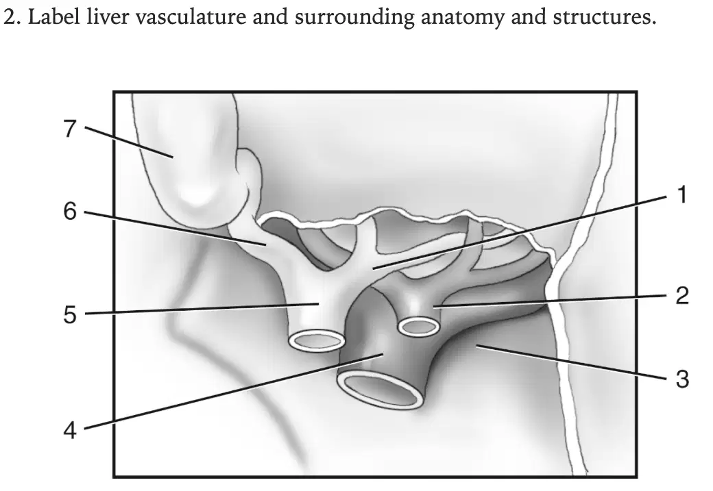
In this image, what is labeled 7?
(a)
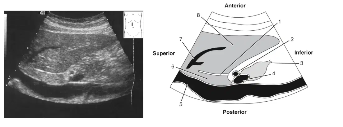
In this image, what is labeled 1
(a)
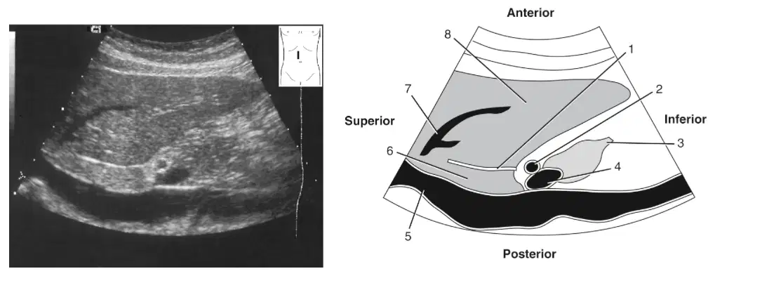
In this image, what is labeled 2?
(a)
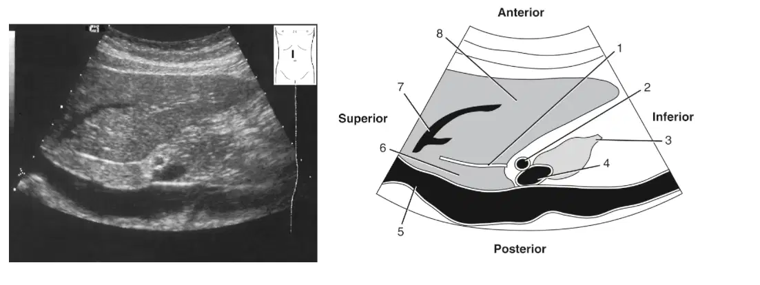
In this image, what is labeled 4?
(a)
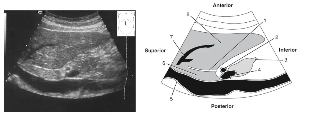
In this image, what is labeled 5?
(a)

In this image, what is labeled 6
(a)
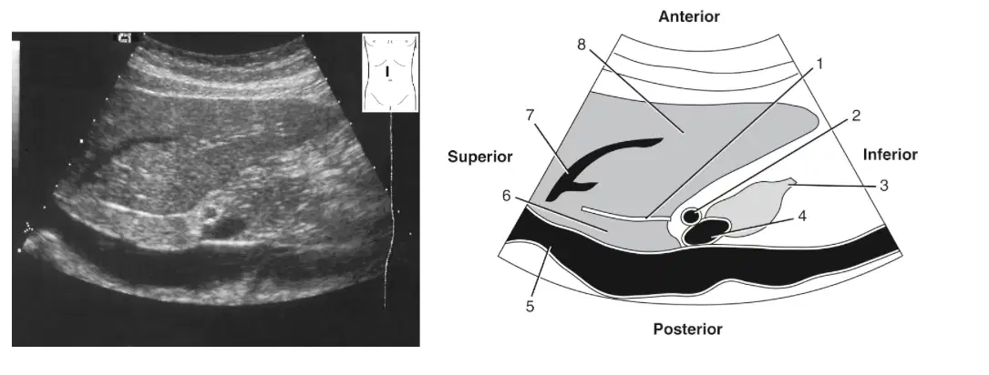
In this image, what is labeled 7?
(a)
In this image, what is labeled 8?
(a)
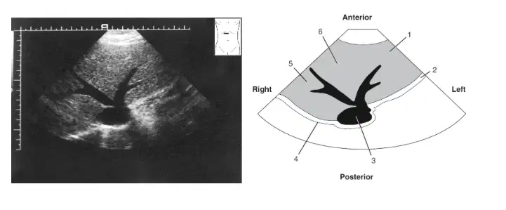
In this image, what is labeled 1
(a)
In this image, what is labeled 2 &4?
(a)
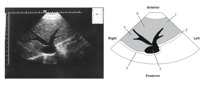
In this image, what is labeled 3?
(a)
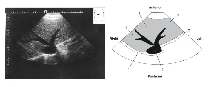
In this image, what is labeled 5?
(a)
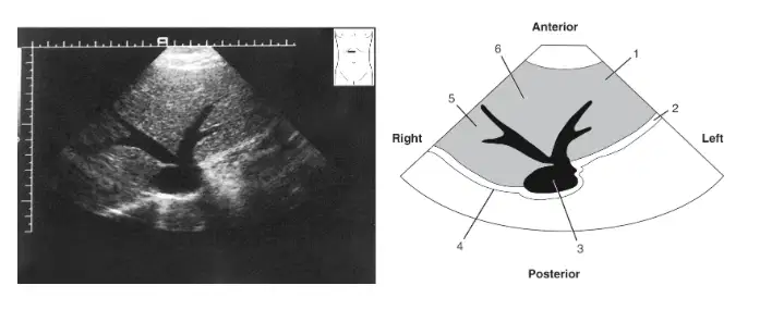
In this image, what is labeled 6?
(a)
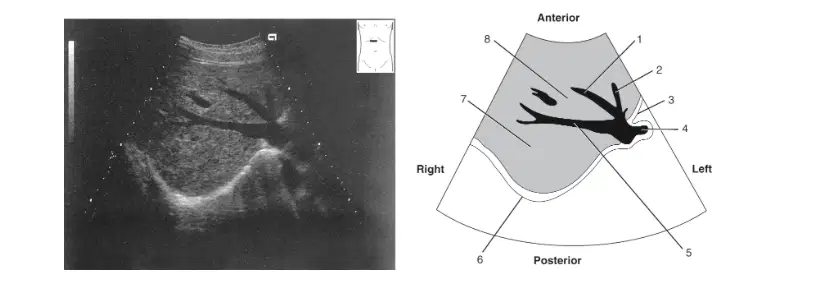
In this image, what is labeled 1?
(a)
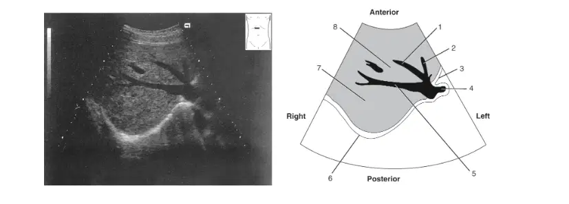
In this image, what is labeled 2?
(a)
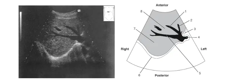
In this image, what is labeled 3&6?
(a)
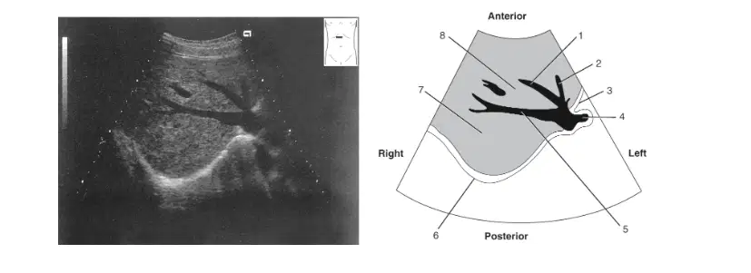
In this image, what is labeled 4
(a)
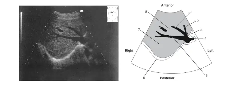
In this image, what is labeled 5
(a)
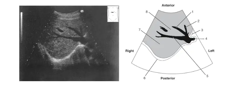
In this image, what is labeled 7?
(a)
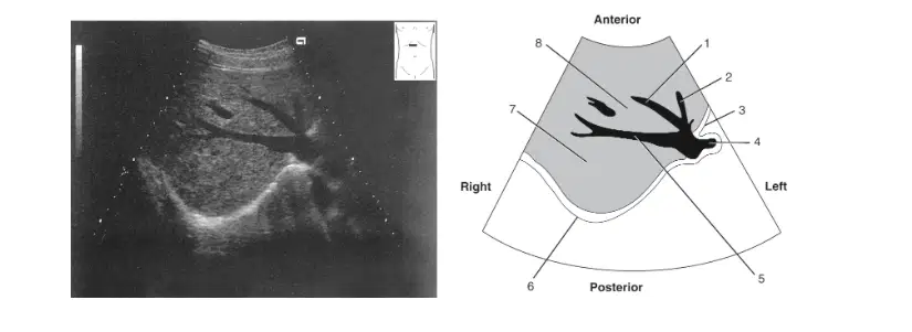
In this image, what is labeled 8
(a)
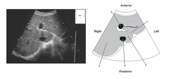
In this image, what is labeled 1
(a)
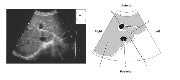
In this image, what is labeled 2?
(a)
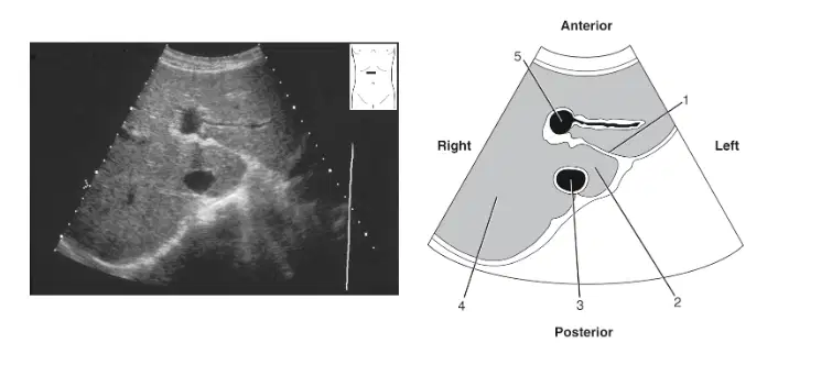
In this image, what is labeled 3?
(a)
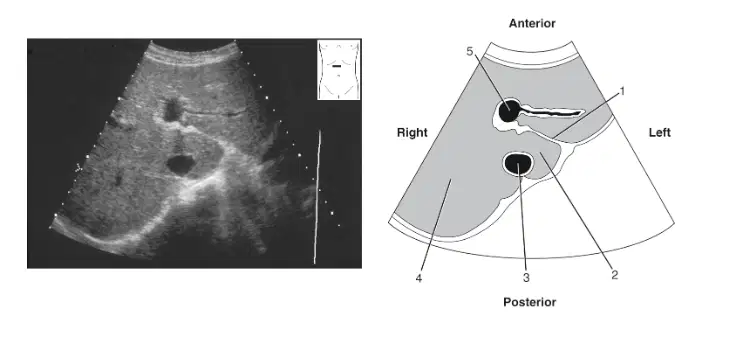
In this image, what is labeled 4?
(a)
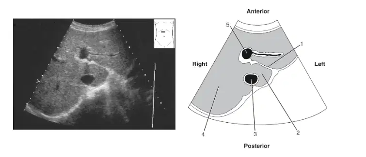
In this image, what is labeled 5?
(a)
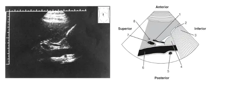
In this image, what is labeled 1?
(a)
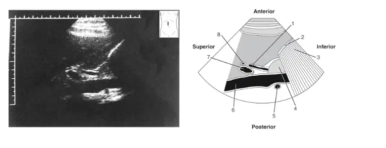
In this image, what is labeled 2?
(a)
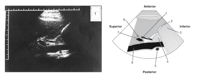
In this image, what is labeled 4?
(a)
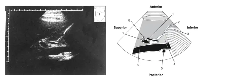
In this image, what is labeled 5?
(a)
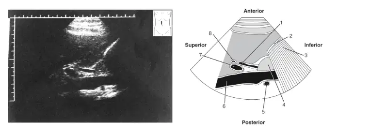
In this image, what is labeled 6?
(a)
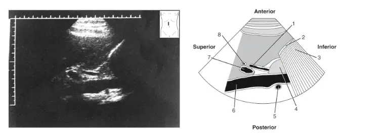
In this image, what is labeled 7?
(a)
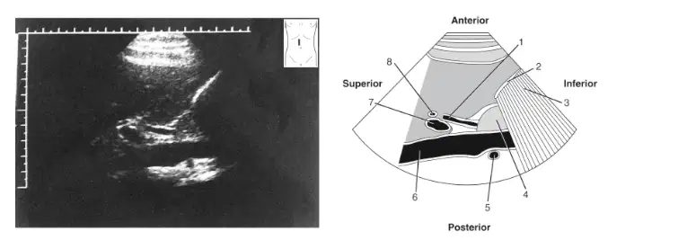
In this image, what is labeled 8?
(a)
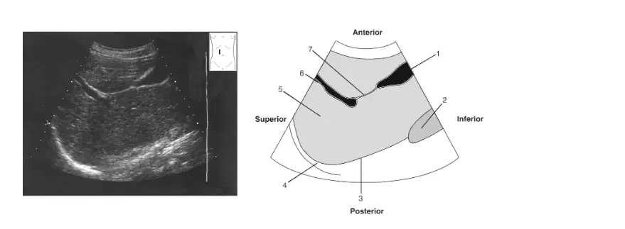
In this image, what is labeled 1
(a)
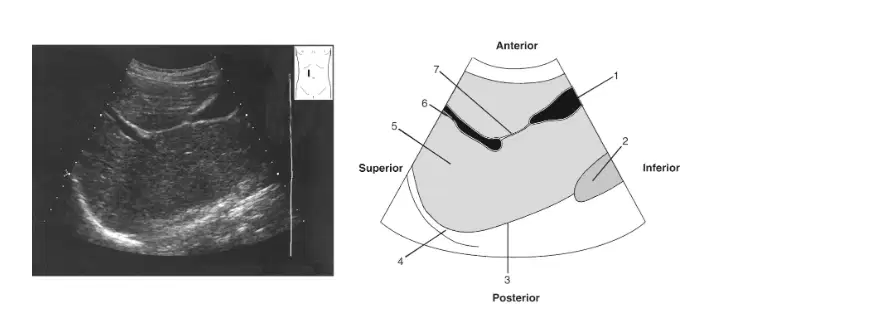
In this image, what is labeled 2?
(a)
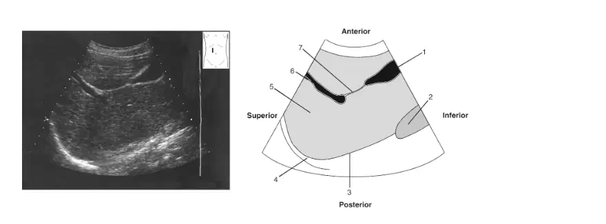
In this image, what is labeled 3?
(a)
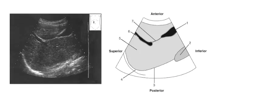
In this image, what is labeled 4?
(a)
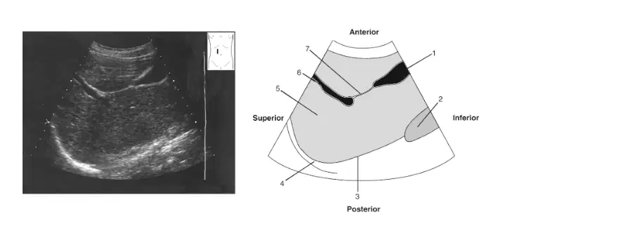
In this image, what is labeled 5?
(a)
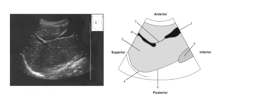
In this image, what is labeled 6?
(a)
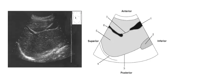
In this image, what is labeled 7?
(a)

In this image, what is labeled 1?
(a)
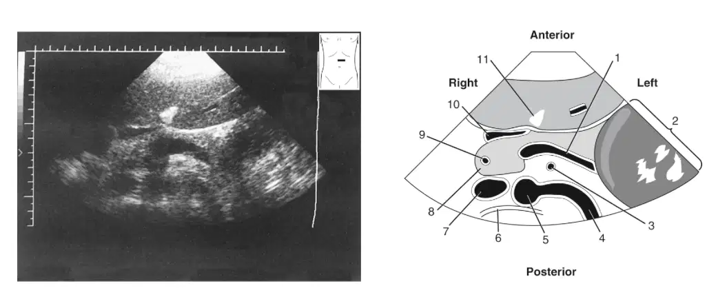
In this image, what is labeled 2?
(a)
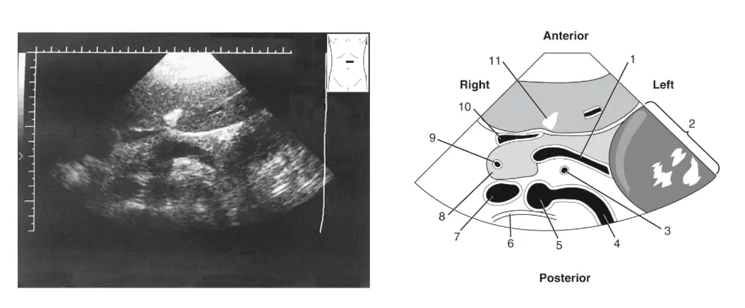
In this image, what is labeled 3?
(a)
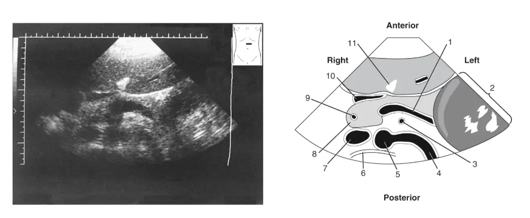
In this image, what is labeled 5?
(a)
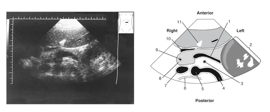
In this image, what is labeled 6?
(a)
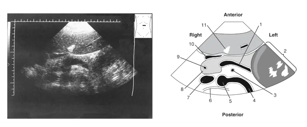
In this image, what is labeled 7?
(a)
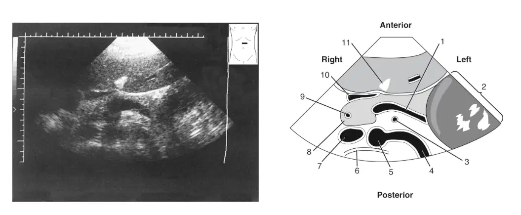
In this image, what is labeled 8?
(a)
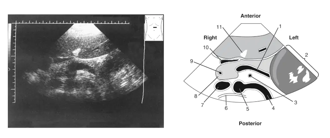
In this image, what is labeled 9?
(a)
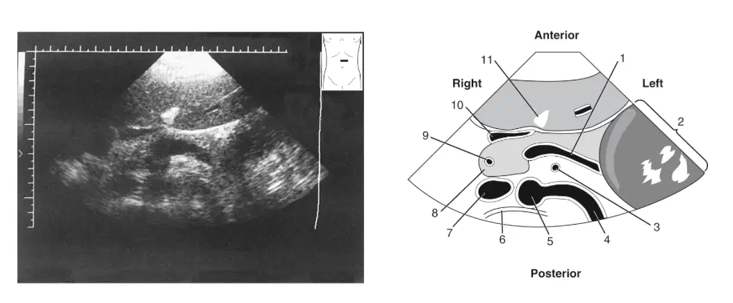
In this image, what is labeled 10
(a)
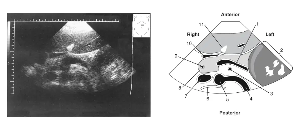
In this image, what is labeled 11?
(a)
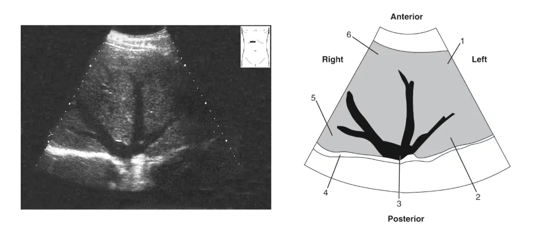
In this image, what is labeled 1
(a)
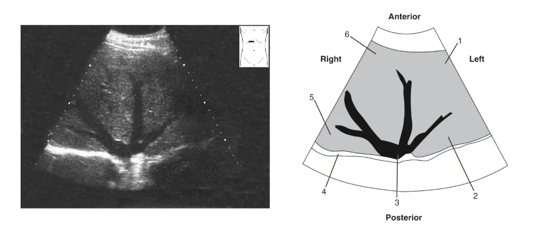
In this image, what is labeled 2
(a)
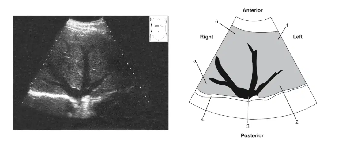
In this image, what is labeled 3
(a)
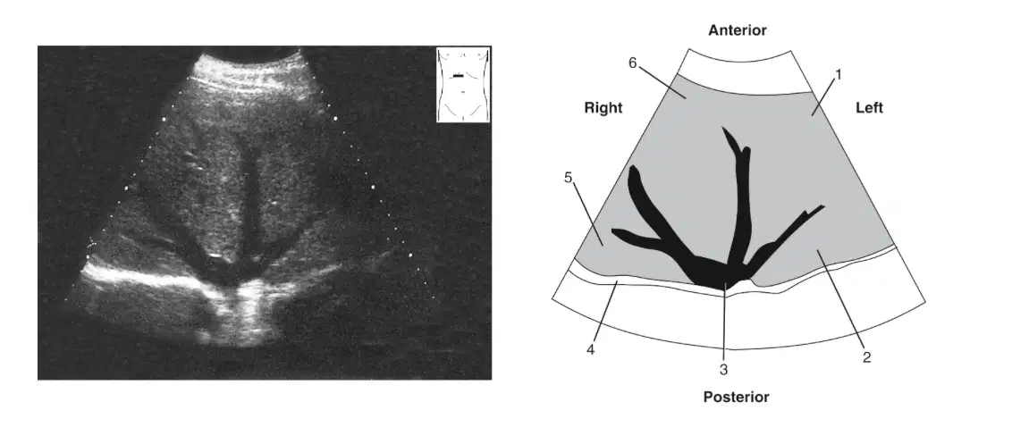
In this image, what is labeled 4?
(a)
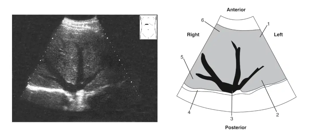
In this image, what is labeled 5?
(a)
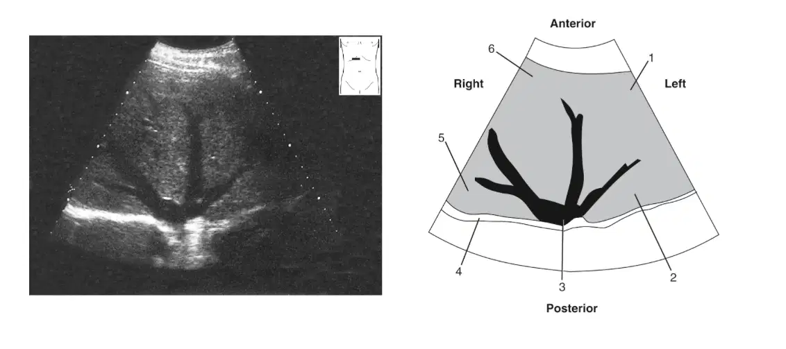
In this image, what is labeled 6
(a)
The section of liver that is not covered by peritoneum is called the
Caudate lobe
Bare area
Mid clavicular line
Medial left lobe
The omental bursa is identified by its location posterior to the stomach and anterior to the pancreas and transverse colon. Another name for this bursa is the
Falciform ligament
Greater sac
Lesser sac
Morrison's pouch
The area of the liver hilum where the portal vein enters and the common bile duct exits is called the
Porta hepatis
Portal confluence
Subhepatic space
Subphrenic space
Liver structures such as the hepatic ligaments and fissures, arteries, portal and hepatic veins and ducts are easily identifiable structures that help divide the liver into segments.
True
False
Which of the following liver surfaces would not be in contact with the diaphragm?
The superior surface
The inferior surface
The anterior surface
Bare area
The surface of the liver marked with indentations and in contact with abdominal organs is the
Superior surface
Inferior surface
Anterior surface
Bare area
Which of the following ligaments or fissures marks the left lateral boundary of the medial portion of the left liver lobe?
Hepatoduodenal Ligament
Coronary ligament
Falciform ligament
Main lobar fissure
A large left lobe of the liver would be helpful in visualizing which of the following structures?
Pancreas
Ascending colon
Veriform appendix
Right portal vein
The right and left lobes of the liver are divided into four segments, medial and lateral on the right and anterior and posterior on the left
True
False
Which of the following is another term for the medial segment of the left lobe?
Caudate lobe
Quadrate lobe
Reidels lobe
Posterior portion of the inferior surface
Only area of the liver not covered by peritoneum
(a)
Smallest lobe of the liver, bordered by fossa for the inferior vena cava (IVC), falciform ligament, and lesser omentum.
(a)
Branch of the celiac axis that supplies the liver and divides into the gastroduodenal artery and proper hepatic artery.
(a)
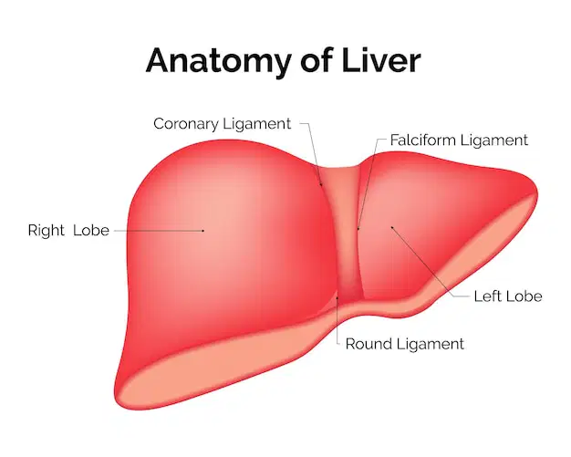
Anterosuperior surface of liver that runs superiorly, then posteriorly on the right to the anterior leaf of the coronary ligaments.
(a)
Division of liver segments based on hepatic or portal venous anatomy; used for dividing the liver into eight segments.
(a)
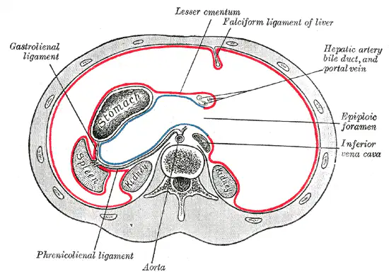
Communication between the greater and lesser sacs of the peritoneum
(a)
Divides right and left lobes; ends at the ligamentum teres or round ligament inferiorly
(a)
Portion of the lesser omentum that extends across the transverse fissure for the ligamentum venosum at the porta hepatis of the lesser curvature of the stomach.
(a)
Tight, fibrous capsule covering the liver
(a)
Fold of omentum that extends from the lesser curvature of the stomach and covers the intestines.
(a)
Protective, thin layer that encloses most of the abdominal organs
(a)
Right or left half of the liver; a division based on Couinaud liver segmentation system.
(a)
Portion of the lesser omentum that extends as the right free border of the gastrohepatic ligament to the proximal duodenum and the right flexure of the colon
(a)
One of three main veins draining the liver via the IVC; drains the left lobe
(a)
Branch of the main portal vein that marks the anterior border of the caudate lobe and carries blood from the gastrointestinal tract to the left lobe.
(a)
Anterosuperior surface of the liver that runs superiorly, then posteriorly on the right to the left triangular ligament.
(a)
Double layer of omentum that extends from the liver to part of the duodenum
(a)
Small sac posterior to the stomach and anterior to the pancreas and part of the transverse colon; also known as the omental bursa
(a)
Marks the left anterolateral border of the caudate lobe; travels within the transverse fissure.
(a)
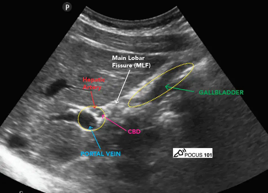
Echogenic line connecting the neck of the gallbladder to the right portal vein; also referred to as the plane associated with the Rex-Cantlie (RC) line in Couinaud liver segmentation system. The RC line runs from the gallbladder fossa to the IVC along the plane of the main lobar fissure.
(a)
Formed by the splenic, superior, and inferior mesenteric veins; drains blood from the gastrointestinal tract to the liver to be processed.
(a)
One of three main veins draining the liver via the IVC; drains a portion of the right and medial left lobes of the liver.
(a)
Space between the posterior subphrenic and posterior subhepatic space; should be free of fluid.
(a)
Normal variant of the caudate lobe. Process can extend distally from the lobe and mimic a lesion.
(a)
Area of the liver hilum where portal vein and hepatic artery enter and common bile duct exits.
(a)
Union of the splenic, superior, and inferior mesenteric veins near the head of the pancreas that forms the portal vein before entering the liver
(a)

Portion of the portal vein, biliary duct, and hepatic artery that are disbursed throughout the liver; can be seen microscopically.
(a)
Division of the common hepatic artery that supplies the liver.
(a)
Fourth” lobe of the liver, which is actually the medial portion of the left lobe.
(a)
Normal variant of the right lobe in which the right lobe extends caudally into the abdomen and toward the iliac crest.
(a)
One of three main veins draining the liver via the IVC; drains the right lobe of the liver.
(a)
Largest lobe of the liver, occupying most of the right hypochondrium
(a)
Branch of the main portal vein that carries blood from the gastrointestinal tract to the right lobe of the liver.
(a)
Helps to form the boundary of the bare area of the liver.
(a)
Terminal end of the falciform ligament.
(a)
Fissure that conveys the ligamentum venosum.
(a)

