
 Worksheets
Vascular Final
Total questions: 96
Worksheet time: 52mins
What distinguishes venous Doppler signals in the upper extremities from the lower extremities?
Lack of augmentation with distal compression
Continuous waveform is normal
Pulsatility is common in the upper extremeties
Spontaneous signals are not present in the lower extremities
Which vessel is most commonly affected in chronic mesenteric ischemia?
IMA
SMA
Renal artery
Aorta
What is a key symptom of chronic mesenteric ischemia?
Weight gain
Leg pain during walking
Sitophobia
Chest Pain
What defines an acute condition?
Pain over weeks or months
Slow onset with mild symptoms
Sudden pain or bleeding
Minimal symptoms and no urgency
What condition does continuous venous Doppler flow in the lower extremity suggest?
Valvular incompetence
Pulmonary embolism
Proximal DVT
Peripheral arterial disease
As the superficial femoral artery passes through Hunter’s canal, it becomes which artery?
Profunda femoris
Popliteal
Peroneal
Tibial
Which of the following is a deep vein of the lower extremity?
Basilic
Small saphenous
Popliteal
Great saphenous
What is a TIA?
Stroke w/ permanent damage
Neurological dysfunction resolving in <72 hours
Temporary neurological dysfunction resolving within 24 hours
Sudden cardiac death
What Doppler signal is seen at the site of a significant arterial stenosis?
Tardus parvus
Decreased flow velocity
Spectral broadening and high PSV
Reversal of flow
Which of the following is a clinical indication for an abdominal aortic ultrasound?
Pain after eating
Pulmonary edema
Pulsatile abdominal mass
Fever
At what size is the abdominal aorta considered aneurysmal?
>2.0 cm
>2.5 cm
>3.0 cm
>4.5 cm
Surgical repair is required for an abdominal aortic aneurysm when it reaches:
4 cm
4.5 cm
5 cm
6 cm
The most common location for an aortic aneurysm is:
Thoracic
Suprarenal
Infrarenal
Renal
he most common shape of aortic aneurysm is:
Saccular
Fusiform
Dissecting
Irregular
What maneuver is used to evaluate venous insufficiency?
Trendelenburg
Deep inspiration
Valsalva maneuver
Leg elevation
Which vessel has high resistance and extracranial branches?
ICA
ECA
Vertebral
Basilar
A pressure drop of 20 mmHg between segments during segmental pressure testing suggests:
No stenosis
Arterial ulcer
Subclavian stenosis
Venous insufficency
Which is a feature of acute DVT?
Echogenic thrombus
Collateral formation
Loosely attached thrombus
Recanalization
Which feature helps rule out acute DVT?
Collateral veins
Aliasing
Vein compression
Reverse flow
May-Thurner Syndrome is caused by compression of:
Right iliac vein by the left iliac artery
Left iliac vein by the right iliac artery
Femoral artery
IVC
Renal arteries are considered to be:
Low resistance
High resistance
Pulsatile
Triphasic
What happens in subclavian steal syndrome?
Brachial artery reverses flow
Vertebral artery reverses flow
CCA occludes
ICA tap is negative
A PSV greater than 200 cm/s in the aorta suggests:
10% stenosis
30% stenosis
>50% stenosis
Occlusion
Fibromuscular dysplasia appears as:
Fusiform dilation
Atherosclerotic narrowing
String of beads on angiogram
Collateral vessel
What is the first branch of the External Carotid Artery (ECA)?
Opthalmic
Maxillary
Lingual
Superior Thyroid
What muscle action describes the function of the calf muscle pump?
Relaxation of the quads
Soleal and gastrocnemius muscle contraction
Calf flexion and extension
Store blood in the calf
Systolic blood pressure reflects:
Heart relaxation
Diastolic runoff
Heart contraction
Valve closure
The main risk factor for primary varicose veins is:
Pregnancy
Heredity
DVT
Inactivity
What is EVAR?
External vascular artery rotation
Endovenous aneurysm resection
Endovascular aneurysm repair
Emergency vascular aneruysm repair
The most common site for plaque formation in the carotid system is:
Proximal CCA
Mid ICA
Carotid Bifurcation
Vertebral Artery
What is the normal spectral Doppler pattern for the SMA after eating?
High resistance
Bidirectional flow
Low resistance
Pulsatile with reversal
What resistance pattern does the abdominal aorta typically show?
High resistance throughout
Low resistance due to visceral branches
Biphasic flow
Retrograde flow
The external carotid artery ultimately terminates as the:
Facial A.
Maxillary A.
Superficial Temporal A.
Occipital A.
Normal venous Doppler waveform in the lower extremities shows:
Continuous flow
Phasic respiratory variation
High resistance triphasic
Retrograde flow
Which feature distinguishes a venous ulcer from an arterial ulcer?
Regular deep wound
Near toes
Mild pain near medial malleolus
Hairless, shiny skin around the wound
Minimum appropriate vein diameter for AV fistula creation is:
1mm
1.5mm
2mm
2.5-3mm
A renal artery stenosis of 60% or greater shows what findings?
PSV <100 cm/s and RAR >3.5
PSV >100 cm/s and RAR <2.5
PSV > 180 cm/s and RAR >3.5
PSV > 180cm/s and RAR <2.5
What does a continuous waveform suggest in venous doppler?
Proximal DVT
Distal DVT
Superficial Thrombus
Valve insufficency
Select the superficial veins of the upper extremity:
Basilic
Ulnar
Cephalic
Radial
Median Cubital
Select the veins of the lower extremity:
Great saphenous
Iliac Veins
Small saphenous
PTV / ATV
Soleal Sinus
What is observed proximal to a stenosis in the arterial system?
Normal flow
Tardus parvus
Possible increase in resistance, very low diastolic flow
Aliasing
Increased PSV and EDV
What is observed at the stenotic site in the arterial system?
Increase in PSV and EDV
Turbulent flow
Aliasing
Decreased pulsatility
Decrease in PSV and EDV
What is observed distal to a stenosis in the arterial system?
Increased velocity
Turbulent flow
Rounded peak systole
Decreased velocity
Normal flow
Aortic diameter >2cm but <3cm
(a)
Select the characteristics of Venous ulcers:
Near medial malleolus
Mild pain
Severe pain
Thickened toenails
Near Tibial area, toes and bony areas
"stopcocks" of the vascular system, control contraction and resistance
(a)
Smallest vessel in the body, primary place of nutrient exchange in the body
(a)
What is the first branch of the Aortic Arch?
Innominate artery
Left CCA
Right Subclavian
Right CCA
Right renal artery is ___ and ___ than the left
Longer and higher
Shorter and lower
Longer and lower
Shorter and higher
How does the right renal artery course to the IVC
anteriorly
posteriorly
laterally
distally
The Tibial-peroneal trunk bifurcates into the?
Posterior tibial and Peroneal arteries
Anterior tibial artery and Posterior Tibial artery
Peroneal artery
Medial and lateral plantar and doralis pedis
The popliteal artery bifurcates into the ?
Anterior tibial artery and Tibioperoneal trunk
Tibioperoneal trunk
Anterior tibial and Posterior Tibial artery
Peroneal artery
Anterior circulation symptoms:
Aphasia
Amaurosis Fugus
Hemiplegia
Parethesia
Dysphagia
Most common site of thrombus formation in the LEV
Soleal sinus
PTV/ATV
Peroneals
Gastrocnemius
Select the correct phasicity of the upper extremity veins:
Thoracic cavity pressure decreases during inspiration
Thoracic cavity pressure increases during inspiration
Thoracic cavity pressure increases during expiration
Flow in veins increases during inspiration
Flow in veins increases during expiration
How is flow in the lower extremities during inspiration and expiration?
Increased flow during expiration
Decreased flow during expiration
Decreased flow during inspiration
Increased flow during inspiration
Superficial Thrombophlebitis usually causes __ pain than DVT
More
Less
What is the first branch of the abdominal aorta?
Celiac artery
SMA
IMA
Splenic
Where is the celiac artery in relation to the pancreas
Superior
Inferior
Lateral
Proximal
In SAG, how is the Celiac artery identified in comparison to the Aorta and SMA
Superior to the SMA
Inferior to the SMA
Anterior to the Aorta
Posterior to the Aorta
How is the SMA identified in relation to the pancreas
Posterior
Anterior
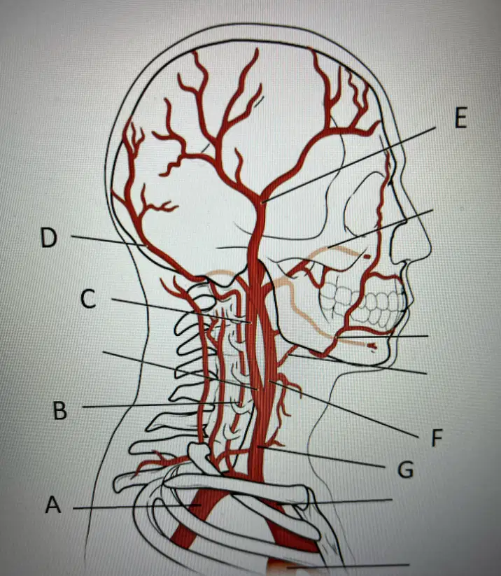
A
Right Subclavian
Right vertebral
Right ICA
Right ECA
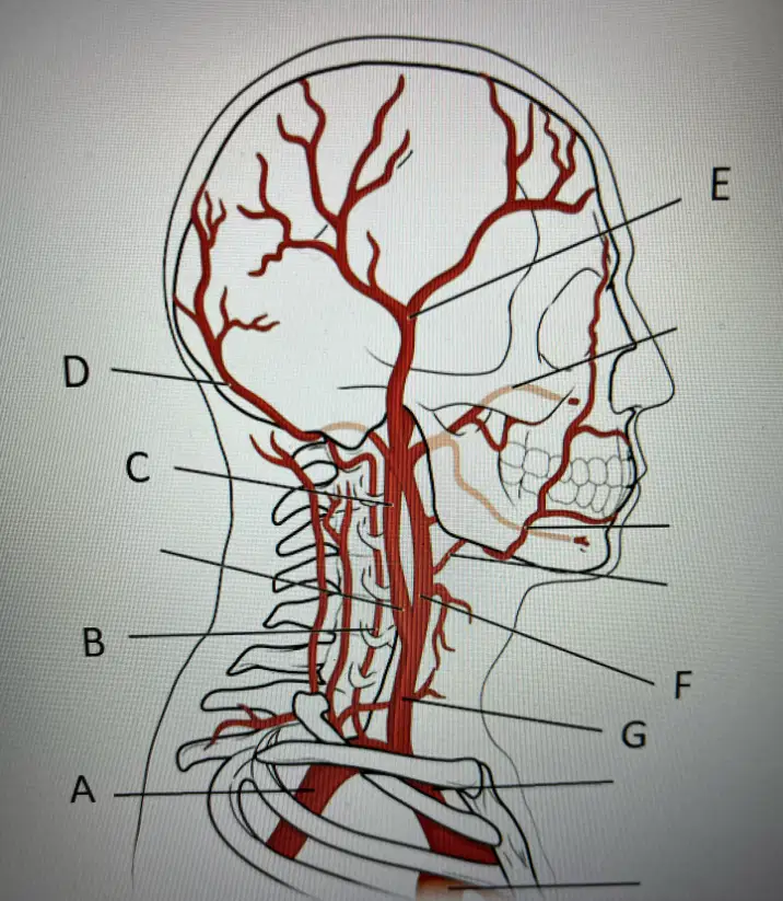
B?
Right Subclavian
Right Vertebral
Right ICA
Superficial Temporal
Right CCA
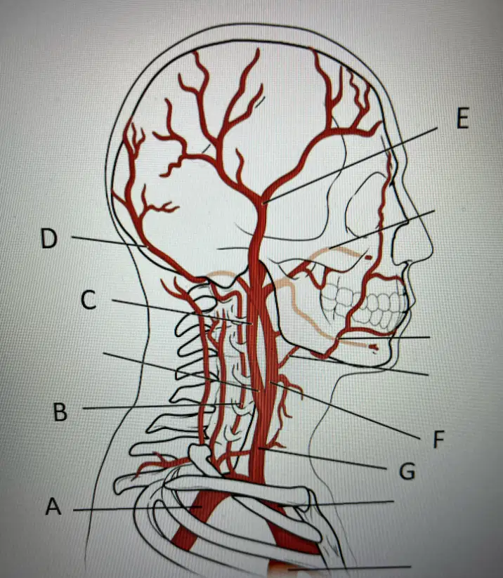
C
Right ICA
Right ECA
Occipital
Right CCA
Right Vertebral
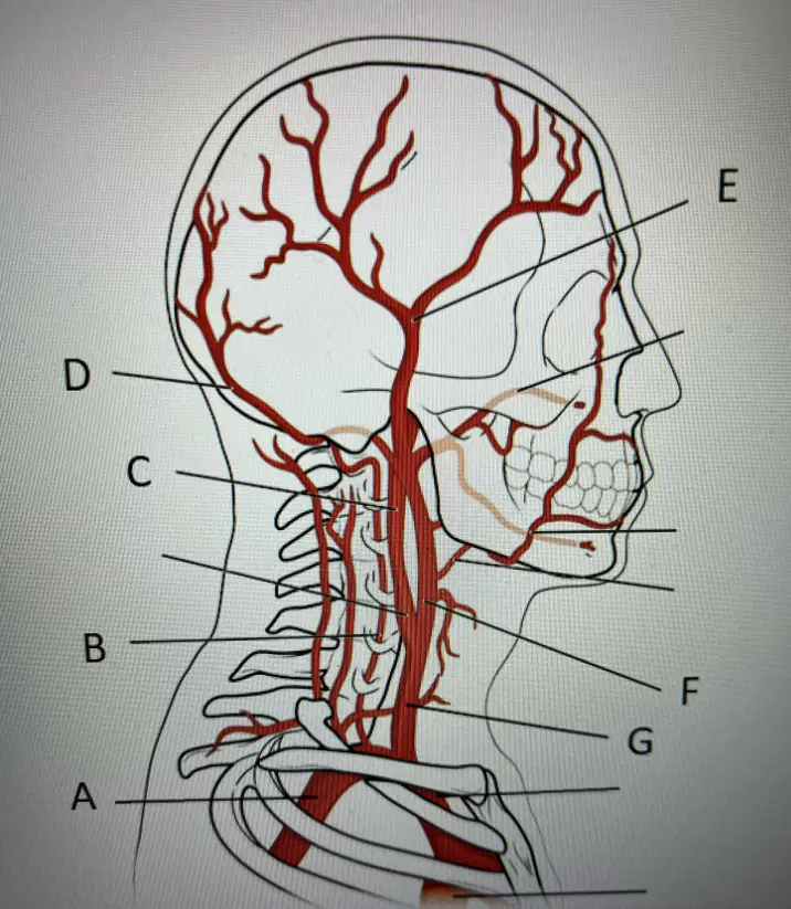
D
Occipital
Superficial Temporal
ECA
Subclavian
CCA

E
Superficial Temporal
ECA
ICA
CCA
Occipital
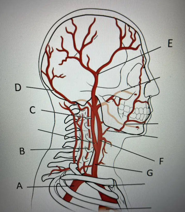
F
ECA
ICA
CCA
Vertebral
An ICA occlusion may cause the contralateral ICA velocities to be which one of the following?
Same
Reversed
Increased- compensatory flow
Decreased
The sound heard upon auscultation of a vessel within a stenotic area is called
a bruit
a tick
a thrill
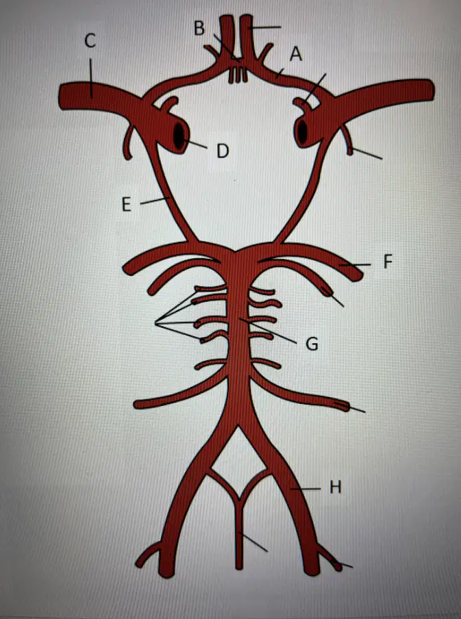
A
Anterior cerebral
Anterior communicating
Posterior communicating
Posterior cerebral
Middle cerebral
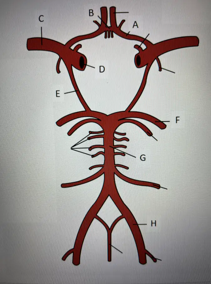
Select the Anterior Communicating Artery
A
B
C
D
E
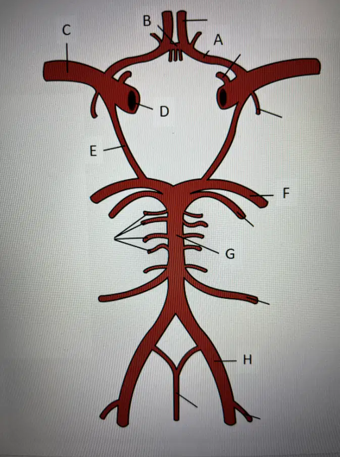
Select the Middle Cerebral Artery
B
C
D
E
F

Select the Posterior Communicating Artery
A
C
E
H
G
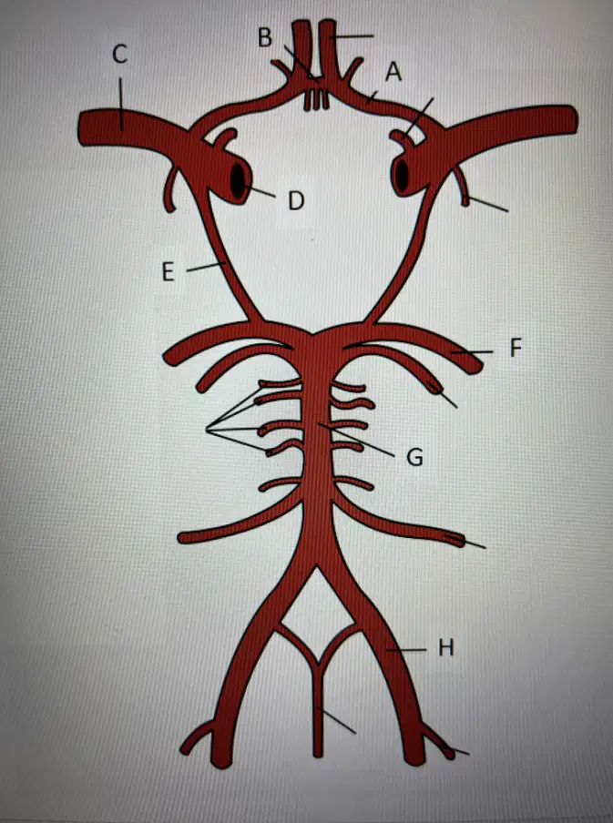
Select the Posterior Cerebral Artery
A
C
F
E
B
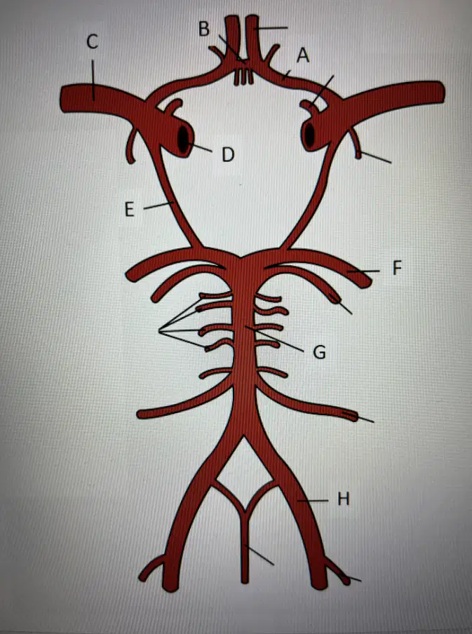
Select the Basilar Artery
G
B
A
H
C
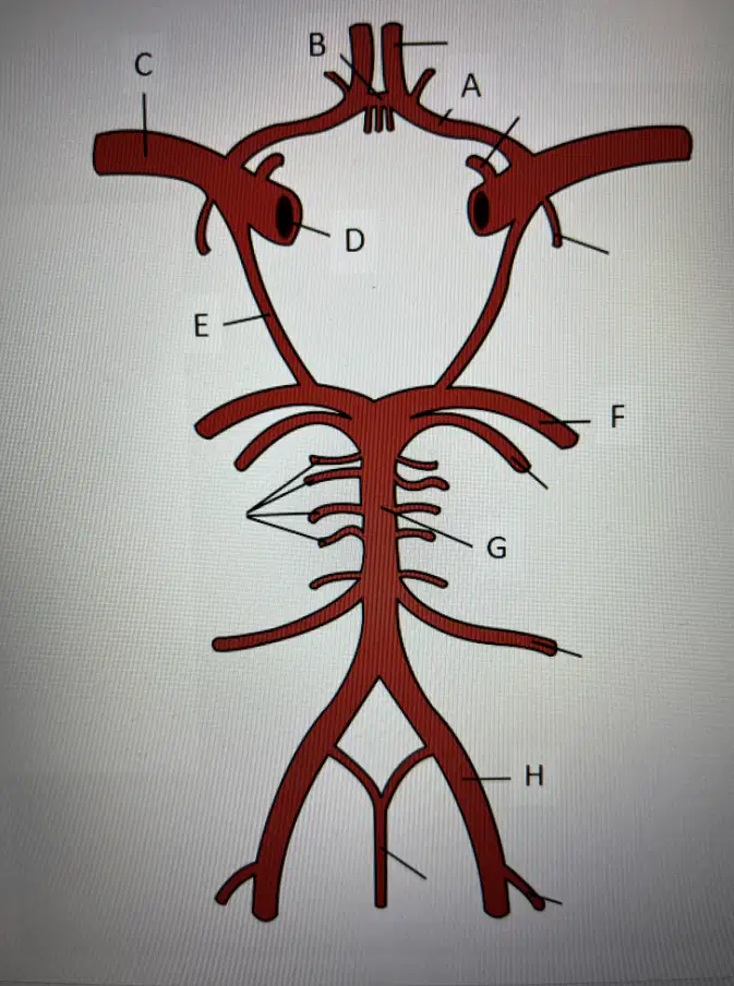
Select the Vertebral Artery
A
B
H
G
D
What type of flow would you expect to see in a normal carotid bulb?
Laminar
Disturbed
Turbulent
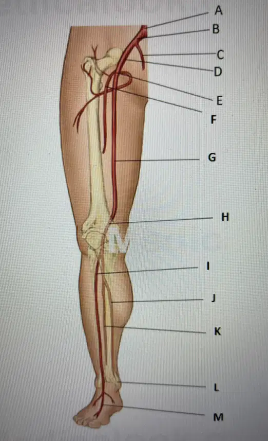
Right Common Femoral Artery is?
e
f
g
h
i
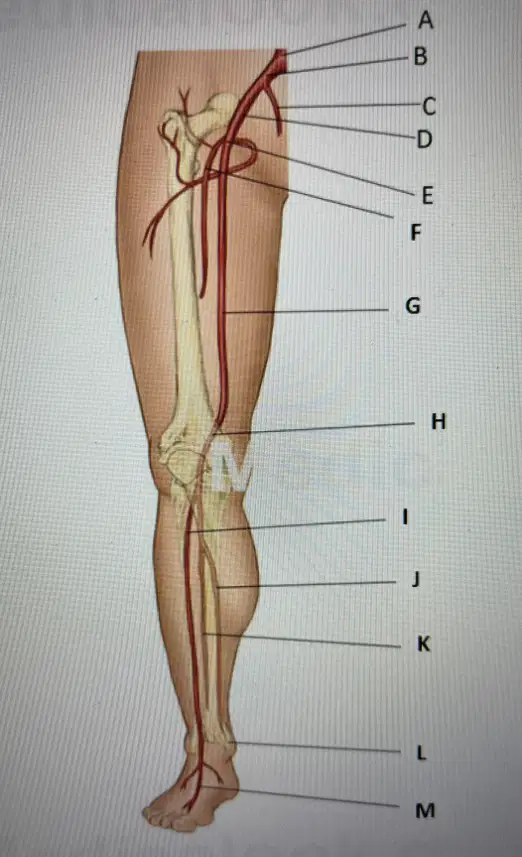
Right Profunda Femoral Artery
F
G
H
I
M
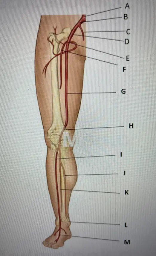
Right Superficial Femoral Artery
G
H
I
M
E
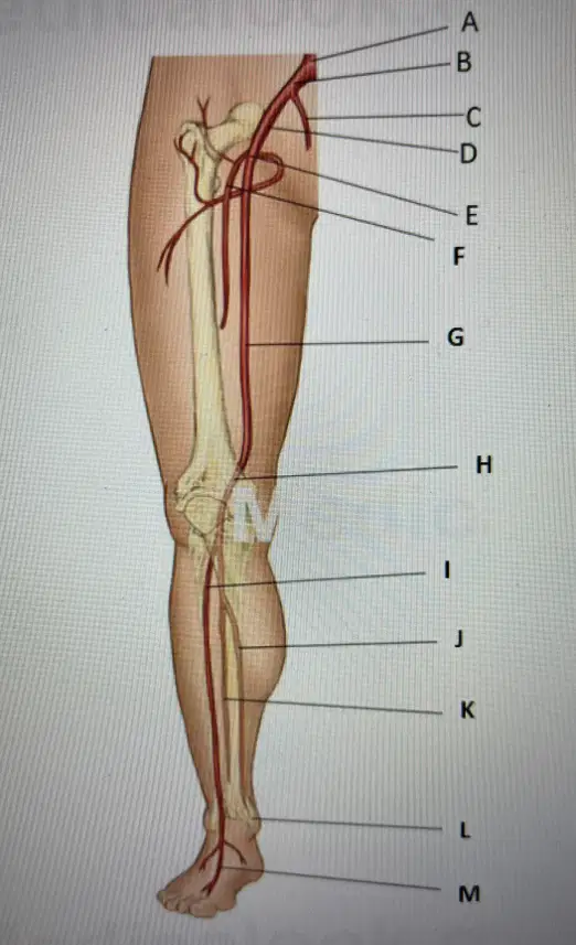
Right Popliteal Artery
H
I
M
F
E
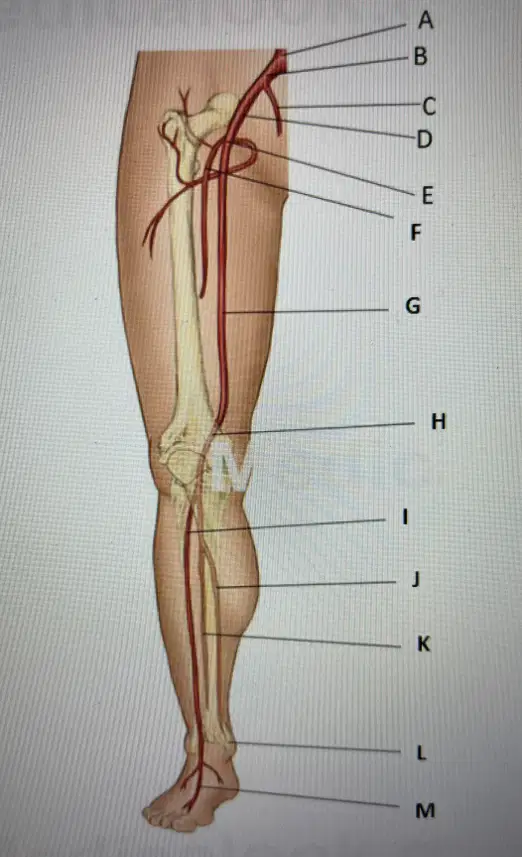
Right Anterior Tibial Artery
I
M
E
F
G
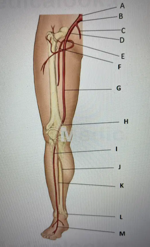
Right Dorsalis Pedis Artery
M
I
H
G
F
The dorsalis pedis artery is the direct continuation of which vessel?
Posterior Tibial
Anterior Tibial
Peroneal
Tibioperoneal Trunk
The two-terminal branches of the internal carotid artery are the:
Middle cerebral and posterior cerebral
Anterior cerebral and middle cerebreal
Middle cerebral and posterior communicating
Anterior cerebral and posterior communicating
The popliteal artery terminates by branching into the:
Posterior tibial and peroneal
Anterior tibial and peroneal
Anterior tibial and Tibiperoneal trunk
DPA and PTA
Flow in a normal, competent perforating vein should flow:
Superficial to deep
Exhibiting reversal
Deep to superficial
In many directions
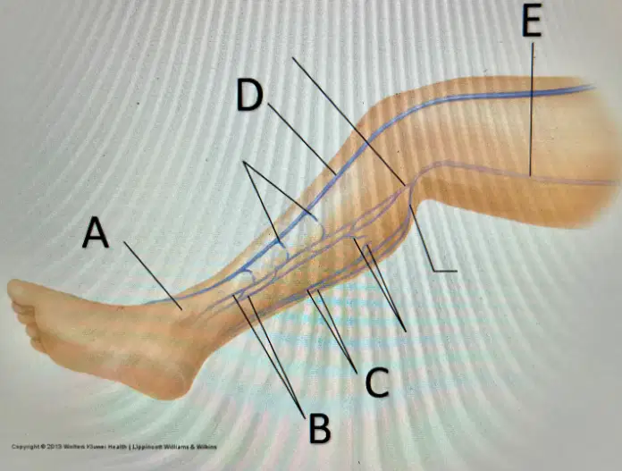
Right medial malleolus
A
B
C
D
E
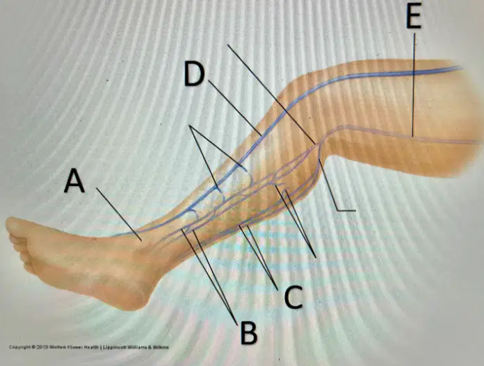
Right Peroneal Veins
A
B
C
D
E
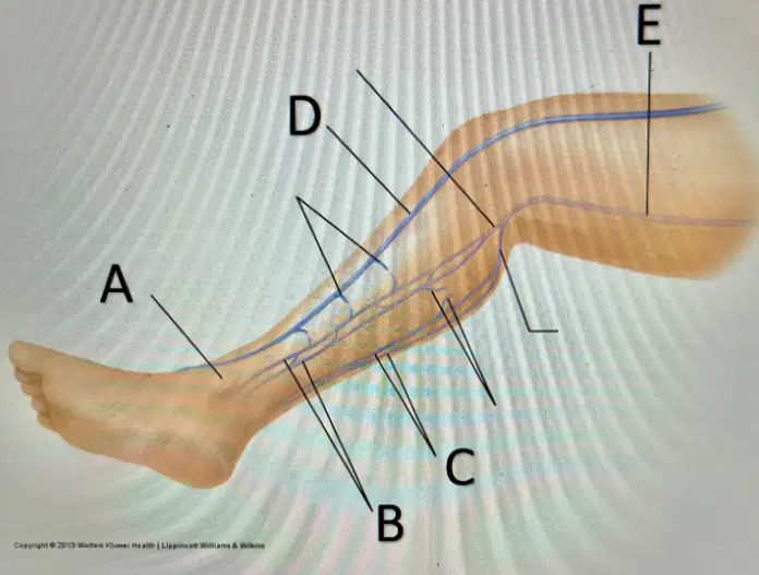
Right posterior tibial veins
a
b
c
d
e
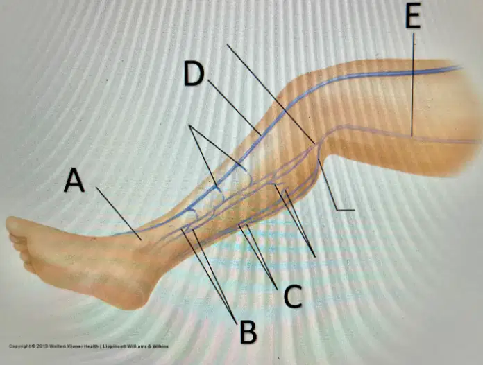
right great saphenous
a
b
c
d
e
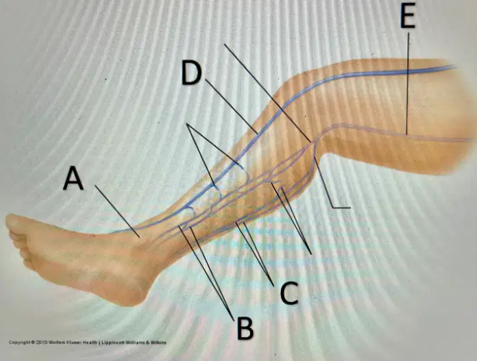
right popliteal
a
b
c
d
e
The small saphenous vein most commonly drains into the:
Popliteal vein
Vein of Giacomini
SFV
GSV
Compressibility of veins is very important in the diagnosis of DVT. Which of the following veins are impossible to compress due to their location?
Distal femoral veins
Iliac veins
Common femoral veins
Popliteal veins
Lower extremity venous flow is aided in its return to the heart by all of the following mechanisms EXCEPT:
Venous valves
Inspiration
Calf muscle contraction
Expiration
The normal flow pattern for the superior mesenteric artery, pre-prandial is:
Low resistance w/ PSV <125 cm/s
High Resistance w/ PSV <125 cm/s
High resistance w/ PSV <110 cm/s
Low resistance w PSV <110 cm/s

