
 Worksheets
Pulser echo instrumentation
Total questions: 128
Worksheet time: 2hrs 49mins
Controls the strength of the transmitted sound: Transducer output
(a)
Determines time between electrical spikes (PRP)
(a)
PRP and PRF are (a) related
Pulser determines the (a)
What are the two major functions of the system?
Preparation and transmission
Doppler
Brightness
Reception of electrical signals
output gain, acoustic power, pulser power, energy out put, transmitter output are synonyms for (a)
Transducer output is determined by the (a) from the pulser. Piezoelectric crystal vibrates with a magnitude related to pulser voltage
The (a) affects the brightness of the entire image
The (a) can be adjusted by the sonographer
When (a) changes, every pulse transmitted to the body changes
When the (a) changes, all reflections from structures in the body also change
When the (a) changes, the brightness of the entire image changes.
Increasing ___ ___ improves signal-to-noise ratio
(a)
Does the transducer output affect patient exposure?
Yes
No
Excessive output (a) axial resolution
Considering transducer output, thermal index and mechanical index are both associated with (a)
When amplification is increased, the electronic signals in the receiver are boosted, and the image will be (a)
Persistent disturbance that blurs a signal
(a)
It fills images with low level undesirable echoes
(a)
Comparison of the meaningful signal in an image compared to the amount of noise
(a)
High s/n ----> Image of (a) quality
Low s/n --> (a) image quality
If the transducer output is low --> (a) signal to noise ratio
If transducer output is increased --> (a) signal to noise ratio
WHAT IS THE MOST COMMON WAY TO IMPROVE SIGNAL-TO-NOISE RATIO?
Decreasing output power
Increasing output power
Part of the transmitted that receives electrical spikes and distributes them to the crystal of the array transducer
(a)
It coordinates the signals to optimize transmission of the sound beam
(a)
It adjusts electrical signals to reduce lobe artifacts (anodization) [We will learn more about this in the lecture on artifacts]
(a)
It receives time delays for dynamic receive focusing.
(a)
It controls the dynamic aperture (# of PZT used during reception and transmission).
(a)
Easy, faster updates (software programming)
Stable system
Versatile
(a)
Transmit-receive regulator
Transmit: high voltages
(a)
Receive: low voltages
Protects the system by handling voltages appropriately
(a)
The signals returning from the transducer are extremely weak
(a)
The receiver boosts the strength of these signals, processes them
and prepares them for display
(a)
Is this order correct for receiver functions:
1. Amplification
2. Compensation
3. Compression
4. Demodulation
5. Rejection
Yes
No
Amplification (receiver functions) step (a)
Compensation (receiver functions) step (a)
Compression (receiver functions) step (a)
Demodulation (receiver functions) is step (a)
Rejection (receiver functions) step (a)
What is another name for amplification;
(a)
Can amplification be adjusted by the sonographer?
Yes
No
What are the units for amplification?
Centimeters
Decibels
Watts
Hertz
The ratio of the output electrical signal strength to the input of the electrical signal strength of the amplifier
(a)
How is the image affected by amplification?
Amplification changes the motion
Amplification changes the TGC
Amplification changes the scale
Amplification changes the brightness
Increasing overall gain (a) create an image with uniform brightness.
Typical values are 60 dB to 100 dB
(a)
What is preamplification?
The process of improving the quality of a signal before it is amplified
The process of improving the quality of a signal before it is gained
The process of improving the quality of a signal before it is not amplified
What is the role in processing signals from the transducer during reception?
Prevent echoes in an image
Prevent brightness on an image
Prevent electronic noise from contaminating the tiny electrical signals
Used to create image of uniform brightness from top to bottom.
(a)
Since attenuation is strongly related to path length, echoes returning from great depths have (a) amplitudes than those returning from shallow depths
How is the image affected by compensation?
Compensation makes all echoes from similar reflectors appear identical regardless of their depth
Compensation treats echoes differently, depending upon the depth at which they arise.
Time gain compensation (TGC)
Depth compensation (DGC)
Which of the following is another name for compensation?
Time gain compensation (TGC)
Depth compensation (DGC)
Amplitude
Motion mode
Can compensation be adjusted by the sonographer?
Yes
No
Compensation makes an image equally (a) at all depths
Is the image of uniform brightness from the top to the bottom?
Yes due to compensation
No due to compensation
If the signal to noise ratio is high, how is the image quality?
Image of low quality
Image of high quality
Image of no quality
If the signal to noise ratio is low, how is the image quality?
Poor
High
Medium
Rare
If transducer output is low, how is signal to noise ratio?
High
Medium
Low
Quality
If the transducer output is increased, how is the signal to noise ratio?
High
Low
Medium
Rare
What is the most common way to improve signal to noise ratio?
Increasing output
Decreasing output
Not adjusting output
Which is the correct order of the receiver functions?
Compensation, amplification, compression, demodulation, rejection
Amplification, demodulation, compression, compensation, rejection
Amplification, compression, compensation, demodulation, rejection
Amplification, compensation, compression, demodulation, rejection
Can you change each echoes brightness with amplification?
YES
NO
Does amplification make an image equally bright at all depths?
YES
NO
Less attenuation
Less TGC needed
(a)
Higher attenuation
More TGC required
(a)
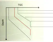
The first arrow points to the:
(a)

The second arrow points to:
(a)
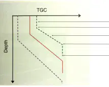
The third line points to:
(a)

The fourth arrow points to the:
(a)
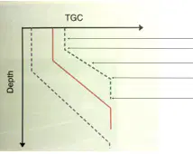
The fifth arrow points to:
(a)
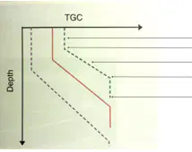
This shows the (a) curve
Reducing the total range, the smallest to the largest signal.
(a)
Keeps signals within the operating range of the system's electronics and the gray scale within the range of what the human eye can see.
(a)
Compression is Accomplished (a) altering the
relative relationships between voltages; largest stays largest, smallest remains smallest.
Compression (a) the dynamic range of the signals
What is the dynamic range?
The ratio of the largest to the largest strengths in a display
The ratio of the smallest to the largest strengths in a display
The ratio of the smallest to the smallest strengths in a display
The ratio of the largest to the smallest strengths in a display
How is the image effected by compression?
The color doppler shifts
The gray scale changes
The spectral changes
The zoom changes
Can the sonographer adjust compression?
YES
NO
It changes the electrical signals within the receiver into a form that fits the TV screen or cathode ray tube (CRT)
(a)
How is demodulation done?
Rectification
Amplifying
Compressing
Smoothing
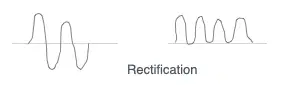
All negatives voltages are converted to positive voltages. Eliminates negative voltages
(a)
Also know as enveloping or smoothing process.
(a)
Can demodulation be adjusted by the sonographer?
YES
NO
How is the image affected by demodulation?
Grayscale changes
Doppler color changes
No visible effect
The depth changes
It controls whether low-level grayscale echoes will appear on the displayed image
(a)
What is another name for rejection?
Wall filter
Overall gain
Compensation
TGC
Threshold and suppression are other names for:
(a)
Can the sonographer adjust rejection?
YES
NO
How is the image affected by rejection?
It changes the grayscale
It does not affect bright echoes.
All noise or other low-level signals in the entire image are eliminated from the displayed image
No visible effect
It allows the system to use only high frequency reflection to display the superficial portion of the image.
(a)
Higher frequency --> (a) axial resolution
Lower frequency are used to display
deeper portions. At this level, higher frequencies have disappeared due to attenuation
(a)
Affects image brightness by adjusting the strength of the sound pulse sent to the tissues.
(a)
When the pulse is more powerful, all of the returning echoes from the body are stronger, and the image is (a)
When the image is too bright due to high output power, the lateral and longitudinal resolution (a)
Affects image brightness by changing the amplification of the electronic signals after returning to the receiver.
(a)
As Low As Reasonably Achievable
(a)

If you see this image, what do you do on the machine?
Decrease receiver gain
Increase output power
Decrease output power
Increase receiver gain
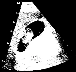
If you see this image, what do you do on the machine?
increase output power
decrease output power
increase receiver gain
decrease receiver gain
Creates and controls electrical signals sent to the transducer that generate sound pulses. The (a) determines the amplitude, PRP, and PRF
The (a) determines the firing delay patterns for phased array systems
Transforms the electrical signals from the transducer
(a)
Presents processed data.
(a)
Archives the ultrasound studies
(a)
Maintains and organizes the proper timing and interaction of the systems components
(a)
When does the pulser function?
After transmission
During the amplitude spikes
During transmission
Between receiver and transducer output time
What is the typical range for the magnitude of the pulsars electrical voltage spike?
1 to 100
0 to 100
3 to 100
10 to 100
Changes in (a) modify the brightness of the entire image
Low voltage of the PZT, (a) image
Higher pulser voltage, (a) image
When transducer output is (a) , noise is more likely to degrade the image
When a sonographer increases output power, the signal to noise ratio (a)
Will a HIGH or LOW signal to noise ratio result in a higher quality image?
High
Low
The (a) protects sensitive electrical components in the receiver from the high voltages created during transmission
(a) is the process of improving the quality of a signal BEFORE it is amplified. This occurs as close to the active elements as practical
(a) are specially designed to prevent electrical noise from contaminating the tiny electrical signals created by the transducers active elements during reception
At superficial depths, reflections undergo a small, constant amount of compensation called the:
(a)
The depth at which variable compensation begins is known as the:
(a)
In the region of the (a) , compensation corrects for the effects of increasing attenuation that result from increasing path length
At the depth of the (a) , reflections are maximally compensation by the ultrasound system

You are using a 4MHz transducer and have adjusted the TGC perfectly, as represented by the red TGC line. You are given a knew transducer with an unknown frequency. The TGC is then adjusted to create the same image as the 4MHz transducer. This new TGC setting is represented by the purple dotted line. Would the new transducer frequency be HIGHER or LOWER than the 4MHz transducer?
Higher
Lower
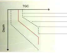
You are using a 4MHz transducer and have adjusted the TGC perfectly, as represented by the red TGC line. You are given a knew transducer with an unknown frequency. The TGC is then adjusted to create the same image as the 4MHz transducer. This new TGC setting is represented by the green dotted line. Would the new transducer frequency be HIGHER or LOWER than the 4MHz transducer?
Higher
Lower
All of the following are components of an ultrasound system EXCEPT:
Transducer
Alternator
Display
Pulser
Synchronizer
This type of pulser generates a constant electrical signal in the form of a sine wave:
Pulsed wave array
Continuous wave
Pulsed wave, single crystal
This type of pulser generates a single electrical spike, which ultimately creates a single sound pulse:
Pulsed wave, phased array
CW
Pulsed wave, single crystal
This type of pulser generates numerous electrical spikes, which ultimately create a single sound pulse:
CW
Pulsed wave, phased array
pulsed wave, single crystal

