
 Worksheets
Kidneys and urinary tract
Total questions: 122
Worksheet time: 2hrs 53mins
Excretory function (forms urine)
(a)
Maintain chemical balance (ph, minerals, iron, salts)
(a)
Detoxification
Blood pressure regulation
(a)
The functional units of the kidney, containing glomeruli (where filtration occurs) and tubules (where reabsorption and secretion take place).
(a)
Which of the following correctly describes the size of an adult kidney?
L: 10-14 cm; Diam: 4-6 cm; A-P: 2.5-4.0 cm
L: 9-12 cm; Diam: 6-7 cm; A-P: 2.5-4.0 cm
L: 10-14 cm; Diam: 5-6 cm; A-P: 3.5-4.0 cm
L: 9-12 cm; Diam: 4-6 cm; A-P: 2.5-4.0 cm
Carry the urine from kidneys to the urinary bladder
(a)
Which of the following correctly describes the size of the ureters?
Length: 26-39 cm (10-12 inches). Diam: 3-4 mm
Length: 25-30 cm (12-14 inches). Diam: 5-6 mm
Length: 25-30 cm (10-12 inches). Diam: 3-4 mm
Length: 29-31 cm (11-12 inches). Diam: 3-4 mm
Stores the urine temporarily
(a)
Carries urine from the urethra to outside the body
(a)
Which of the following best describes the length of the urethra in male and female?
Length: 5 cm (fem); 40 cm (male)
Length: 3 cm (fem); 30 cm (male)
Length: 4 cm (fem); 20 cm (male)
Length: 4 cm (fem); 40 cm (male)
Endocrine hormones (cortex) → corticoids (mineralocorticoids, glucocorticoids, sex hormones supplements)
(a)
Epinephrine and norepinephrine (medulla)
(a)
Kidneys are (a)
Which of the following best describes the location of the kidneys in relation to the spine?
On one side of the spine
Level T-12 to L4
On both sides of the spine
Level L4 to L5
The following structures are (a) to the kidneys:
diaphragm, psoas muscle, transverse muscle, and the quadratus lumborum muscle.
Which kidney is displaced inferiorly by the liver?
Left
Right
The following structures are located anterior to WHICH kidney?
Right lobe of liver, second portion of duodenum, hepatic flexure of colon, jejunum
Right
Left
Virtual space between liver and RT KID
(a)
The following structures are located anterior to WHICH kidney?
tail of pancreas, spleen, stomach, splenic flexure of colon, jejunum
Right
Left
Anterosuperior and slightly medial to RT KID
(a)
Anterosuperior and slightly medial to the LT KID
(a)
Ureters are (a) structures
Start at the renal pelvis in the renal hilum on each side and go inferiorly to merge into the urinary bladder posteriorly
(a)
Ureters pass (a) to the psoas muscle and then cross the bifurcation of the common iliac arteries.
Right ureter is typically shorter than left
True
False
In the pelvic cavity, ureters pass (a) to the ductus deferens (males) and uterine artery (females)

1
(a)

2
(a)

3
(a)
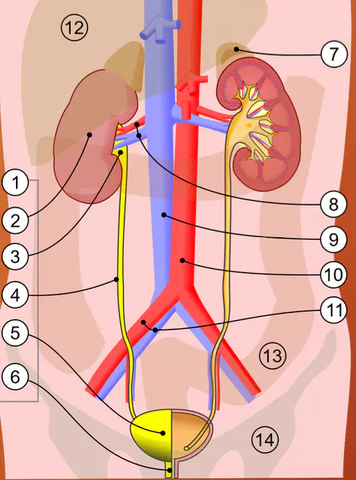
4
(a)
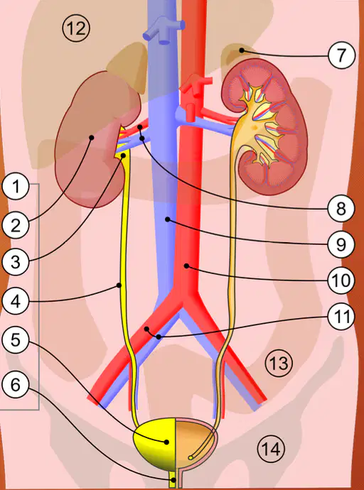
5
(a)

6
(a)
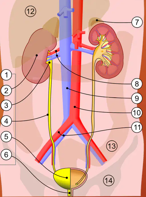
7
(a)
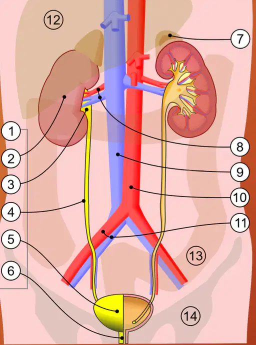
8
(a)

9
(a)
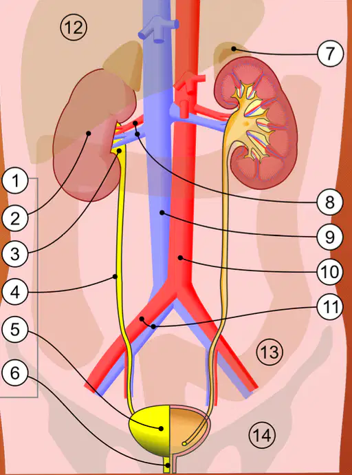
10
(a)
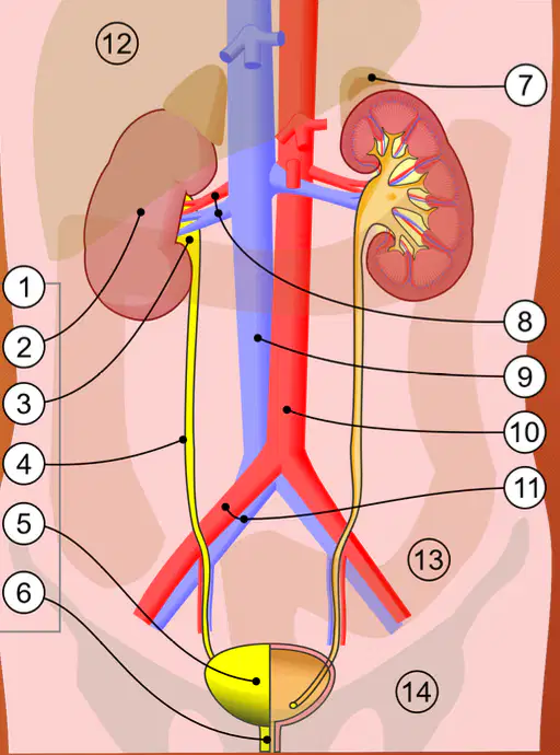
11
(a)

12
(a)

13
(a)

14
(a)
Include apical, upper, middle, and lower segmental arteries.
(a)
Divide into lobar arteries
(a)
Branch into interlobar arteries
(a)
Run between the medullary pyramids
Divide into arcuate arteries
(a)
Form an arch-like structure at the border between the renal cortex and medulla.
(a)
Give rise to the interlobular arteries
(a)
Extend into the cortex
Give off afferent arterioles.
(a)
Carry filtered blood away from kidneys to IVC
(a)
The bladder is a (a) structure
The bladder is (a) to Symphysis Pubis
In the male, the bladder is (a) to the seminal vesicles and rectum
In the male, the bladder is (a) to the prostate gland
Ductus deferens: superior lateral from scrotum; crosses ureter anteriorly into the prostate gland in relation to the (a)
In the female, the bladder is (a) to the vagina, cul-de-sac, and rectum
Membranous canal that originates from the urinary bladder at its inferior border (specifically, at the bladder neck) and extends to the exterior of the body
(a)
Serves as the passageway for urine to exit the body.
(a)
In the (a) , the urethra is much longer
Is divided into several parts (prostatic urethra, membranous urethra, and spongy, aka penile urethra
(a)
Membranous urethra passing through the pelvic floor muscles
(a)
Much shorter
o Passes through the pelvic floor before opening to the exterior.
(a)
focal bulge on the lateral border of LT KID that resembles the hump of a dromedary camel. It is caused by the indentation of the spleen on the LT KID over time. The spleen's pressure creates a bulge in the kidney's outline
(a)
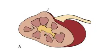
This image shows the __
Dromedary hump
Column of Berlin
Sinus
Pelvis
portion of the renal cortex extends into the renal sinus, potentially mimicking a renal tumor
(a)
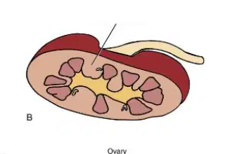
This image shows the __
Dromedary hump
Column of Berlin
Pelvis
Sinus

1
(a)

2
(a)
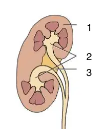
3
(a)
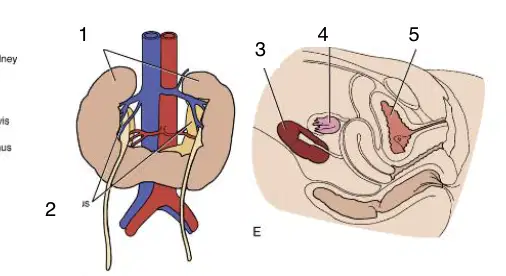
1
(a)
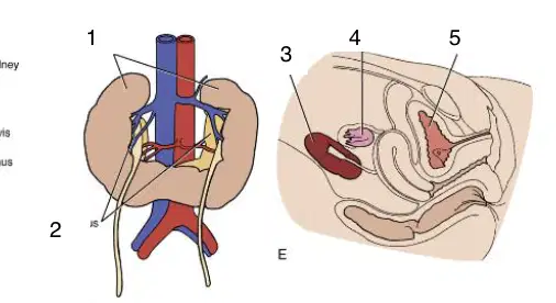
2
(a)
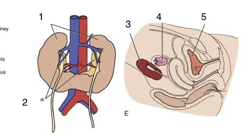
3
(a)
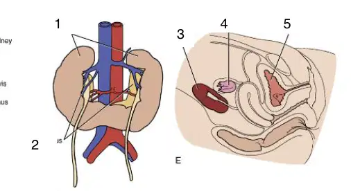
4
(a)
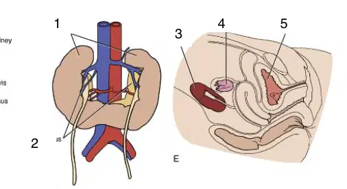
5
(a)
Renal size, length, width, and morphology.
Identify location
Assess vascularity
Assess echogenicity
Differentiate solid vs cystic tumors
Trauma
Hypertension
Kidney disease
(a)
Evaluate for congenital malformations, masses (hematoma, tumors), abscesses, cysts, and hyperplasia.
(a)
Heterogeneous, Smooth, Elliptical (bean shaped) (longitudinal view), Rounded in axial view
(a)
Anechoic, Smooth, Linear tubular, Seen in the hilarious region
(a)
Highly reflective, Thin, continuous, Follows the shape of the kidney, Seen in the periphery
(a)
Medium-gray; low level homogeneous
Lower echogenicity than liver
(a)
Extensions between medullary pyramids→ columns of Bertin
(a)
Anechoic with bright walls, Medullary pyramids, Triangular, round, or blunted areas
(a)
Which of the following best describes the thickness of the renal medulla
1.3 to 1.5 cm thick
1.9 to 1.10 cm thick
1.3 to 1.6 cm thick
1.2 to 1.5 cm thick
Bright-echo dense, Irregular, Ovoid, Contains fat and fibrous tissues,
(a)
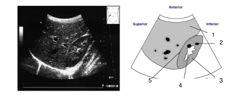
1
(a)

2
(a)
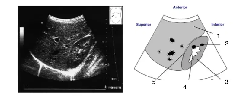
3
(a)

4
(a)

5
(a)

Which kidney is shown in this image?
Right
Left

Which kidney is shown in this image?
Right
Left
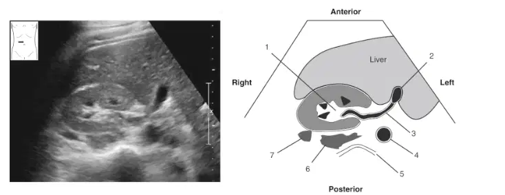
1
(a)
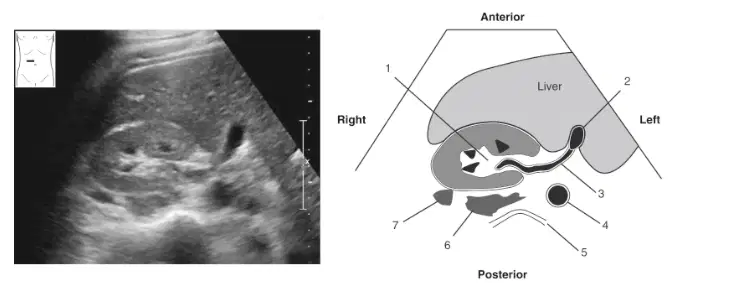
2
(a)

3
(a)

4
(a)

6
(a)
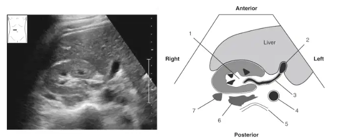
7
(a)
Anechoic lumen Bright walls, Symmetric Smooth
(a)
Axial view: square-like Longitudinal: ovoid or oblong
(a)
Size and shape depends on the volume of urine stored.
Not visible well if empty
(a)
Hyperechoic
May be seen with Color Doppler
Slit-like or oval
(a)
Posterior aspect of bladder (trigone area)
(a)
Hyperechoic, Smooth, Linear tubular, Not always seen
(a)
Anechoic, Smooth, Tubular, Usually not seen; unless distended
(a)
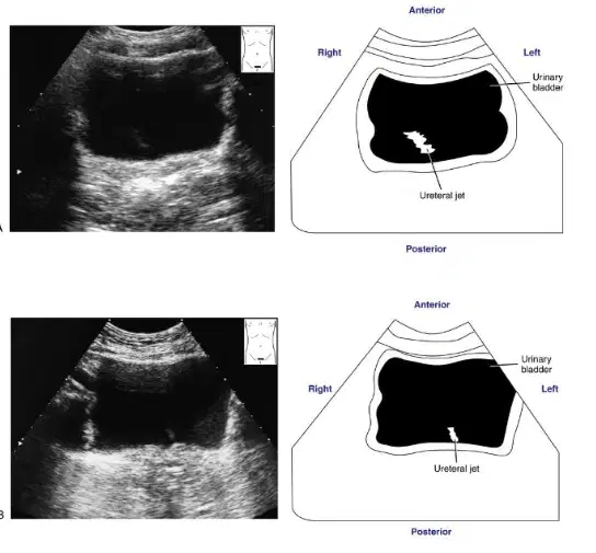
These images show the:
Ureter and urinary jets
Urethra and urinary jet
Urinary bladder and urinary jet
Kidney and urinary jet
Fusion of the lower pole of the kidneys is called a
Lower fusion kidney
Horshoe kidney
Dromedary hump
Bertin
The most echogenic parts of the kidney are the
medullary pyramids
renal cortex
renal sinus
ureters
The left kidney is in contact with the:
liver, stomach, ascending colon
spleen, stomach, and pancreas
pancreas, lungs, descending colon
spleen, stomach, right liver
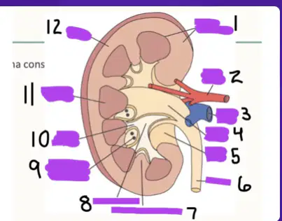
1
(a)

2
(a)

3
(a)

4
(a)

5
(a)
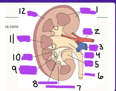
6
(a)
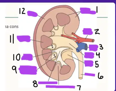
7
(a)
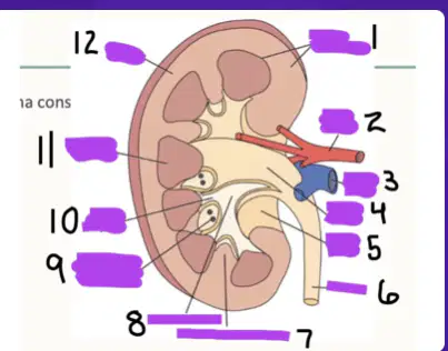
8
(a)

Number 9 is the blood vessel entering the parenchyma
True
False
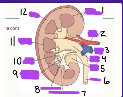
10
(a)
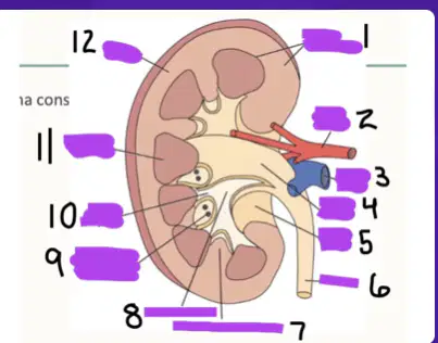
11
(a)
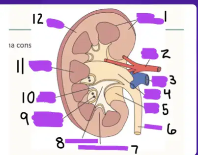
12
(a)
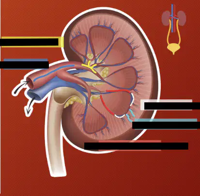
Which arteries are highlighted yellow
(a)

Which artery is highlighted dark blue
(a)

Which artery is highlighted red
(a)

Which artery is highlighted teal
(a)

