
 Worksheets
Radiology Of Back Quiz 17th July
Total questions: 10
Worksheet time: 5mins
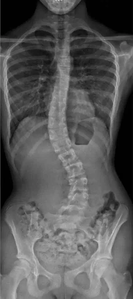
A 13-year-old girl is brought to the clinic by her mother for a routine school physical. On examination, the physician notices that her right shoulder is slightly higher than the left, and her scapula appears more prominent on the right side. There is no pain or neurological deficit. An X-ray of the spine is ordered and reveals a lateral curvature of the thoracolumbar spine.
A. Scoliosis
B. Lumbar disc herniation
C. Spinal tuberculosis
D. Spondylolisthesis

A 72-year-old woman with a history of osteoporosis presents to the emergency department after slipping in the bathroom. She complains of sudden-onset lower back pain. She is unable to stand straight due to pain, but there is no leg weakness or numbness. An X-ray of the lumbar spine is performed.
Sciatica
Spinal Tumor
Vertebral Fracture
Muscle Strain
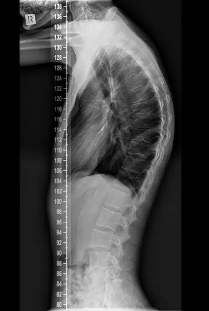
A 15-year-old boy is brought to the clinic by his parents who are concerned about his poor posture. He does not complain of any pain or neurological symptoms. On examination, he has an exaggerated forward rounding of the upper back. An X-ray of the thoracic spine reveals an increased thoracic curvature greater than normal.
Lordosis
Kyphosis
Scoliosis
Vertebral Fracture

A 60-year-old man presents with chronic lower back pain that worsens with activity and improves with rest. He has no numbness, tingling, or weakness in the legs. Physical examination shows reduced lumbar spine flexibility. An X-ray of the lumbar spine reveals decreased disc space and the presence of bony outgrowths (osteophytes) at multiple levels.
Acute lumbar strain
Ankylosing Spondylitis
Spinal Tumor
Degenerative Disc Disease
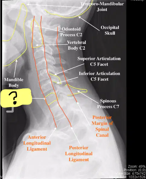
Sagittal View of Cervical Spine X ray is displayed, which part is labelled with a question mark ?
Pedicles
Superior Articular Facet
Vertebral Body
Interverterbral Disc Space
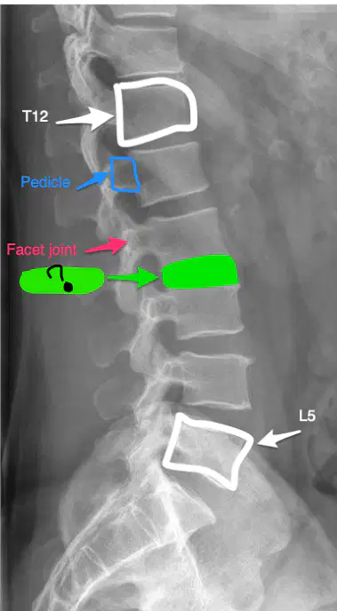
Sagittal View of X ray Lumbar Spine is seen on the image, which part is labelled with a question mark?
Vertebral Body
Intervertebral Disc Space
Sacroiliac Joint
None of the above

You are preparing a patient for an MRI scan. As you position them on the table, the patient appears visibly anxious and says, “I feel scared in closed spaces and I’m really nervous about this test.”What is the most appropriate initial step to help calm this patient?
Explain the procedure, reassure the patient, and offer relaxation techniques
Proceed quickly to finish the scan before the patient panics
Administer a sedative immediately without explanation
Cancel the MRI and refer the patient to psychiatry
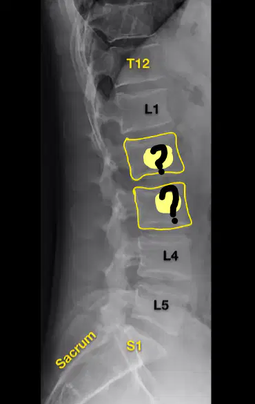
X ray of Spine is shown, Which Vertebras are marked with question marks?
T1, T2
L1, L2
L2, L3
C1,C2
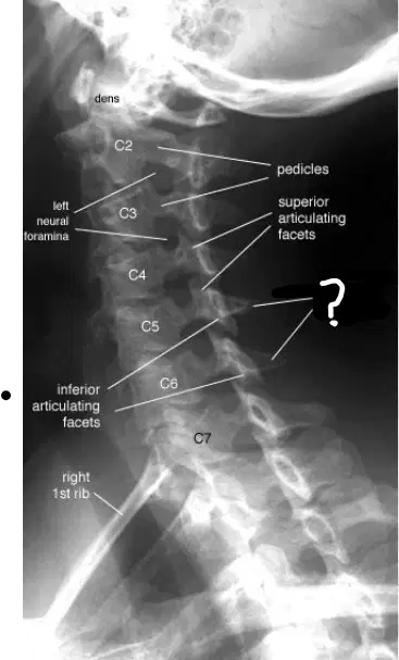
X ray of Cervical Spine Sagittal view is seen, which structures are marked with question mark ?
Spinous Process
Transverse Process
Vertebral Body
Intervertebral Disc Space

Identify the part labeled 9
Inferior articular facet
IV disc space
Spinous process
Transverse process

