
 Worksheets
Pathology Supple Set B
Total questions: 18
Worksheet time: 9mins
Describe the pathogenesis of hepatocarcinoma caused by Hepatitis C.
- HCV core protein (E1, E2) and non structural proteins will inhibit tumour suppressor gene, TP53, TP73 and RB1(retinoblastoma associated protein) - Hence, HCV core AND NS5A proteins participate in development of HCC
- Point mutation in KRAS and p53, and expression of c-MYC, c-MET (the receptor for hepatocyte growth factor), inhibition of TGF-a, and activated insulin like growth factor 2 leads to carcinogenesis
- HBV DNA integration into host genome during regeneration of hepatocytes may activate proto-oncogenes - leads to tumorigenicity / carcinogenicity
- HCV is a positive-strand RNA virus that replicates in cytoplasm and no integration into host DNA - Repeated cycles of cell death and regeneration cause accumulations of mutations and may damage DNA repair mechanisms, eventually leads to carcinogenesis
Describe the pathogenesis of hepatocarcinoma caused by chronic Hepatitis B.
- repeated regeneration of hepatocytes - point mutations in KRAS, p53, increased expression of c-MYC, c-MET TGF-a and insulin-like growth factor 2 leads to carcinogenesis
- Point mutation in KRAS and p53, and expression of c-MYC, c-MET (the receptor for hepatocyte growth factor), inhibition of TGF-a, and activated insulin like growth factor 2 leads to carcinogenesis
- HBV DNA integration into host genome during regeneration of hepatocytes may activate proto-oncogenes - leads to tumorigenicity / carcinogenicity
- HCV is a positive-strand RNA virus that replicates in cytoplasm and no integration into host DNA - Repeated cycles of cell death and regeneration cause accumulations of mutations and may damage DNA repair mechanisms, eventually leads to carcinogenesis
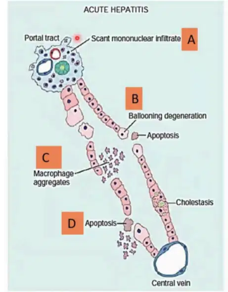
This is infection of?
Acute Hep B
Chronic Hep B
Acute Hep C
Chronic Hep C
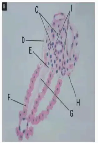
Choose correct labelling.
I : Triaditis
C : Limiting plate eroded
D : Bridging fibrosis
E : Necrosed hepatocytes
F : Proliferated bile duct
G : Portal tract expand
H : Focal necrosis
I : Focal necrosis
C : Limiting plate eroded
D : Proliferated bile duct
E : Necrosed hepatocytes
F : Bridging fibrosis
G : Portal tract expand
H : Triaditis
I : Portal tract expand
C : Triaditis
D : Proliferated bile duct
E : Necrosed hepatocytes
F : Bridging fibrosis
G : Focal necrosis
H : Limiting plate eroded
I : Portal tract expand
C : Focal necrosis
D : Proliferated bile duct
E : Limiting plate eroded
F : Bridging fibrosis
G : Triaditis
H : Necrosed hepatocytes
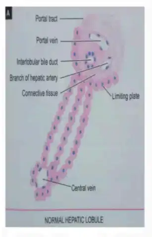
The picture shows normal hepatic lobule (read labelling for revision). Choose complications of viral hepatitis B.
Chronic hepatitis
Cirrhosis
Hepatocellular ca
Liver failure
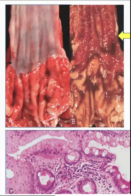
Describe gross and microscopic of Barret Oesophagus.
Esophageal mucosa is smooth and tan white appearance, gastric mucosa has reddish pink rugosities.
Reddish change in mucosal appearance at lower end of esophagus, above the GEJ denoting some epithelial changes.
Squamous cell mucin secreting glandular epithelium with goblet cells at lower end of oesophagus instead of normal tall columnar epithelium. Also metaplastic mucosa containing goblet cells.
Tall columnar mucin secreting glandular epithelium with goblet cells at lower end of oesophagus instead of normal squamous epithelium. Also metaplastic mucosa containing goblet cells.
Arrange pathogenesis of GERD.
• Reflux of gastric juices – resulting in esophagitis through mucosal injury and leading to GERD
• The stratified squamous epithelium of the esophagus is resistant to abrasion from foods but is sensitive to acid.
• Decrease in lower esophageal sphincter LES tone or increase abdominal pressure contribute to GERD
• In severe cases - duodenal bile reflux exacerbate the damage.
• Decrease in lower esophageal sphincter LES tone or increase abdominal pressure contribute to GERD
• The stratified squamous epithelium of the esophagus is resistant to abrasion from foods but is sensitive to acid.
• Reflux of gastric juices – resulting in esophagitis through mucosal injury and leading to GERD
• In severe cases - duodenal bile reflux exacerbate the damage.
• The stratified squamous epithelium of the esophagus is resistant to abrasion from foods but is sensitive to acid.
• Reflux of gastric juices – resulting in esophagitis through mucosal injury and leading to GERD
• In severe cases - duodenal bile reflux exacerbate the damage.
• Decrease in lower esophageal sphincter LES tone or increase abdominal pressure contribute to GERD
• The stratified squamous epithelium of the esophagus is resistant to abrasion from foods but is sensitive to acid.
• Reflux of gastric juices – resulting in esophagitis through mucosal injury and leading to GERD
• Decrease in lower esophageal sphincter LES tone or increase abdominal pressure contribute to GERD
• In severe cases - duodenal bile reflux exacerbate the damage.
Antigen marker to indicate active hepatitis B infection.
HbsAg
HbeAg
serum alpha
serum beta
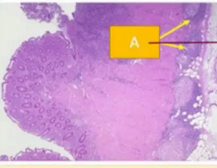
State diagnosis. What is A?
Transmural lymphoid aggregate
Crypt abcess
Ulcerative collitis
Crohn disease
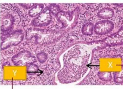
State diagnosis. Label X and Y.
X : Extensive inflammation in lamina propia
Y : Crypt abscess
X : Crypt abscess
Y : Extensive inflammation in lamina propria
Ulcerative collitis
Ischemic enterocollitis
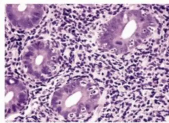
Acute self-limited colitis with patchy neutrophil-mediated crypt injury. This shows microscopic findings of what?
Ischemic enterocollitis
Crohn disease
IBD
Acute infectious enterocollitis
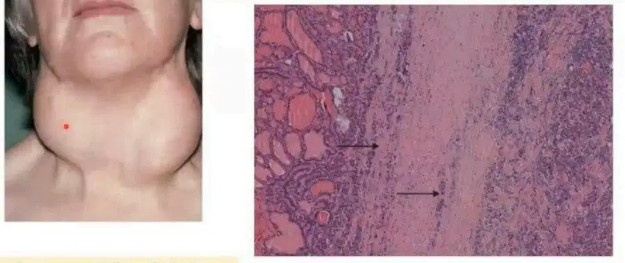
Define Reidel's thyroiditis.
A rare thyroid disorder of unknown etiology, characterized by chronic inflammation and extensive fibrosis causing hypothyroidism with stone -like.
A rare thyroid disorder of unknown etiology, characterized by acute inflammation and extensive fibrosis causing hypothyroidism with stone -like.
A rare thyroid disorder of unknown etiology, characterized by chronic inflammation and extensive fibrosis causing hyperthyroidism with stone -like.
A common thyroid disorder of unknown etiology, characterized by chronic inflammation and extensive fibrosis causing hypothyroidism with stone -like.
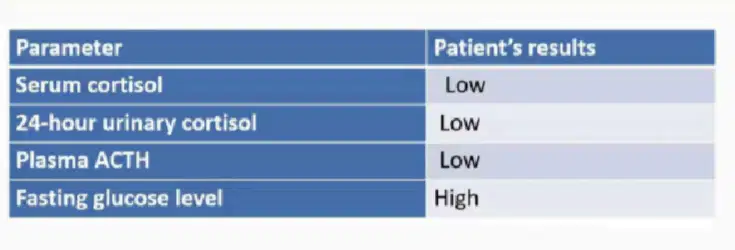
Choose Reidel's thyroiditis.
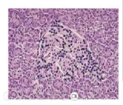
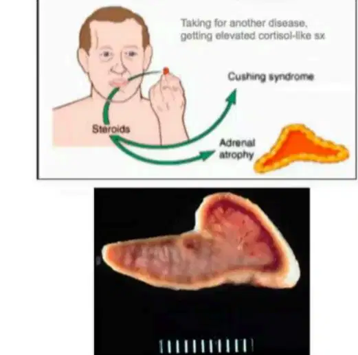

Gross morphology of adrenals in exogenous Cushing syndrome.
Bilateral adrenal hyperplasia
Bilateral adrenal atrophy
Bilateral adrenal hypertrophy
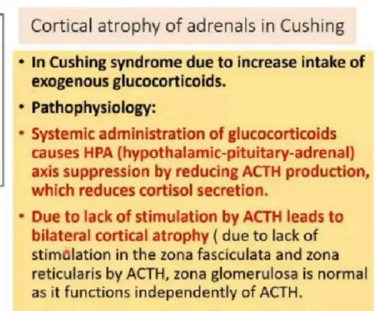
Lab test.
High cortisol, low ACTH with atrophied adrenal cortex
Low cortisol, high ACTH with atrophied adrenal cortex
High cortisol, low ACTH with hypertrophied adrenal cortex
Low cortisol, high ACTH with hypertrophied adrenal cortex
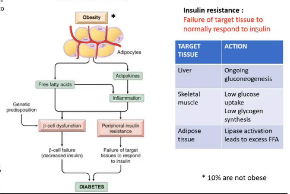
Type of diabetes.
(revise flow chart)
Type 1
Type 2
Acute complication of Diabetes.
DKA
Hyperosmolar hyperglycemic state
Hyperglycemia
Lactic acidosis
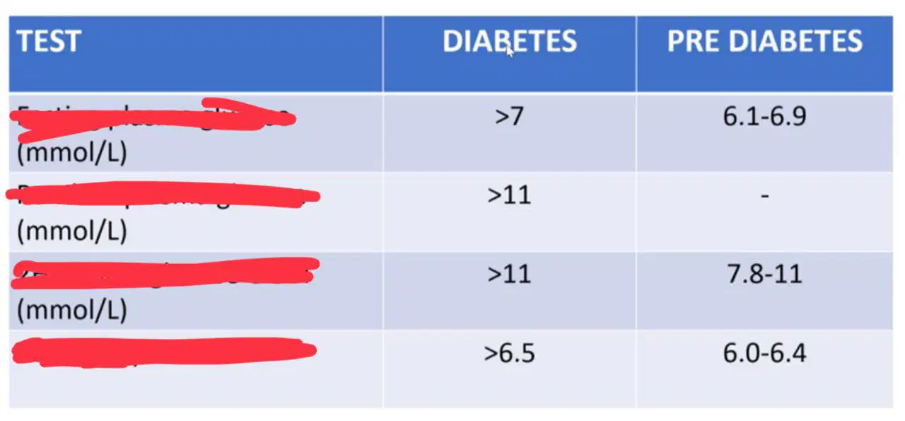
Diagnosis method for DM.
1. Random plasma glucose
2. Fasting plasma glucose
3. 2H plasma glucose OGTT
4. HbA1c (%)
1. Fasting plasma glucose
2. Random plasma glucose
3. 2H plasma glucose OGTT
4. HbA1c (%)
1. HbA1c (%)
2. 2H plasma glucose OGTT
3. Random plasma glucose
4. Fasting plasma glucose
1. 2H plasma glucose OGTT
2. Fasting plasma glucose
3. Random plasma glucose
4. HbA1c (%)

