
 Worksheets
EKG Worksheet Quiz
Total questions: 96
Worksheet time: 8hrs 0mins
Abigail, Emma, and Scarlett are studying EKG waveforms in class. During the P-wave of an EKG, which of the following best describes what is happening in the heart?
Depolarization
Repolarization
Hyperpolarization
Plateau phase
Luna and Aiden are studying EKGs in class. When they look at the QRS interval on the EKG, what is the main event happening in the heart at that moment?
Depolarization
Repolarization
Resting potential
Action potential plateau
Abigail and Charlotte are learning about the heart in science class. After the heart muscle contracts, which process helps the cardiac muscle cells return to their resting state?
Repolarization
Depolarization
Excitation
Contraction
Maya and Charlotte are studying EKG waveforms in class. They notice the T-wave and wonder what it represents. Can you help them out?
What does the T-wave on an EKG waveform represent?
Repolarization
Depolarization
Contraction
Plateau phase
Define depolarization in the context of cardiac physiology.
The process by which cardiac cells become less negative, leading to contraction
The process by which cardiac cells return to their resting negative state
The process of muscle relaxation
The process of oxygen exchange in the heart
Define repolarization in the context of cardiac physiology.
The process by which cardiac cells return to their resting negative state after contraction
The process by which cardiac cells become less negative, leading to contraction
The process of muscle contraction
The process of electrical conduction in nerves
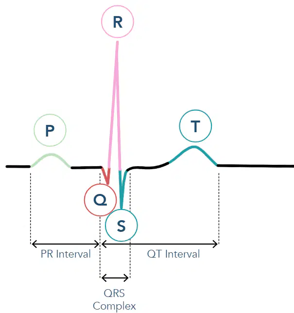
Aria, Ethan, and Zoe are studying for their cardiology quiz! Can you help them out? Which part of the EKG waveform shows when the atria are contracting?
P-wave
QRS interval
T-wave
PR interval
Grace and Benjamin are studying EKGs in class. During the QRS interval of the EKG waveform, what is happening in the heart?
Atrial contracting
Ventricle contracting
Ventricle relaxing
Atrial relaxing
Aria and Nora are studying for their cardiology quiz! Can you help them out? During which phase does repolarization of the ventricles occur in the EKG waveform?
P-wave
PR interval
QRS interval
T-wave
Elijah and Sophia are learning about the heart in class. When their teacher asks, "What does depolarization mean in the context of cardiac physiology?" which answer should they choose?
Relaxing
Decompressing
Refilling with blood
Repolarizing
Which of the following best describes repolarization?
Decompressing
Contracting
Relaxing and refilling with blood
Depolarizing
What does the PR interval represent in an EKG waveform?
The time between ventricular depolarization and repolarization
The time between atrial depolarization and ventricular depolarization
The time between atrial contraction and ventricular relaxation
The time between ventricular contraction and atrial relaxation
Which of the following is associated with depolarization in the EKG waveform?
T-wave
Repolarization
QRS interval
Relaxing
Benjamin and Charlotte are studying heart rhythms for their big test. Which of the following is considered a basic heart rhythm that they should definitely remember?
Normal sinus
Atrial flutter
Ventricular fibrillation
Asystole
Noah and Abigail are studying EKGs in class. Noah asks, "Hey Abigail, do you know what the PR interval measures on an EKG?" Can you help them out?
The time from the start of atrial depolarization to the start of ventricular depolarization
The duration of ventricular depolarization
The time between two consecutive heartbeats
The duration of atrial repolarization
Evelyn and Mason are studying for their EKG exam. Evelyn asks, "Hey Mason, do you remember how many seconds 1 small box on an EKG strip represents?" Can you help them out?
0.04 seconds
0.2 seconds
0.5 seconds
1 second
Nora and James are studying EKG strips together. If they count 5 large boxes on an EKG strip, can you help them figure out the total time represented?
1 second
0.2 seconds
0.04 seconds
2 seconds
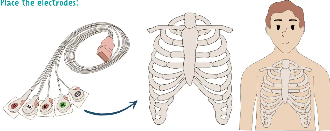
Isla, Harper, and Scarlett are learning how to set up an EKG machine for their science class. Can you help them figure out the correct order for placing the EKG electrodes on the chest?
Right arm, left arm, right leg, left leg, chest
Chest, right arm, left arm, right leg, left leg
Right leg, left leg, chest, right arm, left arm
Left arm, right arm, chest, left leg, right leg
What is the normal range for a sinus rhythm in beats per minute (bpm)?
40–60 bpm
60–100 bpm
>100 bpm
<60 bpm
Abigail and Oliver are racing to see whose heart beats faster! If a heart rate goes above 100 bpm, what is this speedy rhythm called?
Normal sinus
Bradycardia
Tachycardia
Atrial fibrillation
What is the duration of one small box on an EKG strip?
0.20 seconds
0.04 seconds
1 second
0.12 seconds
Which of the following is the correct duration for a normal PR interval?
0.06–0.12 seconds
<0.40 seconds
0.12–0.20 seconds
0.20–0.40 seconds
Olivia and Aiden are learning to read EKG strips in class. If Olivia counts the number of large boxes that equal one second on an EKG strip, how many should she count?
1
2
5
10
Samuel and Avery are learning about ECG electrode placements. According to the memory trick, where should the "WHITE" electrode be placed to help Mason remember?
On the left leg
On the right arm
On the left arm
On the right leg
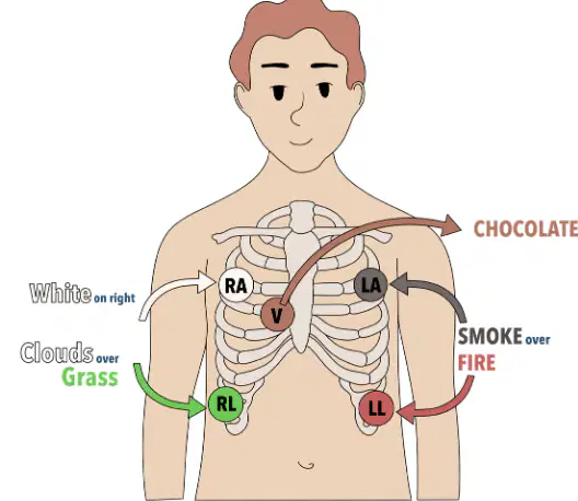
Noah and Ava are helping James set up a standard EKG for their science class. Can you help them out? Which electrode should they place on the left leg?
RA
LA
RL
LL
If a patient’s EKG shows a QRS complex duration of 0.15 seconds, what does this indicate?
The QRS complex is within the normal range.
The QRS complex is shorter than normal.
The QRS complex is longer than normal.
The QRS complex cannot be measured.
What is the maximum normal duration for the QT interval on an EKG?
0.12 seconds
0.20 seconds
0.40 seconds
1 second
Elijah and Anika are trying to remember how to place the "SMOKE" and "FIRE" electrodes using the memory trick. Which of the following best describes where they should put the electrodes?
Smoke over Fire on the left side
Smoke over Fire on the right side
Smoke under Fire on the left side
Smoke under Fire on the right side
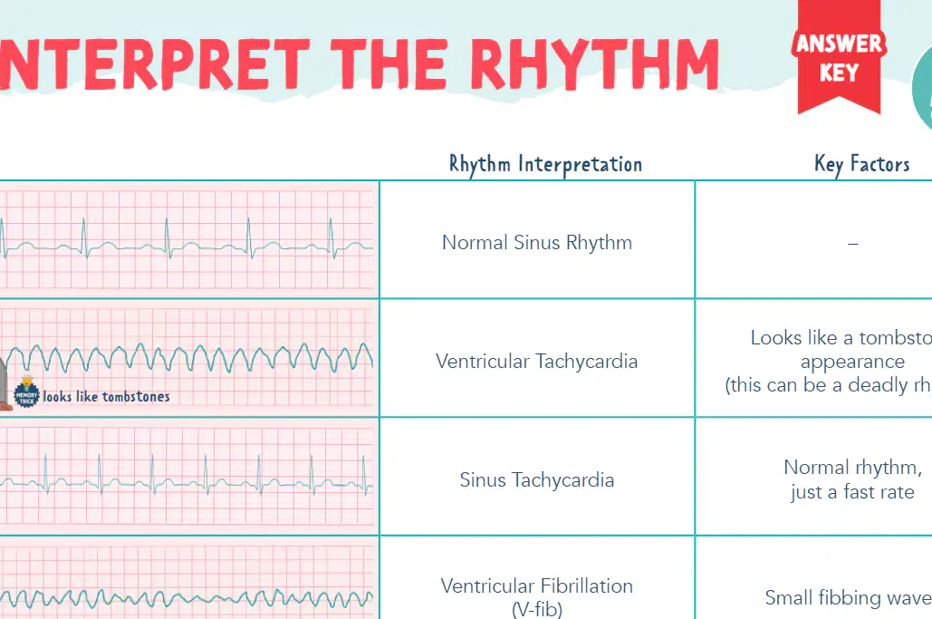
Which rhythm interpretation is characterized by a "tombstone appearance" and can be a deadly rhythm?
Ventricular Tachycardia
Sinus Brady
Atrial Fibrillation
Asystole
Maya and Kai are studying ECGs together. Maya asks, "Kai, do you know the key clue for spotting Atrial Fibrillation (A-fib) on an ECG?" What should Kai answer?
Sawtooth appearance between R-R intervals
Irregular R-R intervals
Flat line
Looks like S's
Scarlett and Aria are reviewing ECG rhythms in class. Which rhythm do they jokingly call the "ultimate nap time" because it looks like a "flat line" on an ECG?
Sinus Tachycardia
Asystole
Ventricular Fibrillation
Premature Ventricular Contraction
Aria and Mason are studying ECGs together. They come across a rhythm that looks like a row of tiny saw blades between the R-R intervals. Which main feature are they most likely seeing that points to Atrial Flutter (A-flutter)?
Small fibbing waves
Sawtooth appearance between R-R intervals
Premature conduction of a QRS complex
Looks like tombstones
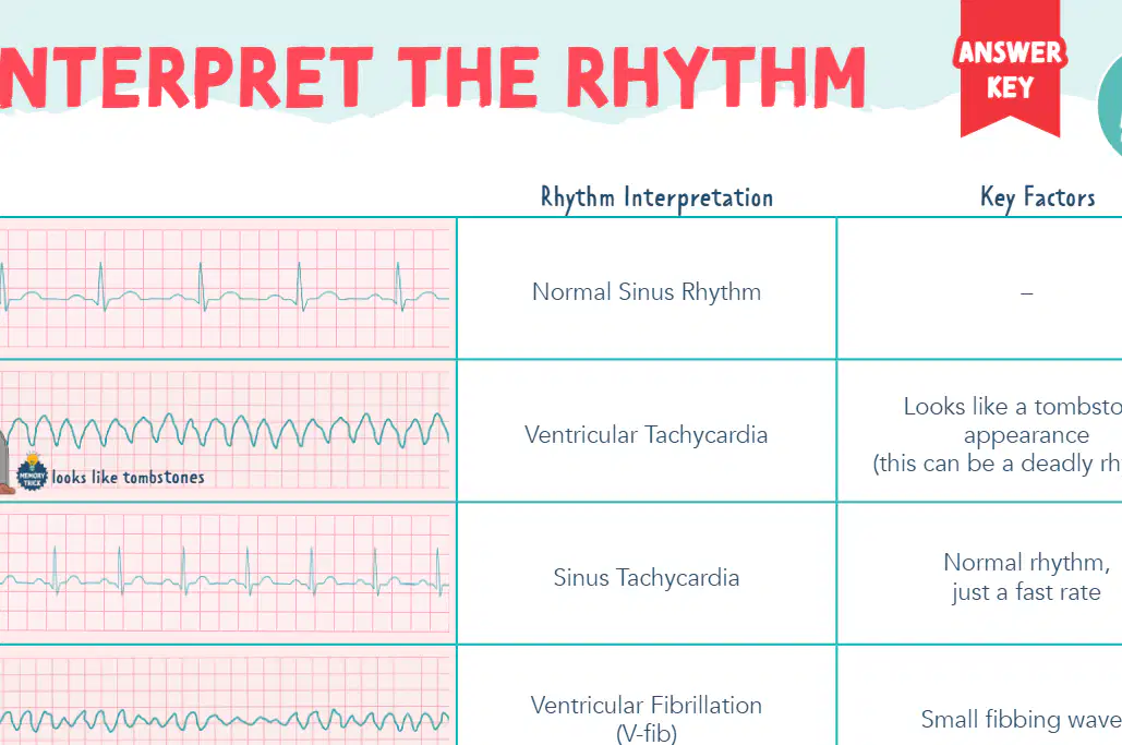
Kai, Anika, and William are studying ECGs together. Suddenly, Kai spots some "small fibbing waves" on the screen and challenges the group: Which rhythm interpretation is associated with these mysterious waves?
Ventricular Fibrillation (V-fib)
Sinus Brady
Atrial Fibrillation (A-fib)
Supraventricular Tachycardia (SVT)
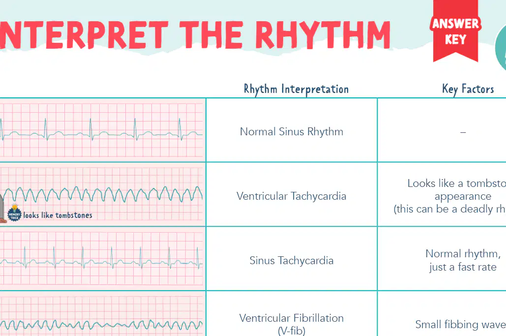
William and Scarlett are reviewing ECGs in class. They spot a patient’s ECG that shows a normal rhythm but at a super speedy rate! Which rhythm interpretation should they choose?
Sinus Brady
Sinus Tachycardia
Atrial Flutter
Asystole
Which rhythm is identified by a premature conduction of a QRS complex?
Sinus Brady
Premature Ventricular Contraction (PVC)
Atrial Fibrillation
Ventricular Fibrillation
If an ECG shows a normal rhythm but at a slow rate, what is the rhythm interpretation?
Sinus Tachycardia
Sinus Brady
Ventricular Tachycardia
Atrial Flutter
Scarlett and Luna are studying ECG rhythms together. Scarlett says, "Hey Luna, which rhythm interpretation is linked to an ECG that looks like S's?" Can you help them out?
Atrial Fibrillation
Supraventricular Tachycardia (SVT)
Ventricular Fibrillation
Sinus Brady

Emma, Lily, and Abigail are studying ECGs together. They come across the rhythm strip below. Which of the following best describes a 1st degree heart block?
1st degree heart block
2nd degree (Wenckebach or Mobitz Type I)
2nd degree (Mobitz Type II)
3rd degree (Complete heart block)
Which heart block is characterized by progressively lengthening PR intervals until a QRS complex is dropped?
1st degree heart block
2nd degree (Wenckebach or Mobitz Type I)
2nd degree (Mobitz Type II)
3rd degree (Complete heart block)
Sophia and Aria are studying ECGs together. They notice a heart rhythm where there are sudden dropped QRS complexes, but the PR interval doesn’t get longer each time. Which type of heart block are they looking at?
1st degree heart block
2nd degree (Wenckebach or Mobitz Type I)
2nd degree (Mobitz Type II)
3rd degree (Complete heart block)
Which heart block is characterized by complete dissociation between P waves and QRS complexes?
1st degree heart block
2nd degree (Wenckebach or Mobitz Type I)
2nd degree (Mobitz Type II)
3rd degree (Complete heart block)
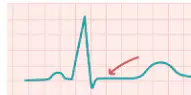
Which electrolyte imbalance is most likely to cause tall, peaked T waves on an ECG?
Hypokalemia
Hypercalcemia
Hyperkalemia
Hypomagnesemia
A patient’s ECG shows a prolonged QT interval. Which electrolyte imbalance is most likely responsible?
Hypercalcemia
Hypocalcemia
Hyperkalemia
Hypermagnesemia
Which electrolyte imbalance is associated with flattened T waves and the presence of U waves on an ECG?
Hypokalemia
Hypercalcemia
Hyperkalemia
Hypomagnesemia
Which of the following electrolyte imbalances is most likely to cause a shortened QT interval on an ECG?
Hypocalcemia
Hypercalcemia
Hypokalemia
Hypomagnesemia
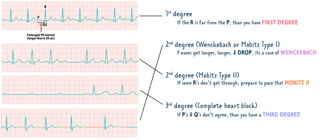
Noah, Emma, and Sophia are studying ECGs together. They notice a heart rhythm where the PR interval is prolonged (longer than 0.20 seconds), and Noah exclaims, "Hey, the R is really far from the P!"
Which type of heart block are they looking at?
1st degree
2nd degree (Wenckebach or Mobitz Type I)
2nd degree (Mobitz Type II)
3rd degree (Complete heart block)
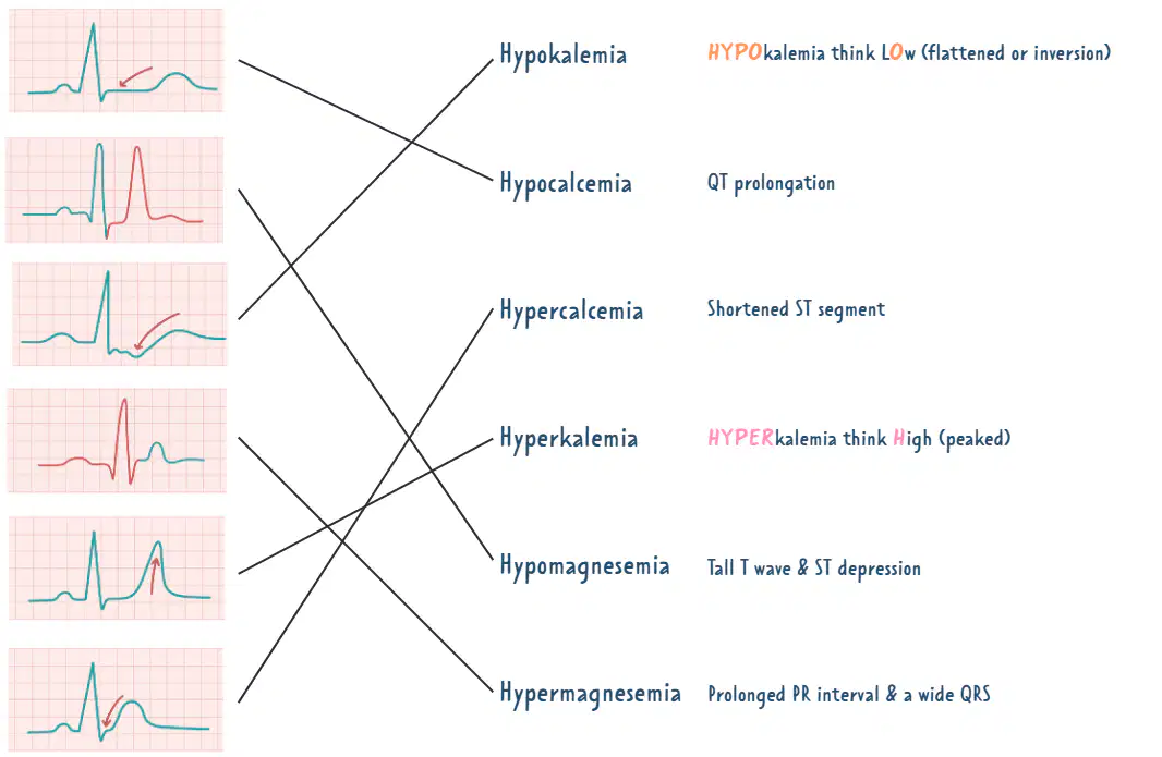
Daniel, Ethan, and Samuel are studying ECGs together. Suddenly, Daniel spots a flattened or inverted T wave on the ECG! Which electrolyte imbalance should he suspect?
Hypokalemia
Hyperkalemia
Hypercalcemia
Hypomagnesemia
What is the main distinguishing feature of a 2nd degree (Wenckebach or Mobitz Type I) heart block on an ECG?
P waves get longer, longer, and then drop
Some R's don't get through
PR interval is prolonged but consistent
P's and Q's don't agree
Which electrolyte imbalance is indicated by a tall T wave and ST depression on an ECG?
Hypomagnesemia
Hypercalcemia
Hypokalemia
Hypermagnesemia
A patient’s ECG shows that some R waves do not get through. Which type of heart block does this indicate?
2nd degree (Mobitz Type II)
1st degree
2nd degree (Wenckebach or Mobitz Type I)
3rd degree (Complete heart block)
Which electrolyte imbalance is associated with a shortened ST segment on an ECG?
Hypercalcemia
Hypokalemia
Hyperkalemia
Hypomagnesemia
Luna and Ethan are studying ECGs together. If they notice that the P waves and QRS complexes just can't seem to agree or sync up on an ECG, which type of heart block are they looking at?
3rd degree (Complete heart block)
1st degree
2nd degree (Wenckebach or Mobitz Type I)
2nd degree (Mobitz Type II)
Which electrolyte imbalance is associated with a prolonged PR interval and a wide QRS complex?
Hypermagnesemia
Hypokalemia
Hypercalcemia
Hypomagnesemia
QT prolongation on an ECG is most likely due to which electrolyte imbalance?
Hypocalcemia
Hypercalcemia
Hyperkalemia
Hypomagnesemia
Which electrolyte imbalance is associated with tall, peaked T waves on an ECG?
Hyperkalemia
Hypokalemia
Hypercalcemia
Hypomagnesemia
What does the acronym STEMI stand for in cardiology?
ST-Elevation Myocardial Infarction
Non-ST-Elevation Myocardial Infarction
Sinus Tachycardia with Elevated Myocardial Index
Standard Test for Electrocardiogram Myocardial Infarction
What does the acronym NSTEMI stand for in cardiology?
Non-ST-Elevation Myocardial Infarction
Normal Sinus Tachycardia with Elevated Myocardial Index
ST-Elevation Myocardial Infarction
Non-Sinus Tachycardia Electrocardiogram Myocardial Infarction
Which of the following best describes the change in biomarkers in both STEMI and NSTEMI?
Biomarkers are elevated in both STEMI and NSTEMI
Biomarkers are decreased in both STEMI and NSTEMI
Biomarkers are only elevated in STEMI
Biomarkers are only elevated in NSTEMI
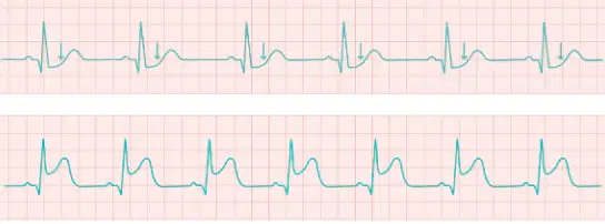
Which ECG finding is characteristic of a STEMI but not of an NSTEMI?
ST segment elevation
ST segment depression
Normal T wave
Prolonged PR interval
What does STEMI stand for?
ST Elevation Myocardial Infarction
Non-ST Elevation Myocardial Infarction
Sinus Tachycardia Myocardial Infarction
Segmental Thrombus Myocardial Infarction
Which type of myocardial infarction is associated with a complete blockage of a coronary artery?
STEMI
NSTEMI
Both STEMI and NSTEMI
Neither STEMI nor NSTEMI
What is a key difference in the ST segment on an ECG between STEMI and NSTEMI?
STEMI shows ST segment elevation, while NSTEMI does not
NSTEMI shows ST segment elevation, while STEMI does not
Both show ST segment elevation
Neither shows ST segment elevation
Which of the following is true about biomarkers in both STEMI and NSTEMI?
Biomarkers are elevated in both
Biomarkers are decreased in both
Biomarkers are only elevated in STEMI
Biomarkers are only elevated in NSTEMI
A patient’s ECG shows ST depression or T inversion, but not ST elevation. Which type of myocardial infarction is most likely?
NSTEMI
STEMI
Both STEMI and NSTEMI
Neither STEMI nor NSTEMI

What does the diagram illustrate about the difference between STEMI and NSTEMI on an ECG?
STEMI shows ST elevation, NSTEMI shows ST depression or T inversion
NSTEMI shows ST elevation, STEMI shows ST depression
Both show ST elevation
Neither shows any ST segment changes
Which two conditions place the patient at risk for blood clots?
Atrial fibrillation & atrial flutter
Ventricular tachycardia & bradycardia
Sinus rhythm & bundle branch block
Myocardial infarction & asystole
What is a racing pulse which originates from above the ventricle called?
Supraventricular tachycardia
Ventricular fibrillation
Atrial flutter
Sinus bradycardia
PEA stands for:
Pulseless Electrical Activity
Primary Electrical Arrhythmia
Partial Ejection Amplitude
Pacemaker Electrical Axis
What is the term for irregular and false readings caused by patient or lead movement?
Artifact
Arrhythmia
Asystole
Fibrillation
NSR stands for:
Normal Sinus Rhythm
Nodal Sinus Response
Non-Sustained Rhythm
Normal Systolic Rate
Which node is the primary pacemaker of the heart?
Sinoatrial (SA) node
Atrioventricular (AV) node
Bundle of His
Purkinje fibers
Ventricular Tachycardia (VT) patients can have a pulse or no pulse.
True
False
A myocardial infarction will always present with ST elevation.
True
False
A bundle branch block (BBB) is not able to be detected by an EKG.
True
False
An implanted pacemaker is a contraindication to performing an EKG.
True
False
Which two heart conditions both cause pooling of blood in the atria, increasing the risk for blood clots?
Atrial Fibrillation (A-fib) & Atrial Flutter
Ventricular Tachycardia & Supraventricular Tachycardia
Normal Sinus Rhythm & Pulseless Electrical Activity
Bundle Branch Block & Myocardial Infarction
What is supraventricular tachycardia?
A racing pulse which originates from above the ventricle
A slow pulse originating from the atria
A pulse with irregular rhythm
A pulse that cannot be detected by EKG
What does PEA stand for in EKG terminology?
Pulseless electrical activity
Primary electrical arrhythmia
Pacemaker electrical activity
Partial electrical arrest
What is the term for irregular and false EKG readings caused by patient or lead movement?
Artifact
Arrhythmia
Block
Flutter
What does NSR stand for in EKG interpretation?
Normal Sinus Rhythm
Nodal Sinus Response
Non-Sustained Rhythm
Normal Systolic Rate
Which node is the primary pacemaker of the heart?
Sinoatrial node (SA node)
Atrioventricular node (AV node)
Bundle of His
Purkinje fibers
Ventricular Tachycardia (VT) patients can have a pulse or no pulse. Is this statement true or false?
True
False
A myocardial infarction will always present with ST elevation. Is this statement true or false?
True
False
A bundle branch block (BBB) is not able to be detected by an EKG. Is this statement true or false?
True
False
An implanted pacemaker is a contraindication to performing an EKG. Is this statement true or false?
True
False
Which treatment is recommended for pulseless ventricular tachycardia?
IV amiodarone, IV lidocaine
Treat with defibrillation
Morphine, aspirin, oxygen, nitroglycerin & cardiac catheterization
Carotid massage, IV adenosine, beta blockers
What is the key point associated with Torsades de pointes?
Appears as twisting patterns; treated with magnesium sulfate
Assess serum K+ and troponins; seen with hyperkalemia or MI
Rearrange and stabilize leads, encourage patient to remain as still as possible
Begin CPR & Code Blue protocol
Which arrhythmia is treated with IV amiodarone or IV lidocaine?
Sinus bradycardia
Ventricular fibrillation
Atrial fibrillation
Recurrent ventricular tachycardia with pulse
What is the recommended treatment for complete heart block?
Placement of pacemaker
Morphine, aspirin, oxygen, nitroglycerin & cardiac catheterization
Carotid massage, IV adenosine, beta blockers
Treated with defibrillation
Which of the following is a key point for artifact on an ECG?
Rearrange and stabilize leads, encourage patient to remain as still as possible
Treated with cardioversion, heparin therapy
Can be caused by certain psychiatric medications or ondansetron
Infrequent occurrence not always concerning; frequent occurrence may lead to weakened cardiac muscle
A patient with peaked T-waves should have which lab values assessed?
Serum K+ and troponins
Blood glucose
Serum calcium
Serum sodium
Which arrhythmia is often seen in endurance athletes at baseline?
Sinus bradycardia
Sinus tachycardia
Paroxysmal atrial fibrillation
Supraventricular tachycardia
What is the initial action for a patient with PVCs (Premature Ventricular Contractions)?
Begin CPR & Code Blue protocol
Treat with defibrillation
Treated with cardioversion, heparin therapy
Assess serum K+ and troponins
Which arrhythmia is associated with anxiety and stimulants, and may be treated with beta blockers?
Sinus tachycardia
Supraventricular tachycardia
Paroxysmal atrial fibrillation
Ventricular fibrillation
What is the key point for paroxysmal atrial fibrillation?
Irregular rhythm which begins and may resolve by itself within 1 week (intermittent)
Treated with defibrillation
Appears as twisting patterns; treated with magnesium sulfate
Morphine, aspirin, oxygen, nitroglycerin & cardiac catheterization
Which treatment is recommended for Complete heart block?
IV amiodarone, IV lidocaine
Carotid massage, IV adenosine, beta blockers
Recommended treatment is placement of pacemaker
Treat with defibrillation

