
 Worksheets
Abdomen WK 1 Powerpoint pt 2
Total questions: 105
Worksheet time: 2hrs 43mins
Male → Rectovesical pouch (between rectum & bladder)
(a)
Female → Rectouterine pouch (pouch of Douglas, between rectum & uterus), and Vesicouterine pouch (between bladder & uterus
(a)
Lowest points in upright posture → common for fluid accumulation
(a)
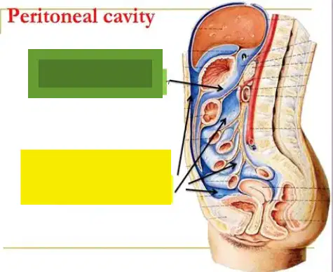
Green
(a)
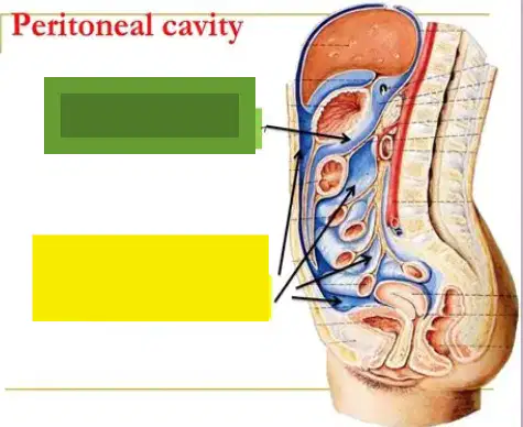
yellow
(a)
Space behind the stomach, connected to the greater sac through the omental foramen (Foramen of Winslow)
(a)
Lesser sac is the Space behind the stomach, connected to the greater sac through the (a)
Extends downward into the fold of the greater omentum (inferior recess).
(a)
Present in infants, usually reduced in adults as folds fuse.
(a)
Anterior, left wall, and right wall are all boundaries of the (a)
(a) boundary is posterior wall of stomach, lesser omentum (hepatogastric ligaments)
the (a) boundary includes the splenorenal and gastrosplenic ligaments
The (a) wall boundary of the lesser sac includes the mental foramen (posterior to hepatoduodenal ligament carrying the portal triad)
are folds of peritoneum formed when the peritoneum (a thin serous membrane) is reflected off the abdominal wall or organs to cover another structure
(a)
connects liver to anterior abdominal wall; contains ligamentum teres (round ligament).
(a)
attach liver to diaphragm; triangulars are at the ends of the coronary ligament.
(a)
connects liver to duodenum; contains the portal triad (hepatic artery, portal vein, bile duct).
(a)
connects liver to stomach; part of the lesser omentum
(a)
part of greater omentum; connects stomach to transverse colon
(a)
suspends transverse colon from posterior abdominal wall; carries vessels/lymphatics
(a)
attaches sigmoid colon to posterior wall; allows mobility of sigmoid.
(a)
fans out from posterior wall to hold jejunum and ileum; carries blood vessels, lymphatics, nerves
(a)
peritoneal folds anchoring organs (e.g., falciform, hepatoduodenal)
(a)
folds suspending bowel segments and providing passage for vessels/nerves (e.g., small bowel mesentery, mesocolons).
(a)
Also called: small omentum, gastrohepatic omentum, or gastrohepatic ligament
(a)
A double layer of peritoneum
(a)
Connects the lesser curvature of the stomach and the first part of the duodenum to the liver (via the fissure for ligamentum venosum and porta hepatis)
(a)
Forms the anterior–superior border of the lesser sac, separating it from the supracolic compartment of the greater sac.
(a)
Contains important structures within the hepatoduodenal ligament:
Portal vein
Hepatic artery proper
Common bile duct
(a)
is a thin peritoneal fold linking the stomach/duodenum to the liver and serving as a key passageway for the portal triad.
(a)
A large apron-like fold of peritoneum.
(a)
Hangs down from the greater curvature of the stomach and the proximal duodenum, covering the intestines
(a)
Then it folds back upward to attach to the transverse colon
(a)
In infants, the anterior and posterior layers are separate, but in adults they usually fuse into a thick sheet
(a)
It contains variable amounts of fat, lymphatics, and blood vessels.
(a)
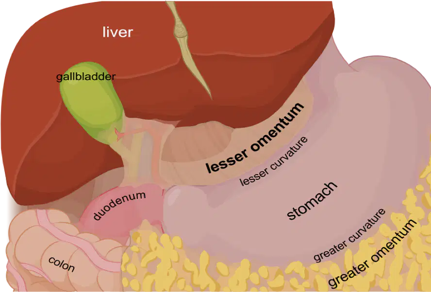
acts like a protective fatty apron covering the intestines, helping with immunity, fat storage, and infection control (sometimes called the “abdominal policeman” because it can wall off infections).
(a)
Area behind the parietal peritoneum and abdominal wall muscles
(a)
Extends from the diaphragm to the pelvis.
(a)
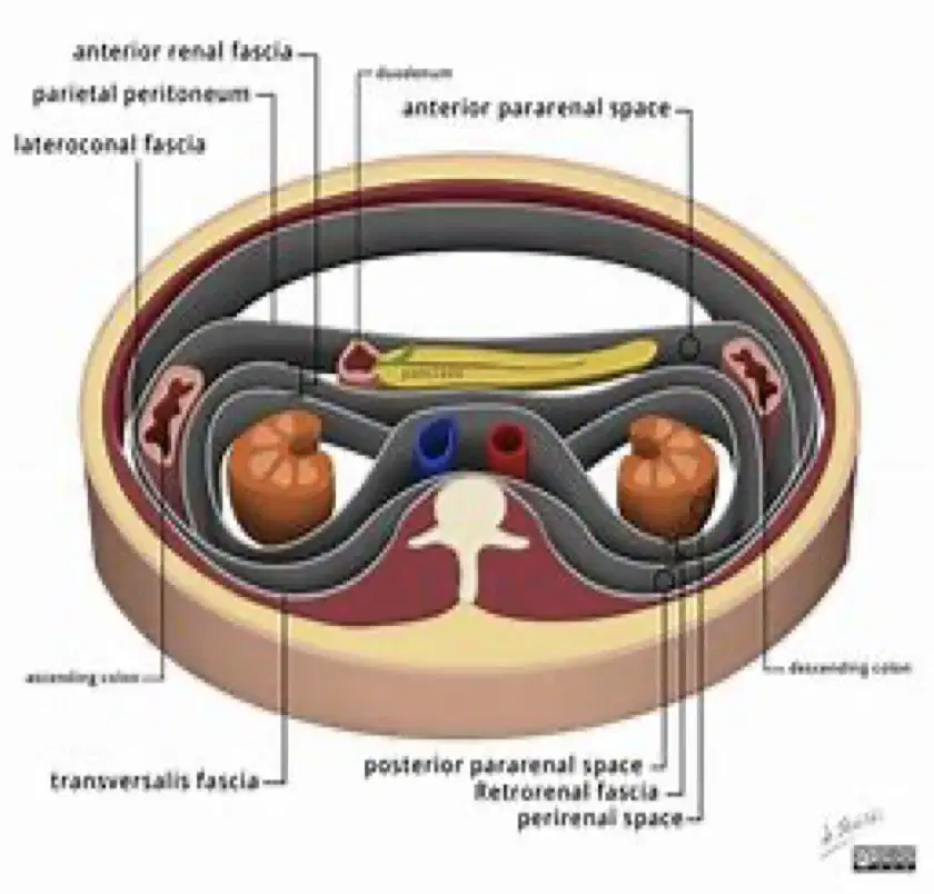
(a) the retroperitoneum extends to the extraperitoneal fat in the transversalis fascia

(a) the retroperitoneum encloses the great vessels
Which of the following are subdivisions of the retroperitoneum
Anterior Pararenal Space
Space of Retzus
Posterior Pararenal Space
Perirenal Space
Between the parietal peritoneum and the anterior reflection of the perirenal fascia.
(a)
Which of the following are organs of the anterior pararenal space?
Liver, duodenum, kidneys
Pancreas, duodenum, and liver
Kidneys, ureters, adrenal glands
Pancreas, duodenum, ascending/descending colon
Largest division of the retroperitoneum
(a)
Which space of the retroperitoneum contains the adrenal glands, kidneys, ureters, and great vessels?
(a)
Bound by:
Anterior perirenal fascia (Gerota’s fascia)
Posterior perirenal fascia (Zuckerkanl’s fascia)
(a)
•Smallest and least clinically significant portion.
(a)
Which organs are within the •Posterior Pararenal Space
Muscles, nerves, lymphatics, psoas
Ureters, kidneys, liver
Blood vessels, lymph nodes, aorta, iliac fossa, ureter and major branches of great vessels
Liver, spleen, brain
•Made up of posterior abdominal wall muscles, nerves,
lymphatics, and areolar tissue behind
the transversalis fascia.
(a)
Which retroperitoneal space contains the psoas, lumbar, and iliac?
(a)
Located posterior to the parietal peritoneum and anterior to the posterior abdominal wall
(a)
Kidneys
Ureters
Adrenal (suprarenal) glands
Pancreas (except tail)
Duodenum (2nd–4th parts)
Ascending and descending colon
Rectum (middle and lower thirds)
Major vessels: Aorta, IVC, renal vessels
Lymph nodes and sympathetic trunks
(a)
retropubic (front of bladder)
(a)
between bladder & rectum for males
pouch of Douglas for females
(a)
beside bladder
(a)
behind rectum
(a)
Space of retzius, rectovesical, Paravesical, presacral, and prerectal are (a) spaces
Female: urinary bladder, distal ureters, uterus, and rectum
(a)
Male: Urinary bladder, distal ureters, prostate, seminal vesicles, rectum
(a)
Suprarenal (adrenal glands), Aorta/IVC, Duodenum, Pancreas (minus the tail), ureters, colon, kidneys, esophagus, rectum
(a)
Anterior, posterior, lateral, superior, and inferior borders are all of the (a) space
Posterior peritoneum (retroperitoneal space)
(a)
Transversalis fascia (retroperitoneal space)
(a)
Quadratus lumborum
Peritoneal leaves of mesentery
(retroperitoneal space)
(a)
Diaphragm (retroperitoneal space)
(a)
Pelvic brim (retroperitoneal space)
(a)
Dome-shaped muscle separating thoracic and abdominal cavities
(a)
forms floor of thorax --> diaphragm
(a)
forms roof of abdomen --> diaphragm
(a)
Right dome (a) (due to liver), central area slightly depressed (due to heart/pericardium
Tendinous structures that extend from the diaphragm to the vertebral column.
(a)
•Longer, larger, and more lobular.
•Located near the anterior lumbar vertebral ligament
(a)
•Bound by the inferior vena cava (in front and to the side) and the right adrenal gland and liver (behind).
(a)
•Runs along the front of the lumbar vertebral bodies and attaches to the central tendon of the diaphragm.
(a)
Both crura help anchor the diaphragm and assist in muscular contraction.
(a)
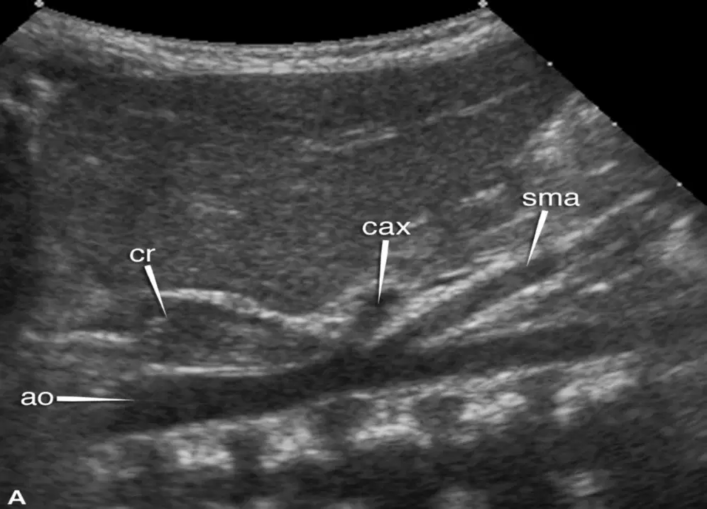
Visible in images that include the right lobe of the liver, kidney, and adrenal gland
(imaging the crus)
(a)
Seen using the spleen and left kidney for reference, located to the left of the aorta. --> more difficult to image in ultrasound
(a)
Upper part of trunk, enclosed by ribs, sternum, vertebrae, and diaphragm
(a)
Right pleural cavity (right lung)
Left pleural cavity (left lung)
Mediastinum (central space → heart, great vessels, trachea, esophagus).
(a)
Each lung sits in its own pleural cavity
True
False
Very thin fluid-filled space that allows lungs to expand smoothly during breathing
(a)
Parietal pleura, visceral pleura, pleural space
(a)
lines chest wall, diaphragm, and mediastinum
(a)
→ covers the lung surface directly.
(a)
Between the two pleura's is the (a)
3 lobes (superior, middle, inferior
(a)
2 lobes (superior, inferior) + cardiac notch.
(a)
Sits in the mediastinum, protected by the pericardium.
Pumps blood to lungs and body.
(a)
Fever, pain, palpable mass, bleeding, trauma, post op, distended abdomen
(a)
Fasting to reduce bowel gas
May be attempted non-fasting
Adults-8 hour
Children- 6 hours
Infants- 4 hour
(a)
Rapid bedside scan for free fluid in peritoneal spaces
(a)
Hepatorenal space (Morrison’s pouch)
Right diaphragm/liver interface
Splenorenal recess (LUQ)
Rectouterine pouch (Douglas pouch) in females
(a)
Sonographic appearance of the (a) thin, echogenic (bright line at surface)
Sonographic appearance of the (a) variable echogenicity, usually hypoechoic relative to skin and muscle, often with thin internal echogenic septations
Sonographic appearance of the (a) typically hypoechoic relative to subcutaneous fat, with fine echogenic striations (fibers).
Sonographic appearance of the (a) seen as thin echogenic lines separating muscle groups.
Normally a potential space, not visible unless there is free fluid
(a)
Free fluid: appears anechoic (black), outlining bowel loops or organs
(a)
occasionally seen as thin echogenic lines
(a)
Normally obscured by overlying bowel gas, but major organs visible:
Kidneys, pancreas, aorta, IVC, adrenal glands with their characteristic echogenicities.
(a)
Fat planes: relatively hyperechoic, separate retroperitoneal structures.
Abnormality suspicion: disruption of fascial planes, masses, or free fluid tracking along spaces.
(a)
normally only a thin echogenic line of the visceral pleura sliding against parietal pleura (“lung sliding”
(a)
strongly echogenic, produces reverberation artifacts (A-lines)
(a)
pleural space is not visible.
(a)
anechoic or hypoechoic collection above diaphragm, separating lung from chest wall/diaphragm.
(a)

