
 Worksheets
Venous WK 2 pt 1
Total questions: 125
Worksheet time: 2hrs 5mins
Venous anatomy is divided into (a) systems
A primary characteristic of the (a) veins is that they run alongside an arteries, and as such, often share the same name.
Blood flows from the superficial veins into the deep veins.
True
False
(a) veins act as communicating veins between the deep and superficial systems
Veins are unique, they have venous valves, which allows unidirectional flow (a) the heart
Deep, superficial, perforating
(a)
lie just below the skin and are easily seen on the surface
(a)
as their name implies, are located deep within the muscles of the leg.
(a)
Deep, Superficial and Perforator veins have one-way (a) within them that allow blood to flow in the direction of the heart
• The main trunk of the Saphenous vein (also known as the Great Saphenous vein (GSV)) lies within the (a)
The SC is a subcompartment of the (a) (SupC) located
• lie below the muscular fascia
(a)
•lie between the muscular fascia and the dermis (skin).
(a)
Carries 85% of limb blood volume
(a)
Veins are short connections between the deep and superficial venous systems in the legs. These connections allow blood to pass between them and return to the heart.
(a)
Considered deep veins
(a)
Gastrocnemius and soleal veins
(a)
Major pathway of blood drainage.
More blood flow
Deep vein thrombosis (DVT)
(a)
Subcutaneous veins that are NOT accompanied by arteries. They eventually join deep veins.
(a)
Venous drainage and thermoregulation.
Less blood flow
Varicose veins/ Superficial thrombophlebitis
(a)
▪ the outside layer. It is composed of collagen, connective tissue, elastin, and fibroblasts.
(a)
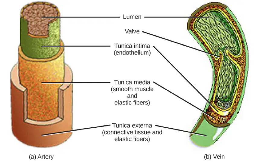
▪- is the middle layer. It is composed of smooth muscle
(a)
▪is the innermost layer. It is made up of endothelial cells which provide a non-thrombogenic surface for flowing blood.
(a)
The (a) are formed from folds of the intima
•Translates to "vessel of a vessel"
(a)
•They are a network of small blood vessels that supply and drain the walls of larger arteries and veins.
(a)
•They are made up of endothelial cells, a thin basement membrane, and pericytes
(a)
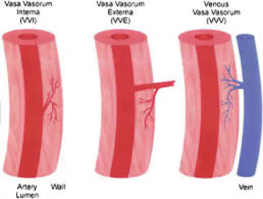
•connects with a branch of the same vessel or a neighboring vessel to form a vascular network.
(a)
•The have many functions including:
-Oxygen and nutrient supply
-Regulating blood flow
(a)
▪Have an adjacent artery.
▪ Are paired in the calf
(a)
Common iliac, internal iliac, external iliac, CFV, FV, PFV, Pop, Tibial/peroneal trunk, PTV, ATV and PV
(a)
This is a central vein. It is the confluence of the common iliac veins
(a)
The right and left common iliac veins join at the level of the umbilicus to form the
(a)
It is a large vein that receives blood from the lower extremities, pelvis and abdomen
(a)
This vein then delivers the blood to the right atrium of the heart.
(a)
The (a) is formed by the union of the external and internal iliac veins at the brim of the pelvis
It passes upward and posterior to the internal iliac artery then merges veering toward the right side of the body at the level of the (a) lumbar vertebra
When the CIV’s join they forms the (a)
The left common iliac vein is often compressed by the (a) common iliac artery against the vertebral column. This may result in partial obstruction of the vein.
come from deep in the pelvic region and rise to the lower portion of the abdomen. There they join with the right and left external iliac veins and form the common iliac veins
(a)
starts at the groin and runs along the edge of the pelvic area. When combined with the internal iliac vein it forms the common iliac vein at the tailbone
(a)
The external iliac vein can cause complications, such as
(a)
This is a venous disease that occurs when the right common iliac artery crosses over the vein. The compression that occurs can result in DVT
(a)
In order to image the iliacs you should use the (a) probe instead of the linear for better penetration -- abdominal region
is formed after the external iliac crosses the inguinal ligament
(a)
courses more superficially and is generally very easy to image. .
(a)
joins the common femoral in the groin, just proximal to the profunda vein and femoral vein (PV/FV) junction in the upper thigh
(a)
long vein segment that courses medially down the leg and eventually becomes the popliteal vein after it dives at the level of the adductor (hunters) canal in the distal thigh
(a)
, is a large deep vein in the thigh. It receives blood from the inner thigh and courses superiorly and medially running alongside the profunda femoris artery to join with the femoral vein
(a)

femoral veins (aka femoral vein duplication) are not uncommon
(a)
FV duplicity occurrence in the thigh is (a) in some studies.
carries the blood from the knee joint and muscles in the thigh and calf back to the heart. Its origin is defined by the junction of the posterior tibial, anterior tibial and peroneal veins.
(a)
of anterior tibial vein with popliteal vein. A very common variant that is often mistaken for a "bifed" popliteal vein.
(a)
•2 veins originate at the plantar arches
(a)
•Course cephalad anterolateral to the tibial bone and the interosseous membrane to reach the tibioperoneal trunk
(a)
•Travels between the tibial head and fibula head.
(a)
•Best scan approach is anterior calf with slightly lateral probe position
(a)
This short vein segment is located in the upper calf, where the anterior/posterior tibials and peroneal veins join
(a)
2 veins originate at the confluence of the plantar arches.
(a)
At the ankle the PTVs course posterior to the medial malleolus and anterior to the Achilles' tendon then course cephalad through the calf posterior to the tibial bone
(a)
The best scan approach is to place probe on the medial calf, angling through to the lateral side; PTVs are the most anterior vessel on image and peroneals are visible beneath them
(a)
In the lower calf/ankle, 2 veins course lateral to the PTVs and medial to the fibula.
(a)
Course up (cephalad) to the midline of the posterior calf, posterior to the fibula
(a)
2 peroneal veins and 2 posterior tibial veins merge to form a single tibio-peroneal trunk in the (a)
The best scan approach is to place probe on the medial calf, angling through to the lateral side; PTVs most anterior on the image and the peroneals are visible beneath them.
(a)
Dilated vessels between soleal and gastrocnemius muscles of the calf.
(a)
Serve as blood reservoirs for the legs.
Important for calf muscle pump and lower extremity venous flow.
(a)
Have an accompanying artery with each set.
Normally contain two pairs
(a)
Positioned laterally and medially.
Can possibly visualize three pairs.
Most commonly drain into the popliteal vein.
(a)
thick-walled reservoirs within the soleal muscle
(a)
They don’t have an accompanying artery.
Do NOT contain valves
(a)
Drain into either the posterior tibial or peroneal veins.
(a)
lie in the deeper soleal muscle and drain into the:
•Posterior tibial veins
•Peroneal veins
(a)
Scanning in this area is normally not included in the protocol. However, if the patient complains of pain/tenderness in the calf region, scan and look for thrombosed soleal vein
(a)
Located within 2cm of the skin surface.
(a)
Course within the subcutaneous fat layer of the leg.
Veins of the superficial system always course between the superficial fascial layers.
(a)
▪ Is the longest vein in the body.
▪ Joins the deep system at CFV.
▪ Connects to deep system through multiple perforating veins
(a)
▪Carries approx. 15% of venous blood volume in leg.
(a)
▪Often has double systems (8%).
▪ No adjacent artery
(a)
▪Originates on the dorsum of the foot travelling anterior to
the medial malleolus.
(a)
▪Ascends the medial thigh with the great saphenous nerve.
▪ Commonly used as a bypass graft for cardiac and arterial bypass surgery.
(a)
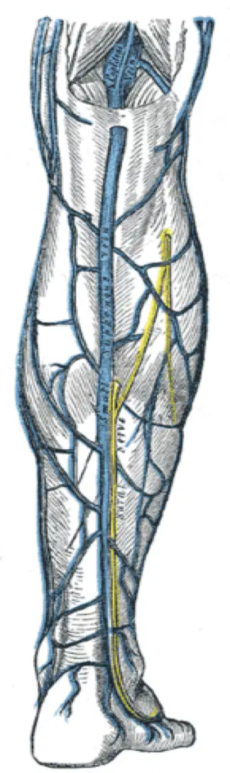
•The dorsal vein of the small toe joins the dorsal venous arch to form the SSV.
•Originates posterior to lateral malleolus.
(a)
•Ascends along the midline aspect of posterior calf.
•Usually joins the popliteal vein in knee/distal thigh area but there are several variations.
(a)
•Course of the vessel in the area of the posterior calf is nicknamed as the "stocking seam
(a)
•Numerous perforators connect the SSV to the GSV in the calf.
•Like the GSV, the LSV also has a number of branches.
(a)
Named after the Italian anatomist Carlo Giacomini (1840-1898).
(a)
A continuation of the SSV above the saphenopopliteal junction (SPJ), it courses along the posterior area of the thigh.
(a)
facilitates communication between the great saphenous vein (GSV) and the small saphenous vein (SSV).
(a)
courses the posterior thigh as either a trunk projection, or tributary of the SSV.
(a)
▪It can terminate at the GSV in the thigh or in the groin through the posteriomedial thigh vein
(a)
▪It can course up the back of the thigh ending near the femoral vein or tributaries in the internal iliac vein
(a)
▪ Most easily identified at the groin.
▪ Ascends along the thigh to join the GSV near the groin
(a)
▪ Differentiated from the GSV by its course anterior to the femoral artery instead of medial to it (GSV).
(a)
▪Not visible in all patients; can be a sign of dilated superficial system and reflux
(a)
Extensions of the intimal layer
(a)
Valves contain two leaflets ( (a) )
Allow unilateral flow direction in veins.
(a)
Helps keep flow moving from superficial to deep system and from peripheral to deep veins.
(a)
IVC, Internal iliac, common iliac, external iliac (75%), and soleal sinuses
(a)
1 valve
(a)
1-3 valves
(a)
<12 valves
(a)
6-12 valves
(a)
12-14 valves
(a)
SVC, innominate veins
(a)
Jugular, subclavian
(a)
Connect superficial and deep systems to equalize pressure between the two systems
(a)
PTV connected to distal GSV near (a)
•Lateral perforator connects to SSV near the __ __
(a)
Each leg normally has about (a) perforators
Normally <2mm diameter. >4mm usually has (a)
Flow is normally from superficial system to deep system.
(a)
Ankle perforator damage/dilatation involved in stasis ulcers
(a)
Dilated perforators can occur in response to DVT formation
(a)
are located in the proximal thigh; connecting the GSV to SFV
(a)
are located in distal thigh; connecting GSV to SFV.
(a)
are located in the knee area; connect GSV to POP
(a)
Posterior arch vein extends superior from the ankle to the GSV in the mid calf. They plays a major role in venous stasis; These veins are connected to 3 ankle perforators called
(a)
▪They act as a “clean-up” system to pick up fluid, protein, and other debris left behind by the veins
(a)
▪They filter and clean the fluid before returning it to the heart.
(a)
▪ Blood must then return through the veins to the right side of the heart where it can enter the lungs and pick up oxygen
(a)
The (a) this is where the end of the arterial system connects to the beginning of the venous system
The blood vessels in the “capillary bed” are very tiny, thin walled vessels. This allows for easy release of oxygen and nutrients (sugars, fats, etc.) into the tissues.
(a)
A pressure gradient exists across all capillary beds with high pressure occurring in the arterial side (arterioles) and (a) in the venous side (venules).
This phenomenon allows blood to perfuse through tissue. If venous pressure in the venules is equal to or higher than pressure in the arterioles, we’re in trouble, as little or no perfusion will occur in the capillary bed.
(a)

