
 Worksheets
Flow and Hemodynamics Concepts Edelman ch 18
Total questions: 125
Worksheet time: 2hrs 51mins
Also called volume flow rate, indicates the volume of blood moving during a particular time
(a)
Units of (a) are volume divided by time such as liters/min
Volume flow rate
Volume of blood moving per unit of time
Units of volume over time
(a)
(a) is the study of blood moving through the circulatory system
(a) indicates speed or swiftness of a fluid moving from one location to another
The units for (a) are distance divided by time, such as cm/s
Speed of blood from one point to another.
Units of velocity. Distance over time
(a)
(a) occurs when blood moves with variable velocity. Commonly appears in the arterial circulation
Variable velocity along with HEART CONTRACTION.
Arterial
(a)
(a) also occurs when blood moves in variable velocity. Blood accelarates and decelerates as a result of respiration. Appears in the venous circulation
Variable velocity along with RESPIRATION
Venous
(a)
Occurs when a fluid moves at a constant speed or velocity. Steady flow is present in the venous circulation
(a)
Constant speed
Venous
During brief absence of respiration
(a)
Is when flow streamlines are aligned and parallel.
(a)
Is characterized by layers of blood that travel at individual speeds.
(a)
Laminar flow patterns are commonly found in (a) physiologic states
Which of the following are the two types of flows related to laminar flow?
Plug
Parabolic
Stenosis
Diastole
(a) flow occurs when all the layers and wood cells travel at the SAME velocity
(a) flow as a bullet shaped profile. Velocity is highest in the center of the lumen and gradually decreases to its minimum at the vessel wall
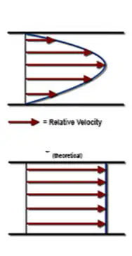
Which image shows a parabolic flow profile?
Top
Bottom
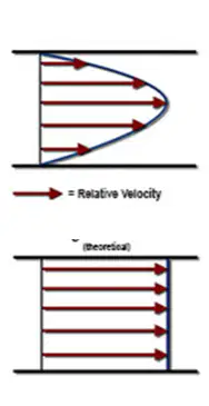
Which image shows a plug flow profile?
Top
Bottom
The (a) predicts whether the flow is laminar or turbulent
Reynolds number < 1500
(a)
(a) flow is characterized by chaotic flow patters in many directions at at many speeds
Swirling, eddy currents
(a)
Seen with cardiovascular pathology
(a)
Associated to stenosis (narrowing) of a vessel.
(a)
Produces sound (murmur or bruit), vibration (thrill)
(a)
Reynolds number > 2000
(a)
Re = Inertia / viscosity = (r V dV/dx) / (mu * d^2V/dx^2)
(a)
Energy produced by the contraction of the heart during systole moves blood from one location to another.
(a)
Blood moves from regions of higher energy to lower energy
(a)
What event provides energy to the circulating blood?
Diastole
Systole
Heart, capillaries, arteries, venues, arterioles, veins
(a)
Object’s mass
Moving speed
(a)
Heavy, swiftly moving objects have lots of kinetic energy. While light, slowly moving objects have little kinetic energy
True
False
Potential
Stored
Ability to do work
(a)
MAJOR FORM for circulating blood
(a)
Potential
Stored
Elevational
(a)
Is a form of stored or potential energy associated with any elevated object
(a)
is the tendency to remain in the same state.
(a)
Applied to the moving blood, is the tendency of the blood to resist changes in its velocity
(a)
(a) describes the thickness of a fluid
Measured in units of poise
(a)
(a) is determined by the hematocrit (% of RBCS)
More viscous fluid moving = (a) energy loss.
(a) occurs when flow energy is converted to heat as one object rubs against another
(a) in the circulatory system occurs in: arterial system (pulsatile), venous system (Phasic), and stenotic areas
Velocity (a) as the vessel narrows
Velocity (a) as blood flows out of the stenosis and into a vessel segment of normal diameter
A (a) is the narrowing in the lumen of a vessel
Change of direction of flow
(a)
Increased velocity at the narrowest section
(a)
Post-stenotic turbulence
(a)
Pressure gradient across stenosis
(a)
Pulsatile flow converts to steady flow
(a)
Relationship between velocity and pressure of a moving fluid.
(a)
Principle of conservation of energy.
(a)
The sum of kinetic energy and pressure energy remains constant.
(a)
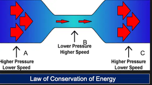
Kinetic energy of blood increases as it speeds up though the stenosis from A to B
(a)
If kinetic energy increases, then pressure energy (a) , so the sum of energy remains constant.
(a) is about the behavior of a fluid – a gas or a liquid – as it flows through a pipe or tube. It states that when the pipe narrows or widens, the fluid pressure and speed vary.
High pressure areas = (a) speeds
Lower pressure = (a) speeds
(a) = flow x resistance
Pressure gradient increases when:
Flow increases
Flow decreases
Resistance decreases
Resistance increases
Flow increases when:
Pressure gradient decreases
Pressure gradient increases
Resistance increases
Resistance decreases
f = pressure gradient / resistance
(a)
(a) flow is multifactorial and complex:
Heart contraction, elastic wall, blood composition, pulsatility, blood pressure
Describes the relationship between the current, voltage, and resistance in a circuit.
(a)
Voltage = current x resistance
(a)
Pressure gradient = flow x resistance
(a)
If resistance is increased and voltage remains the same, the current will (a)
If voltage is increased and resistance remains the same, the current will (a)
Pressure (fluids) = (a) (electricity)
Flow (fluids) = (a) (electricity)
Resistance (arterioles) = (a) (ohms)
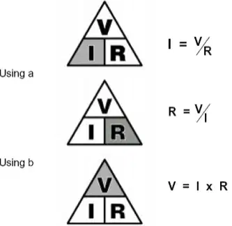
These relationships represent:
(a)
(a) are collapsible, contain low pressure, are partially filled and have low resistance.
(a) accommodate large volume of blood with small increase in pressure
Body Position
Respiratory Changes
Cardiac Contraction
Arterial Pressure
Blood Viscosity
(a)
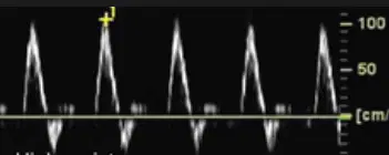
This image shows:
High resistance
Low resistance
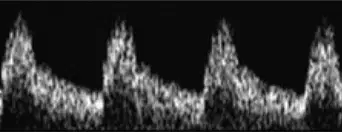
This image shows:
High resistance
Low resistance
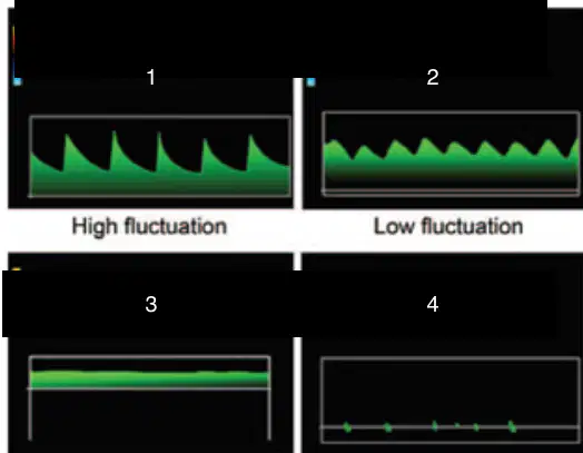
1
Pulsatile
Phasic
Nonphasic
Aphasic
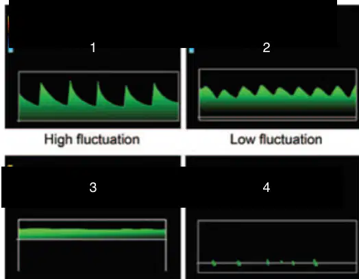
2
Pulsatile
Phasic
Nonphasic
Aphasic
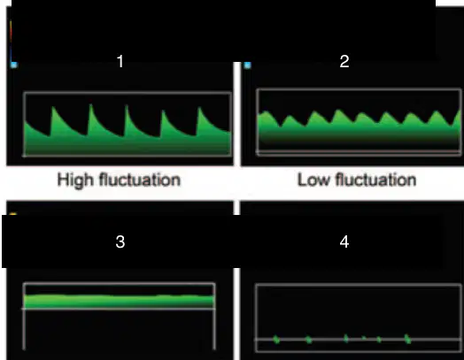
4
Pulsatile
Phasic
Nonphasic
Aphasic
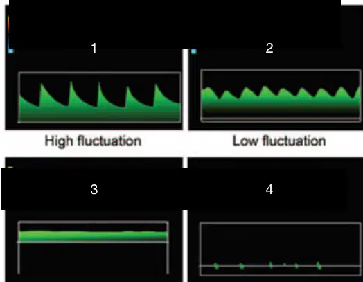
3
Pulsatile
Phasic
Nonphasic
Aphasic
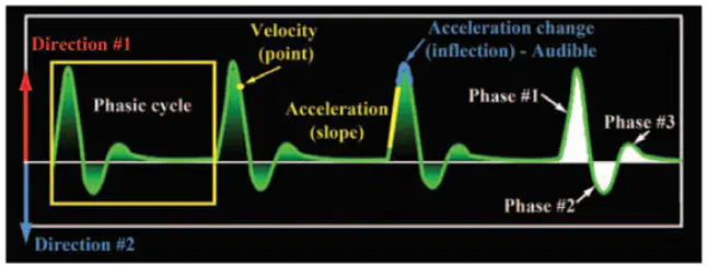
This image shows features of the (a)
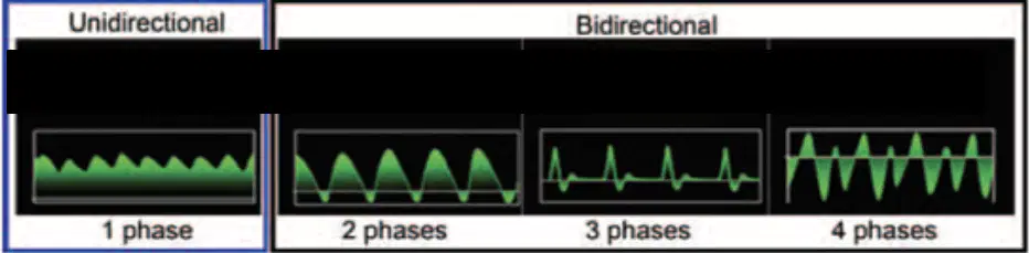
One phase
(a)
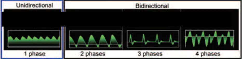
Two phases
(a)
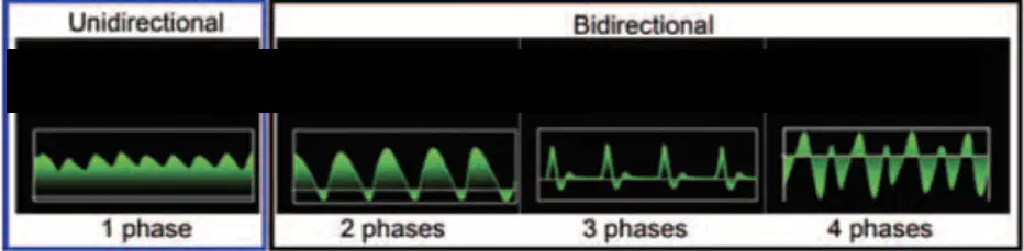
3 phases
(a)
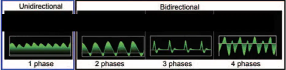
4 phases
(a)
External carotid arteires
Extremity arteries
Fasting mesenteric arteries
(a)
Internal carotid arteries, hepatic arteries, renal arteries, testicular arteries
(a)
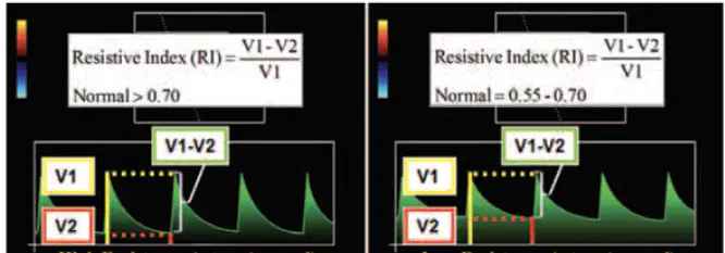
Which image shows high resistance artery?
Left
Right
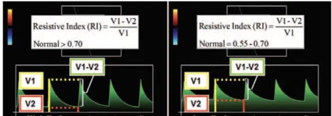
Which image shows a low resistance artery
Left
Right
Pressure related to the weight of blood pressing on a vessel measured at the height above or below the heart
(a)
(a) reported in units of mmHg, same as blood pressure
When a person is (a) , all parts of the body are at the same level as the heart and the hydrostatic pressure is zero everywhere
Knee, ankle, mid chest, top of heart blood pressure
140 mmHg
heart
0mmHg
What is the hydrostatic pressure at all locations within the arterial circulation?
0mmHg
140mmHg
7mmHg
What is the arterial pressure at the knee of the supine patient
140mmHg
0mmHg
Ankle hydrostatic pressure far below the level of the heart
140mmHg
100mmHg
75mmHg
50mmHg
Knee hydrostatic pressure somewhat below the heart
100mmHg
140mmHg
75mmHg
50mmHg
Waist hydrostatic pressure slightly below the heart
75mmHg
50mmHg
100mmHg
140mmHg
Midchest hydrostatic pressure at the heart level
0mmHg
100mmHg
140mmHG
30mmHg
Top of the head hydrostatic pressure above the heart (BP = 140mmHg)
100mmHg
75mmHg
50mmHg
-30mmHg
In normal individuals, respiration affects venous flow for two reasons:
Muscles responsible for respiration alter pressures in the legs
The venous system is high pressure
The venous system is low pressure
Muscles responsible for respiration alter pressures in the thorax
Breathing affects two venous flows:
Venous flow in the legs
Venous flow in the arms
Venous return to the heart, which comprises venous flow from the IVC
Venous return to the heart aorta
During (a) the chest cavity expands creating negative thoracic pressure that increases venous return to the heart
During (a) the diaphragm also presses the abdomen which increases abdominal pressure and decreases venous flow in the legs
During expiration, the diaphragm moves upward. This increased thoracic pressure (a) venous return to the heart
Venous flow from the head, arms, and vena cava all (a) during expiration
During expiration, decreased abdominal pressure (a) venous blood flow in the legs
Thoracic pressure decreases
Abdominal pressure increases
Venous return to the heart increases
Venous flow in legs decreases
(a)
Thoracic pressure increases
Abdominal pressure decreases
Venous return to the heart decreases
Venous flow in legs increases
(a)
Normally, what happens to venous flow in the legs during inspiration?
Increases
Decreases
No change
All of the following occur during expiration except:
Venous return to the heart decreases
Venous flow in the legs increases
Abdominal pressure increases
The diaphragm rises into the thoracic cavity
Volume of blood ejected each minute during ventricular contractions
(a)
Amount of blood ejected during ventricular systole
(a)
Percentage of blood pumped out of the LV during systole
(a)
The cardiac output is corrected for differences in body size
(a)
What is the AVERAGE value for cardiac output?
4-8 liters/min
5-10 liters/min
6-8 liters/min
3-5 liters/min
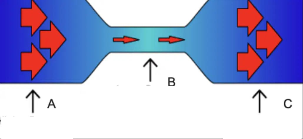
A and C represent:
High pressure, low speeds
Low pressure, high speeds
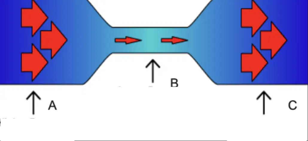
B represents
Low speed, high pressure
High speed, low pressure

