
 Worksheets
Gallbladder Pt 1
Total questions: 114
Worksheet time: 2hrs 52mins
The gallbladder develops early in embryogenesis from the
(a)
an outgrowth of the foregut endoderm (around the 4th week of gestation).
(a)
This diverticulum divides into (a) parts
forms the liver
(a)
gives rise to the gallbladder and cystic duct.
(a)
The connection between the hepatic and cystic ducts eventually joins with the duodenum, forming the
(a)
· By the (a) week of gestation, the gallbladder has its characteristic pear shape and continues to elongate and mature
Gallbladder: Pear-shaped sac in the (a) in the abdomen
The gallbladder is an (a) organ
It lies in the gallbladder fossa on the (a) of the liver.
Located on the __ and __ visceral surface of the liver
(a)
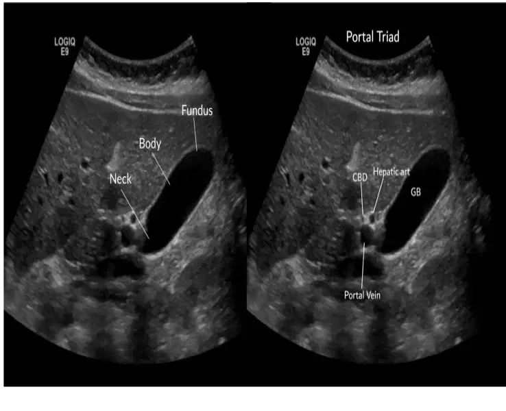
Neck, body, fundus
(a)
The gallbladder generally measured __ and __ in diameter
7 to 10cm
2.5cm to 4cm
5cm to 8cm
3cm to 4cm
The gallbladder measures __ to __ in length
7cm to 10cm
2.5cm to 4cm
3cm to 4cm
5cm to 8cm
The GB walls are less than _ mm thick.
(a)
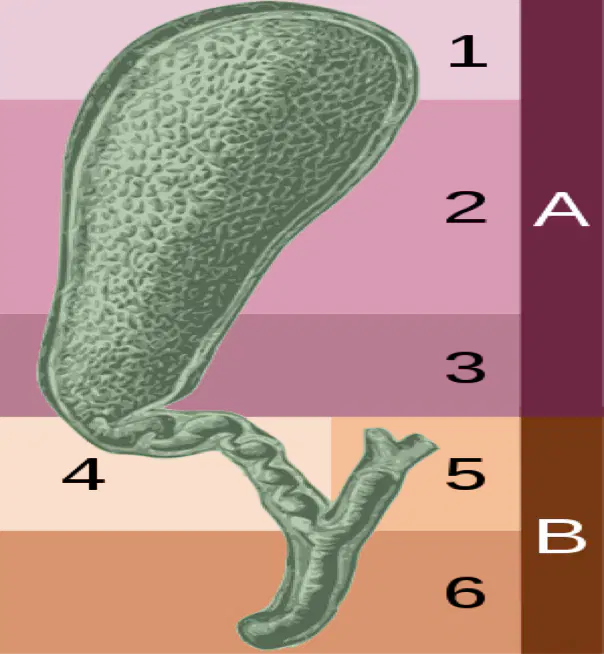
1
(a)
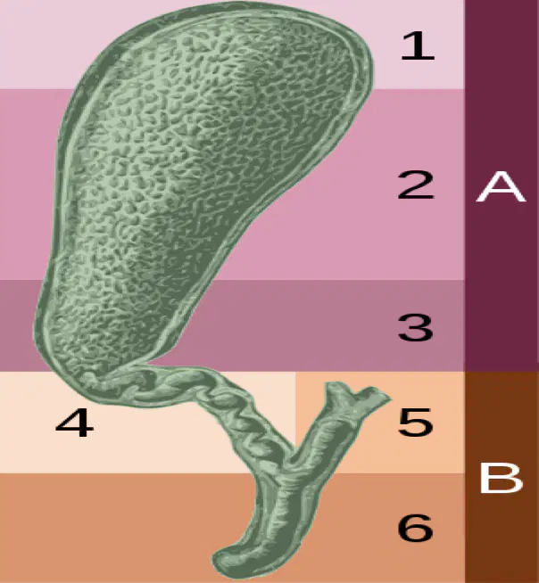
2
(a)
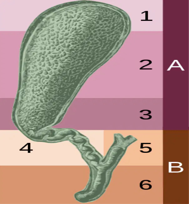
3
(a)
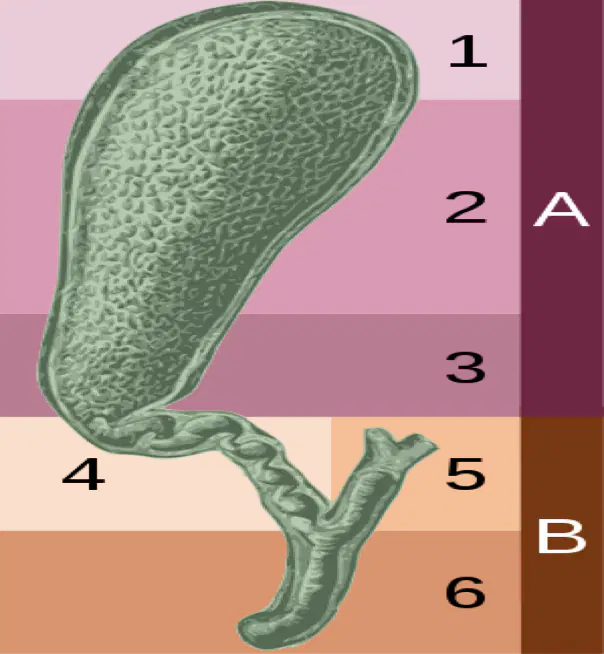
4
(a)
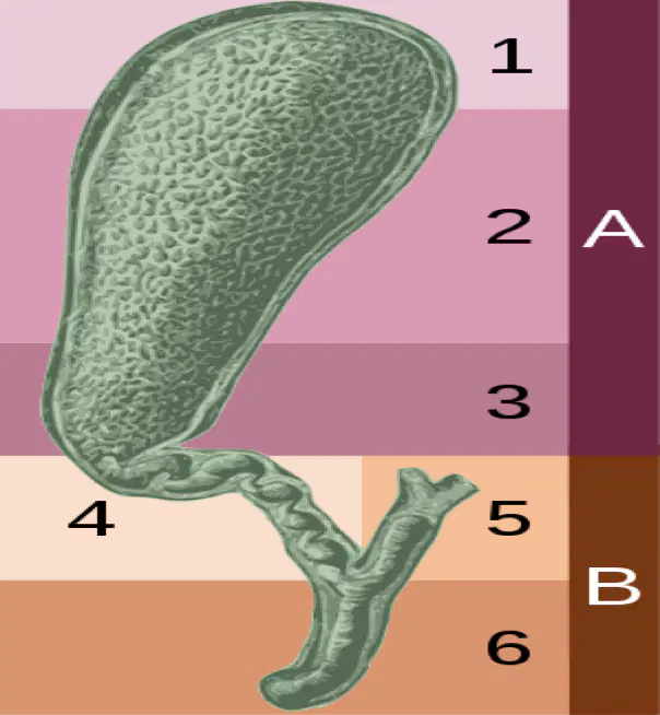
5
(a)
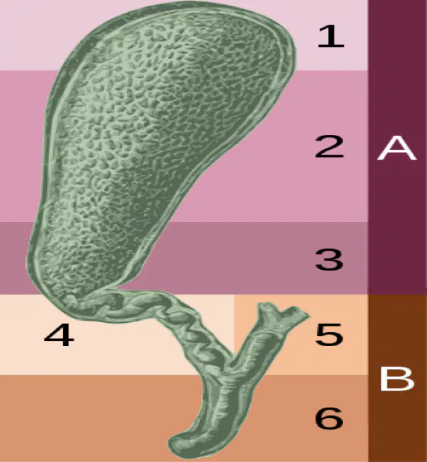
6
(a)
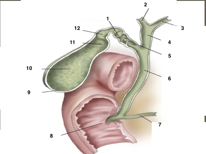
1
(a)
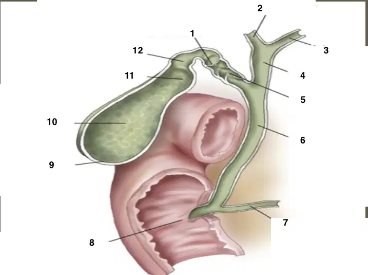
2
(a)
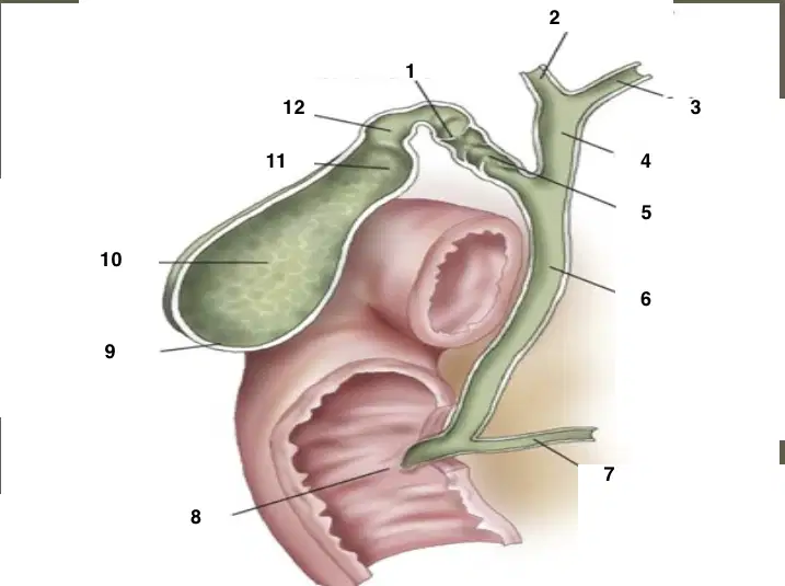
3
(a)
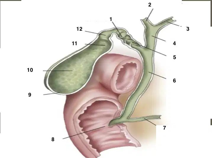
4
(a)
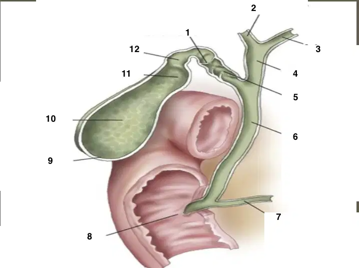
5
(a)
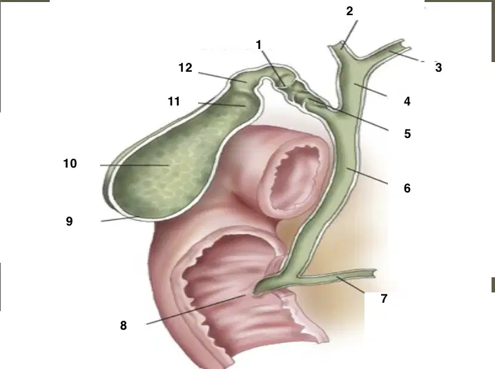
6
(a)
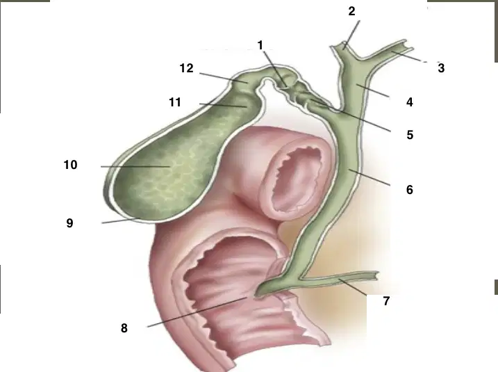
7
(a)
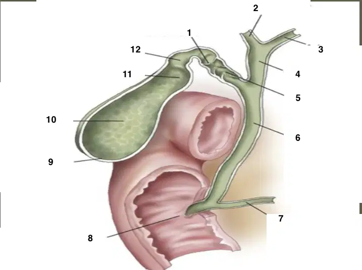
8
(a)
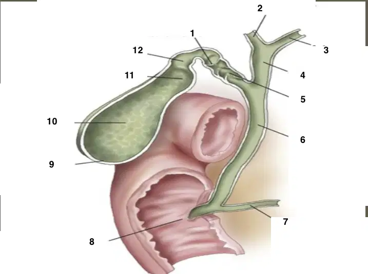
9
(a)
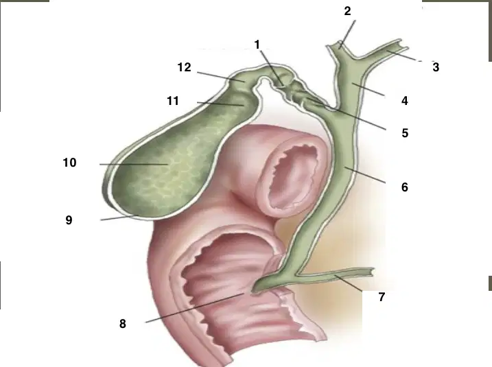
10
(a)
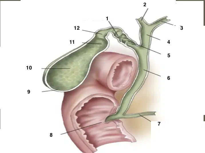
11
(a)
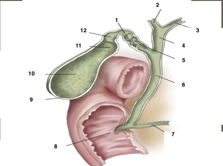
12
(a)
Lined by simple columnar epithelium with apical microvilli, specialized for absorption of water and electrolytes
(a)
Folds of mucosa form (a) (like in the stomach) that flatten as the gallbladder fills
Made of irregularly arranged bundles of smooth muscle Contraction (stimulated by cholecystokinin) expels bile into the cystic duct and onward to the duodenum.
(a)
Loose connective tissue between muscular layer and serosa.
Contains blood vessels, lymphatics, and nerves.
(a)
Provides support and allows the gallbladder to expand
(a)
Outermost covering made of visceral peritoneum (simple squamous mesothelium).
(a)
Epithelial layer, Muscular layer, subserous layer, serosal layer
(a)
Lies in the (a) on the visceral (inferior) surface of the liver, between the right and quadrate lobes.
attached to the liver (not covered by peritoneum).
(a)
covered by peritoneum, related to the first part of the duodenum and the transverse colon.
(a)
projects below the inferior border of the liver, in contact with the anterior abdominal wall (at the level of the 9th right costal cartilage).
(a)
rests against the duodenum and transverse colon.
(a)
narrows into the cystic duct, located near the porta hepatis, in close relation to the common hepatic duct and cystic artery (within Calot’s triangle).
(a)
Located within the liver parenchyma
(a)
Begin as bile canaliculi between hepatocytes.
Canaliculi drain into intralobular ducts → interlobular ducts → segmental ducts.
(a)
These progressively unite to form the right and left hepatic ducts.
(a)
Found outside the liver, they carry bile toward the duodenum.
(a)
Right and left hepatic duct
Cystic duct
CBD
(a)
unite at the porta hepatis to form the common hepatic duct.
(a)
from gallbladder, joins the common hepatic duct.
(a)
formed by the union of cystic duct and common hepatic duct.
(a)
Runs in the hepatoduodenal ligament.
(a)
Courses posterior to the first part of the duodenum and through (or behind) the head of the pancreas.
(a)
Opens into the second part of the duodenum at the major duodenal papilla, along with the main pancreatic duct, regulated by the sphincter of Oddi.
(a)
Common Bile Duct (CBD) - Opens into the (a) part of the duodenum at the major duodenal papilla, along with the main pancreatic duct, regulated by the sphincter of Oddi
is formed by the union of the right and left hepatic ducts
(a)
Measurement of CHD
< 4mm =.4cm
< 5mm =.5cm
< 6mm =.6cm
< 7mm =.7cm
Formed where the Common Hepatic duct and the Cystic duct join
(a)
Within the liver parenchyma, the bile ducts follow the same course as the (a) and _
The hepatic and bile ducts are encased in a common collagenous sheath, forming the (a)
Measurement of the CBD
< 4mm =.4cm
< 5mm =.5cm
< 6mm =.6cm
< 7mm = .7cm
How many parts does the CBD have?
(a)
lies in the right free edge of the lesser omentum
(a)
is situated posterior to the first part of the duodenum
(a)
lies in a groove on the posterior surface of the head of the pancreas
(a)
CBD Terminates by piercing the medial wall of the (a) part of the duodenum
CBD is joined by the pancreatic duct where they open through a small ampulla into the duodenal wall; called the
(a)
The ends of both ducts and ampulla are surrounded by circular muscle fibers know as the
(a)
Two ducts that merge together --> pancreatic and CBD
(a)
(a) surrounds the ampulla of vater
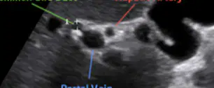
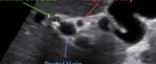
Green
(a)
Red
(a)
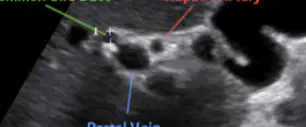
blue
(a)
CBD, Hepatic artery, and Portal vein
(a)
Closest to the right side of the patient
(a)
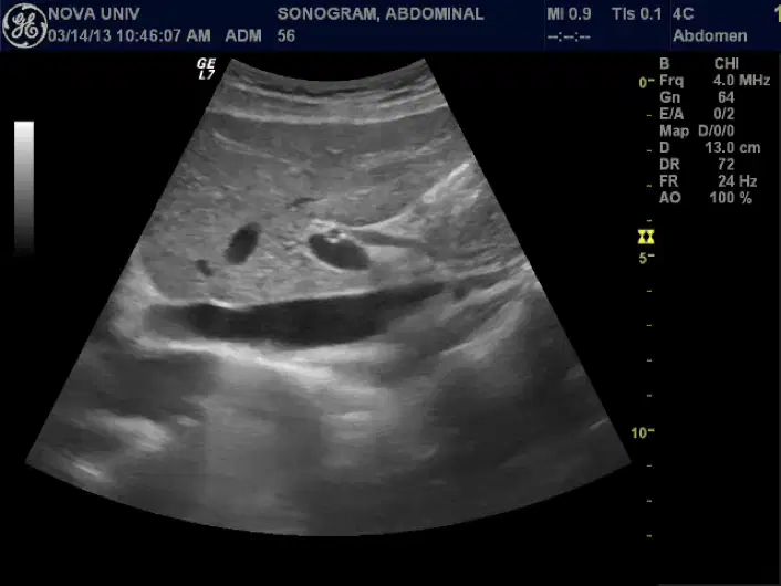
This shows a sagittal
(a)
The cystic duct is ___ long
1cm
2cm
3cm
4cm
Connects the neck of the gallbladder with common hepatic duct to form the common bile duct
(a)
Contains undulating folds or valves called the Valves of Heister
(a)
Somewhat spiral
Help to regulate the flow of bile in and out of the gallbladder
(a)
Lateral to hepatic artery
Anterior to portal vein
(a)
The CBD moves more posteriorly after it descends behind the (a)
Distal CBD lies (a) to the IVC
Right hepatic gives rise to the (a)
which supplies blood to the gallbladder and cystic duct
(a)
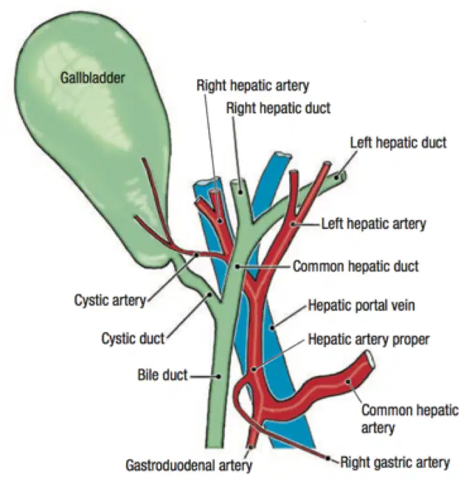
drains the blood from the gallbladder and cystic duct usually ends in the right branch of the portal veins.
(a)
Junctional fold known as Hartman’s pouch
(a)
Phrygian cap
(a)
Congenital anomaly where gallbladder does not develop - rare
(a)
A small outpouching (pathological or acquired dilatation) of the gallbladder wall at the junction of the neck and cystic duct
(a)
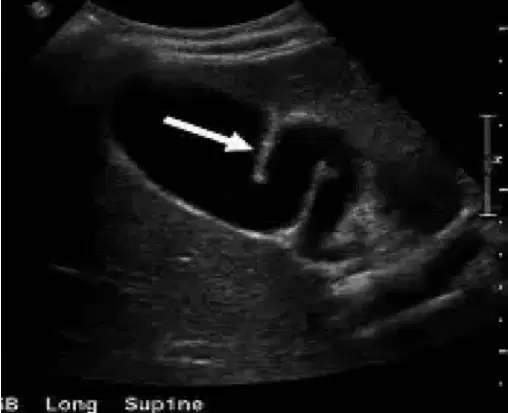
Arrow
(a)
Common site where gallstones can lodge.
Can cause obstruction, cholecystitis, or difficulty during cholecystectomy.
(a)
acquired, pathological, neck of gallbladder, linked to gallstones.
(a)
A Phrygian cap deformity of the gallbladder occur in about _% of the patients
(a)
A congenital anatomical variant of the gallbladder in which the fundus folds back on itself, resembling the cap worn by ancient Phrygians
(a)
Usually an incidental finding on imaging.
Benign and not associated with gallstone disease or symptoms
(a)
congenital, normal variant, fundus of gallbladder, usually harmless
(a)
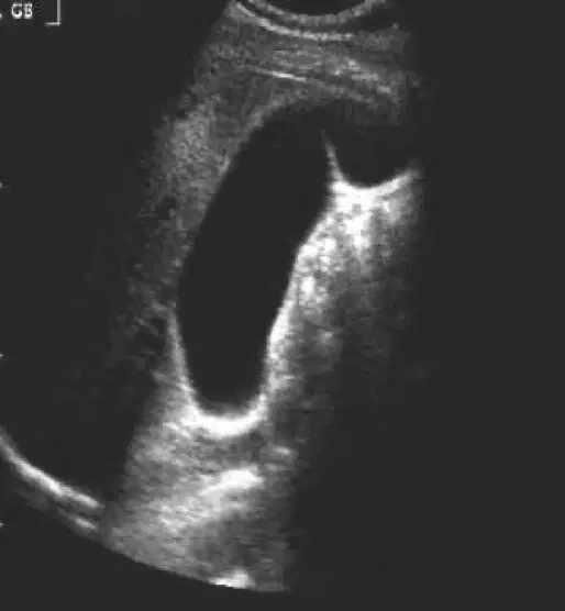
this image shows
(a)
The gallbladder mucosa can form irregular folds and ridges, especially when the organ is empty or contracted
(a)
These are normal anatomical features of the mucosal lining
(a)
They help accommodate changes in gallbladder volume as bile is stored and released.
These folds flatten out when the gallbladder is distended
(a)
internal mucosal ridges (common, functional)
(a)
external folding of the fundus (congenital variant).
(a)
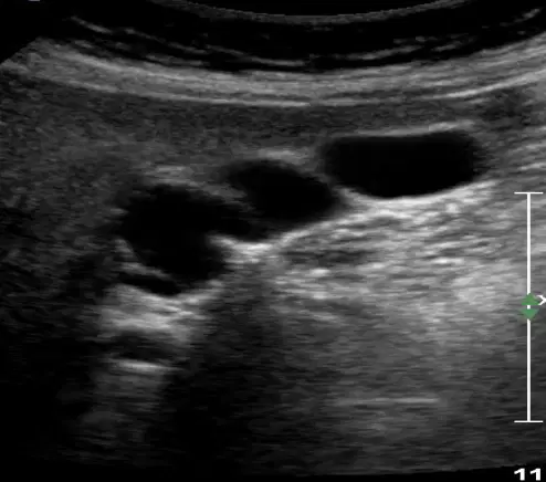
This image shows
(a)
Multi-septations seen within the gallbladder, very rare congenital anomaly of the gallbladder
(a)
characterized by multiple thin septations within the gallbladder lumen
(a)
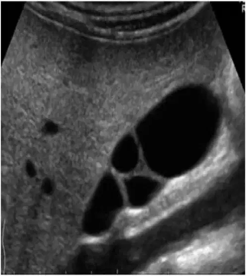
This image shows
(a)
occurs when two separate gall bladder cavities develop, each with separate cystic ducts
(a)
Embryologic abnormality of biliary tract development with a persistence and development of a normal vestigial out pouching of the biliary tree
(a)
Increased incidence of foregut malformations and aberrant (deviating from normal expected pattern) vasculature
(a)
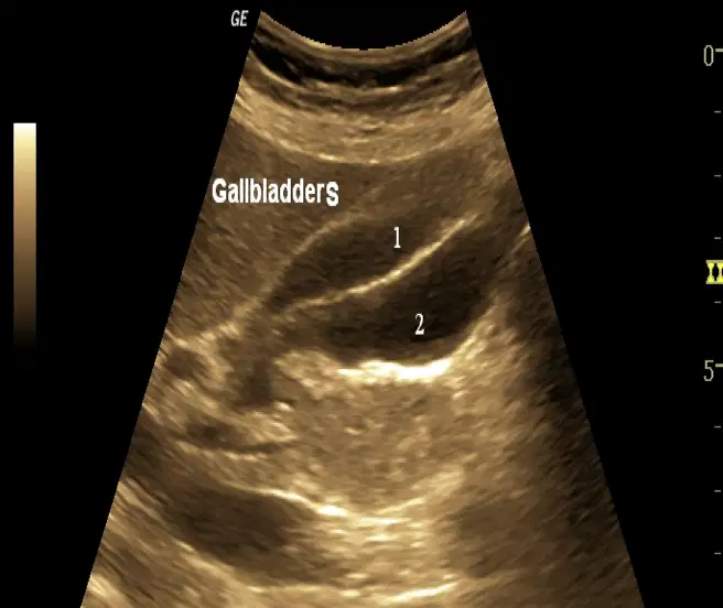
This shows
(a)

