
 Worksheets
Exam 1 Study Guide
Total questions: 49
Worksheet time: 25mins
In anatomical position, the thumb is ___ to the little finger.
lateral
medial
proximal
distal
In anatomical position the shoulder joint is ___ to the hip joint.
superior
inferior
lateral
medial
The directional term for the bottom aspect of the foot is _____.
plantar
dorsal
palmar
superior
Which one of the following best describes the vertical axis?
passes through the body from front to back
passes through the body from side to side
passes through the body from top to bottom
passes through the body on a diagonal
Which movement occurs in the horizontal plane?
torso twist
lateral tilt
leg swing (front)
parallel brush (side)
Which plane does the anteroposterior axis allows movements?
frontal
horizontal
diagonal
sagittal
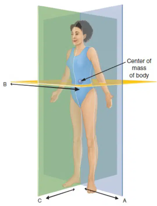
Identify the illustrated basic anatomical planes in the diagram.
Frontal A Horizontal B Sagittal C
Sagittal A Transverse B Coronal C
Oblique A Frontal B Transverse C
Transverse A Sagittal B Oblique C
The three types of attachments of muscle to bone:
tendon, direct, and aponeurosis
ligament, indirect, and fascia
cartilage, indirect, and sheath
membrane, direct, and ligament
What is the definition of an agonist in muscle movement?
A muscle that is primarily responsible for generating a specific movement
A muscle that opposes the action of another muscle
A muscle that assists another in performing a movement
A muscle that stabilizes a joint during movement
Which type of muscle fiber has higher force production but lower resistance to fatigue?
Fast twitch
Slow twitch
Intermediate twitch
Cardiac muscle fibers
Name one bone that is part of the axial skeleton.
spine
femur
humerus
scapula
Myofilament that attaches to the actin filament- ________
myosin
tropomyosin
troponin
collagen
___ term used to describe an increase in muscle fiber size associated with increased training.
Hypertrophy
Atrophy
Dystrophy
Myopathy
List four properties of skeletal muscle tissue.
contractility, irritability, extensibility, and elasticity
rigidity, flexibility, permeability, and opacity
adhesion, cohesion, viscosity, and density
absorbency, transparency, fragility, and solubility
Examples of the following types of joints: • Uniaxial- ________ • Biaxial- ________ • Triaxial joints- ________
Uniaxial- elbow and knee; Biaxial- finger and toes; Triaxial joints- hips and shoulder
Uniaxial- shoulder and hip; Biaxial- elbow and knee; Triaxial joints- finger and toes
Uniaxial- finger and toes; Biaxial- hips and shoulder; Triaxial joints- elbow and knee
Uniaxial- wrist and ankle; Biaxial- shoulder and hip; Triaxial joints- elbow and knee
Name three different types of muscle contractions.
concentric, eccentric, and isometric
static, dynamic, and ballistic
flexor, extensor, and rotator
agonist, antagonist, and synergist
Primary plane and axis in which the roll-down spinal dance movement occurs ________
sagittal plane and mediolateral axis
frontal plane and anteroposterior axis
transverse plane and vertical axis
frontal plane and mediolateral axis
When trying to enhance strength, the goal is to maximize the ____ response of connective tissue.
elastic
chemical
thermal
magnetic
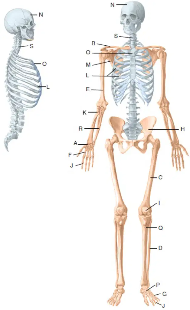
Identify the illustrated bones. letter A
carpals
tarsals
phalanges
metatarsals
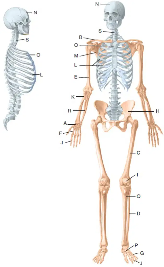
Identify the illustrated bones. B. ______
clavicle
scapula
humerus
sternum
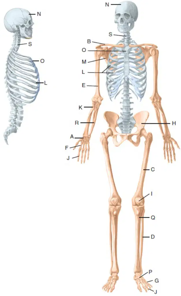
Identify the illustrated bones. C. ______
femur
humerus
tibia
radius
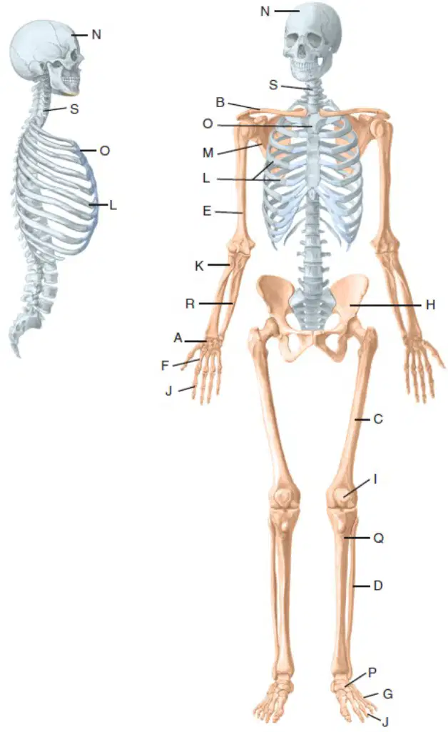
Identify the illustrated bones. D. ______
fibula
tibia
humerus
radius
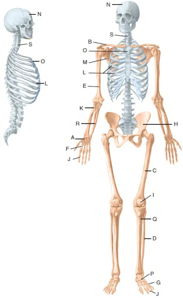
Identify the illustrated bones. E. ______
humerus
femur
radius
scapula
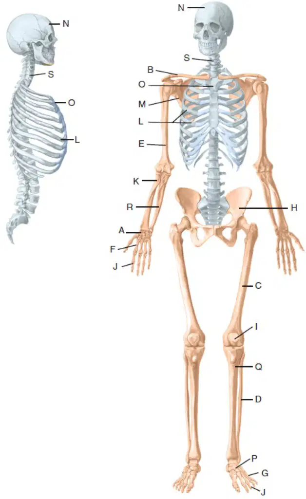
Identify the illustrated bones. F. ______
metacarpals
phalanges
tarsals
clavicle
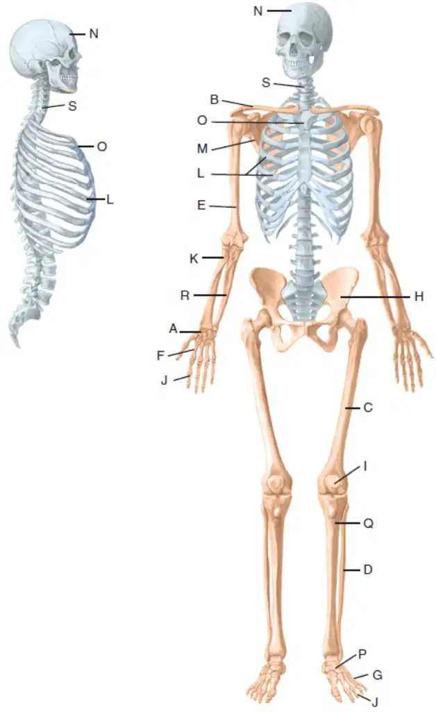
Identify the illustrated bones. G. ______
metatarsals
carpals
phalanges
scapula
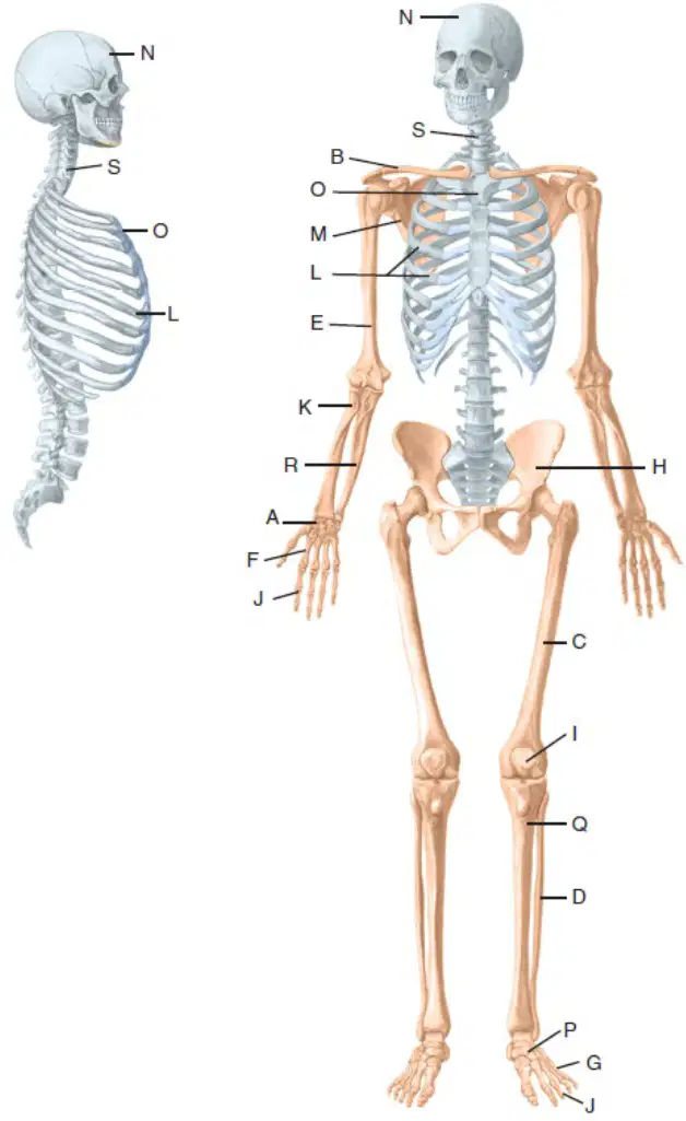
Identify the illustrated bones. H. ______
os coxae (ilium)
scapula
clavicle
femur
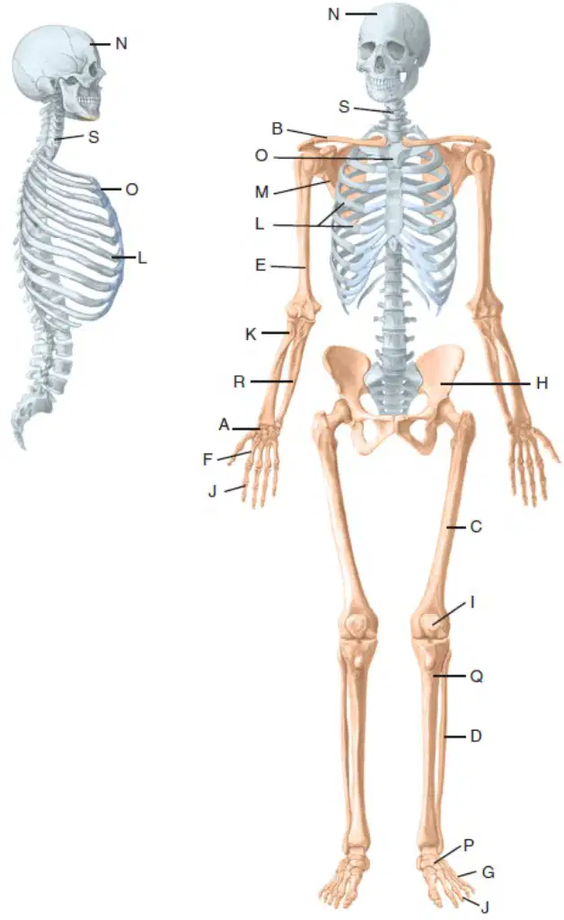
Identify the illustrated bones. I. ______________
patella
scapula
femur
humerus
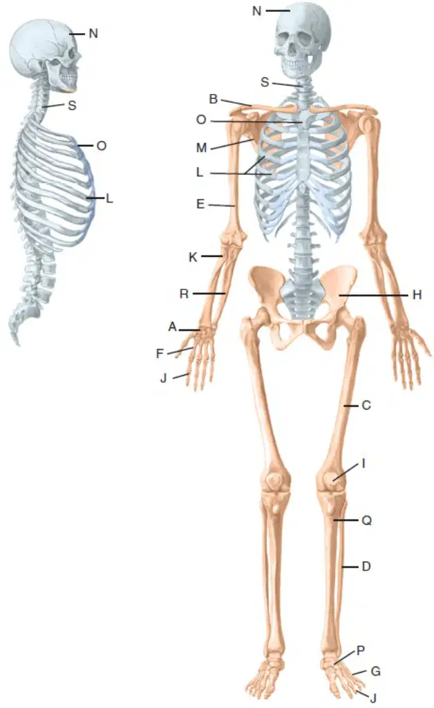
Identify the illustrated bones. J. ______
phalanges
metacarpals
tarsals
clavicle
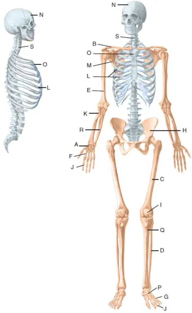
Identify the illustrated bones. K. ______
radius
ulna
femur
humerus
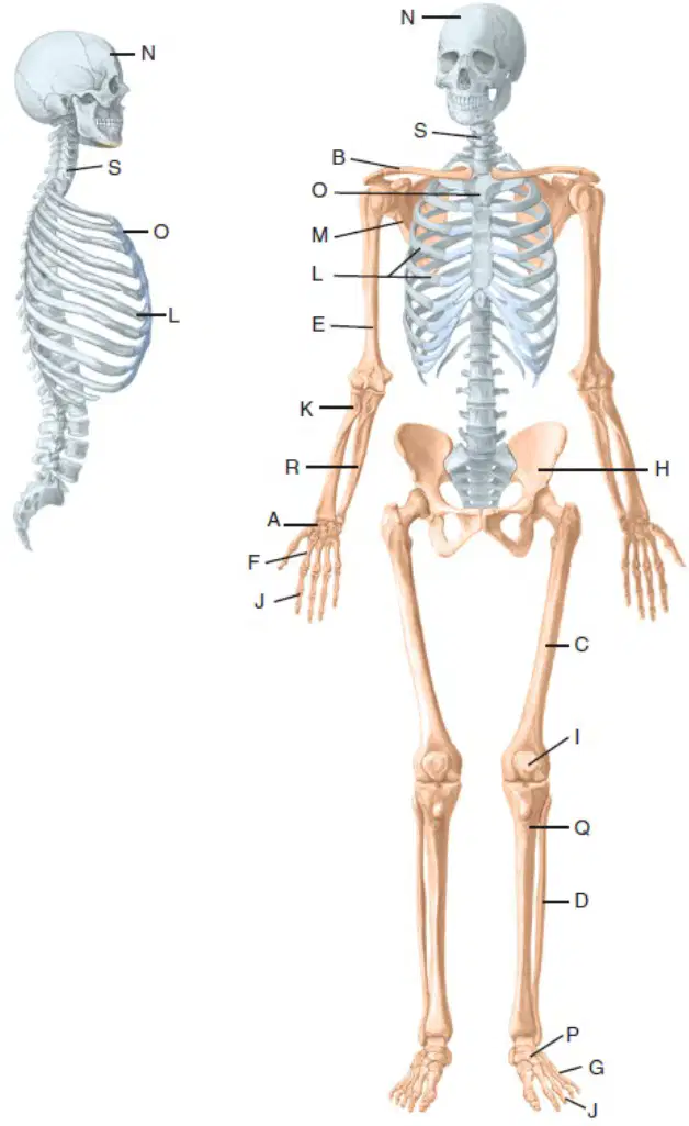
Identify the illustrated bones. L. ______
ribs
femur
scapula
humerus
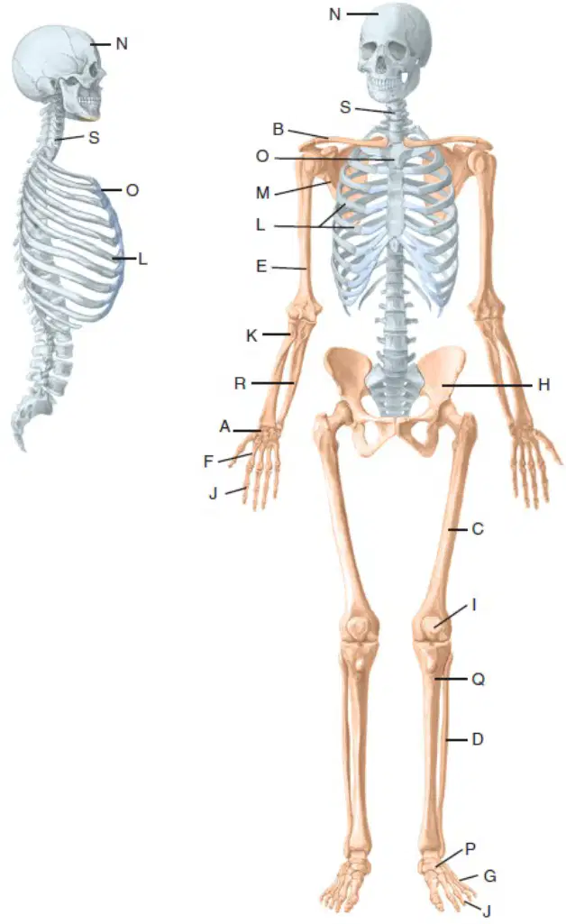
Identify the illustrated bones. M. ______
scapula
femur
humerus
tibia
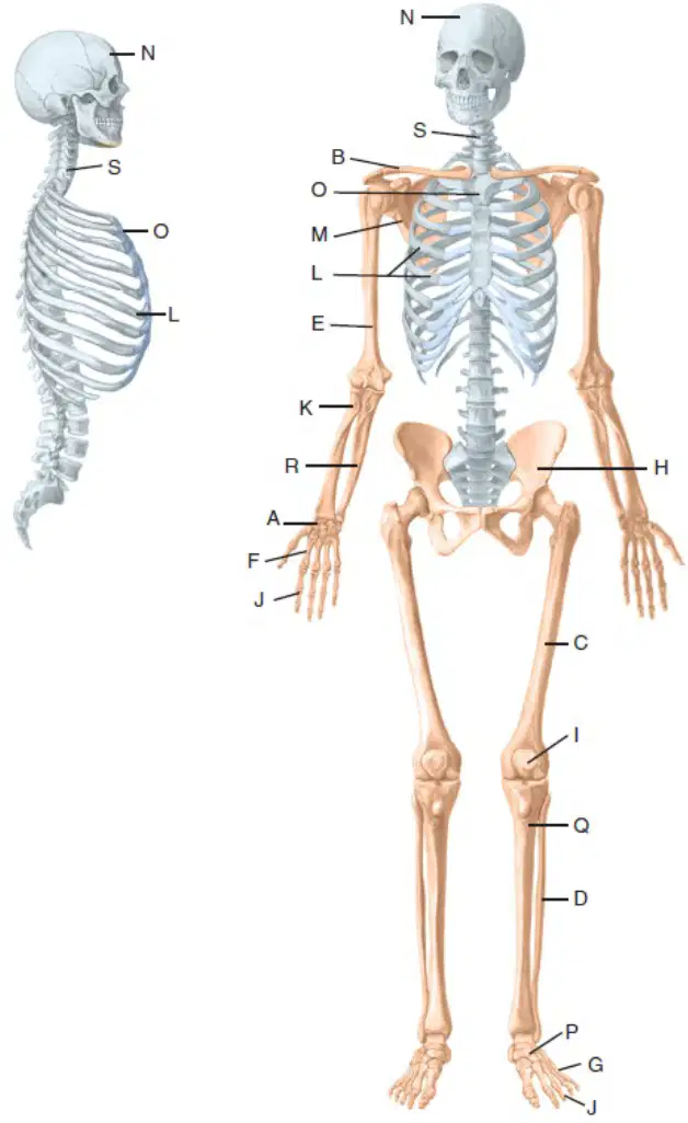
Identify the illustrated bones. N. ______
skull
femur
humerus
pelvis
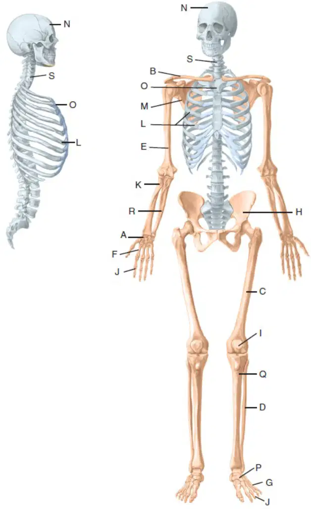
Identify the illustrated bones. O. ______
sternum
clavicle
scapula
humerus
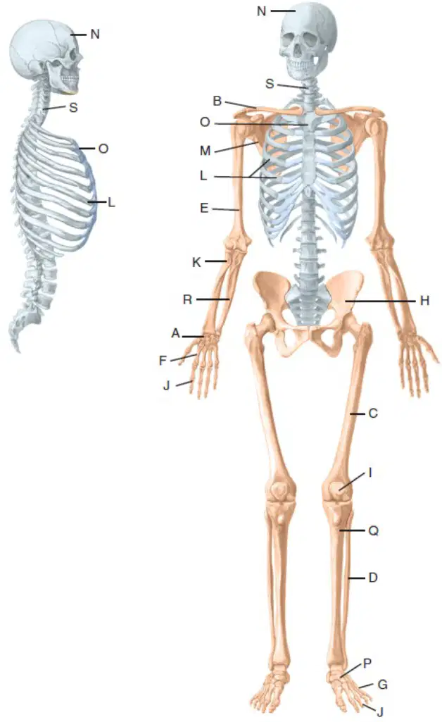
Identify the illustrated bones. P. ______
tarsals
carpals
metatarsals
phalanges
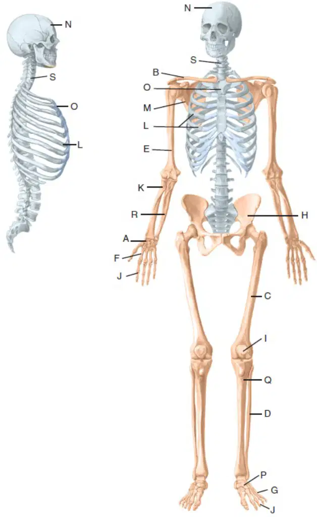
Identify the illustrated bones. Q. ______
tibia
femur
fibula
humerus
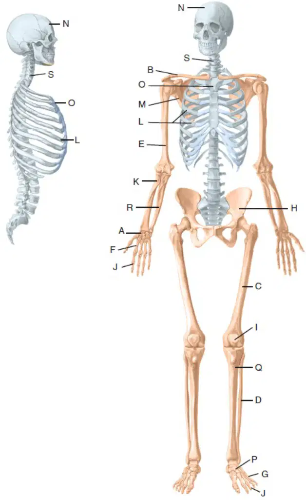
Identify the illustrated bones. R. ______
ulna
radius
femur
tibia
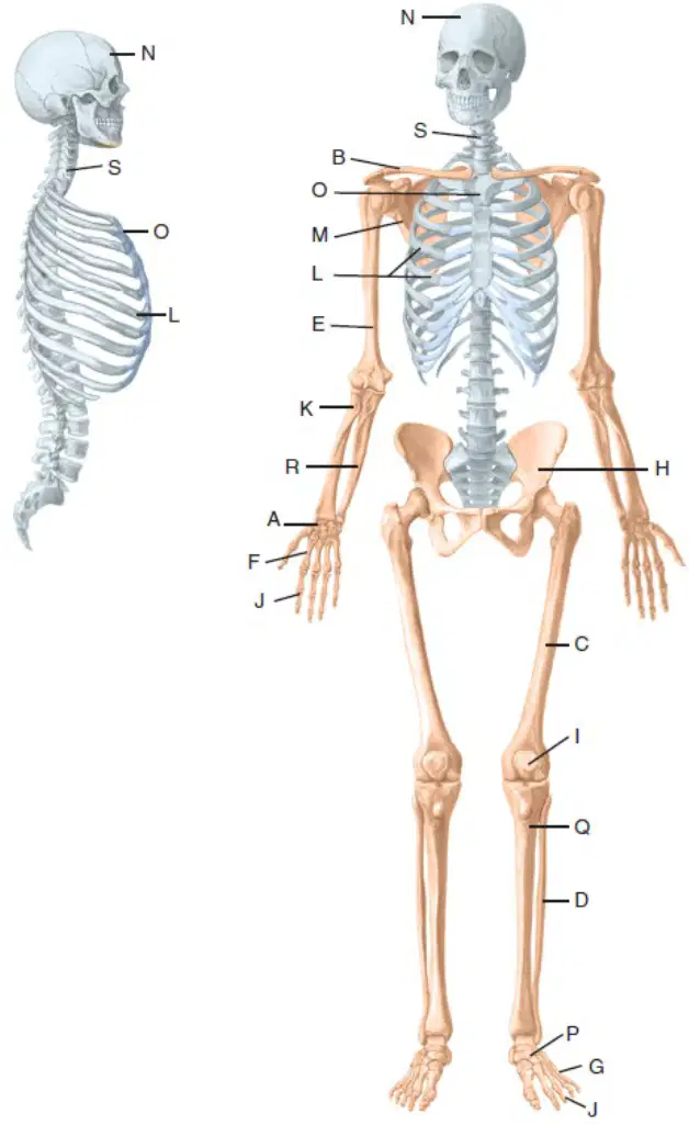
Identify the illustrated bones. S. ______
spine
femur
humerus
rib
Adduction joint motion occurs in which plane?
Horizontal plane
frontal plane
sagittal plane
none
Internal rotation occurs in which plane
horizontal plane
frontal plane
sagittal plane
none
Extension occurs in which plane
frontal plane
horizontal plane
sagittal plane
none
List the appropriate name of the bones with the following bones: patella, femur, tarsal, ilium
long bone, short bone, flat bone, sesmoid
sesmoid bone, long bone, short bone, flat bone
sesmoid bone, short bone, long bone, flat bone
Define Bursa
flattened sac of synovial membrane that reduces friction
band of connective tissue that holds tendons in place
double-layered covering surrounding a tendon that reduces frictio
circumferential fibrocartilage ring that enhances joint contact area
Define labrum
flattened sac of synovial membrane that reduces friction
band of connective tissue that holds tendons in place
double-layered covering surrounding a tendon that reduces frictio
circumferential fibrocartilage ring that enhances joint contact area
Define tendon sheath
flattened sac of synovial membrane that reduces friction
band of connective tissue that holds tendons in place
double-layered covering surrounding a tendon that reduces frictio
circumferential fibrocartilage ring that enhances joint contact area
Define retinaculum
flattened sac of synovial membrane that reduces friction
band of connective tissue that holds tendons in place
double-layered covering surrounding a tendon that reduces frictio
circumferential fibrocartilage ring that enhances joint contact area
List the type of synovial joint classification with the correct number of axes of movement: pivot, gliding, saddle, and ball-and-socket
uniaxial, nonaxial, biaxial, triaxial
nonaxial, uniaxial, biaxial, triaxial
triaxial, nonaxial, biaxial, nonaxial
What is the joint classification for the sutures of the skull?
fibrous joint
cartilaginous joint
synovial joint
What is the joint classification for the knee?
fibrous joint
cartilaginous joint
synovial joint
What is the joint classification for the epiphyseal plate?
fibrous joint
cartilaginous joint
synovial joint

