
 Worksheets
PRACTICE QUESTIONS (EXAM 2)
Total questions: 49
Worksheet time: 25mins
Where is the hyoid bone located?
Head
Neck
Chest
Back
Men with larger laryngeal prominences typically have deeper voices.
The posterior fontanelle typically closes before the anterior fontanelle.
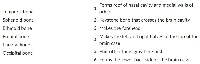
Match each brain cavity bone to its description.
Temporal bone-1
Sphenoid bone-2
Ethmoid bone-3
Frontal bone-4
Parietal bone-5
Occipital bone-6
Temporal bone-3
Sphenoid bone-4
Ethmoid bone-6
Frontal bone-2
Parietal bone-1
Occipital bone-5
Temporal bone-5
Sphenoid bone-2
Ethmoid bone-1
Frontal bone-3
Parietal bone-4
Occipital bone-6
Temporal bone-2
Sphenoid bone-5
Ethmoid bone-6
Frontal bone-1
Parietal bone-3
Occipital bone-4
Temporal bone-4
Sphenoid bone-5
Ethmoid bone-3
Frontal bone-1
Parietal bone-6
Occipital bone-2
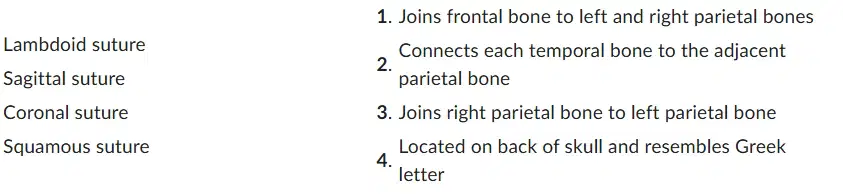
Match each skull suture to the correct description.
Lambdoid suture-1
Sagittal suture-4
Coronal suture-2
Squamous suture-3
Lambdoid suture-3
Sagittal suture-4
Coronal suture-2
Squamous suture-1
Lambdoid suture-2
Sagittal suture-1
Coronal suture-4
Squamous suture-3
Lambdoid suture-4
Sagittal suture-3
Coronal suture-1
Squamous suture-2
The pterion is located at the intersection of the parietal bone, temporal bone, and occipital bone on each side of the brain case.
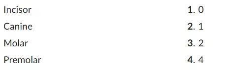
Match each type of tooth to its number of cusps (pointed ends).
Incisor-4
Canine-3
Molar-1
Premolar-3
Incisor-1
Canine-2
Molar-4
Premolar-3
Incisor-4
Canine-2
Molar-3
Premolar-1
Incisor-2
Canine-4
Molar-1
Premolar-3

Classify each bone as belonging to the axial or appendicular skeleton.
Incus (middle ear bone)- 1
Femur (thigh bone)-2
Incus (middle ear bone)-2
Femur (thigh bone)- 1
Which sinuses are the largest and most often involved in sinus infections?
Ethmoid
Frontal
Sphenoid
Maxillary

Match each structure at the base of the skull to the correct description.
Sella turcica-1
Foramen magnum-2
Cribriform plate-3
Sella turcica-3
Foramen magnum-1
Cribriform plate-2
Sella turcica-2
Foramen magnum-1
Cribriform plate-3
The plantar fascia connects the:
Bases of the metatarsals to the calcaneus
Bases of the phalanges to the calcaneus
Bases of the phalanges to the talus
Bases of the metatarsals to the talus
The pain caused by a herniated disc occurs when a weakened anulus fibrosus allows a nucleus pulposus to protrude, compressing a spinal nerve.
How many metatarsal bones are there?
5
6
4
3
How many phalanges are in the thumb?
1
3
2
4
The heel of the foot is formed by the:
Phalanges
Calcaneus
Talus
Metatarsals
The femur attaches to the:
Pubic symphysis
Obturator foramen
Acetabulum
Sacroiliac joint
A Colles fracture is a fracture of the distal:
Scaphoid
Ulna
Humerus
Radius
The ilium, ischium, and pubis fuse to form the adult hip bone.
True ribs have costal cartilages that attach to the costal cartilage of the rib above.
A bunion is a strained muscle caused by wearing high-heeled shoes.

Match each spine disorder to the correct description.
Kyphosis-2
Scoliosis-1
Lordosis-3
Kyphosis-3
Scoliosis-1
Lordosis-2
Kyphosis-1
Scoliosis-2
Lordosis-3
Kyphosis-3
Scoliosis-2
Lordosis-1
Which bone fits into the glenoid cavity?
Humerus
Radius
Sternum
Ulna
Vertebral body size decreases as one goes from cervical vertebrae down to lumbar vertebrae.
How many vertebrae are in the thoracic spine?
12
7
4
5
The foot has three arches, which can fall in some individuals.
The medial malleolus, lateral malleolus, and talus make up the:
Elbow joint
Shoulder joint
Ankle joint
Knee joint
FOOSH stands for Fall On Outstretched Humerus.
Patellofemoral syndrome occurs when the patella moves too far to the medial side of the femur.
The male pelvis has a larger subpubic angle than the female pelvis.

Match each structure to the correct association.
Ulnar nerve- 1
Median nerve-2
Anterior longitudinal ligament-3
Ulnar nerve- 1
Median nerve-3
Anterior longitudinal ligament-2
Ulnar nerve- 3
Median nerve-2
Anterior longitudinal ligament-1
Ulnar nerve-2
Median nerve-1
Anterior longitudinal ligament-3

Match each joint to the correct functional classification.
Intervertebral disc-2
Metacarpophalangeal joint-3
Manubriosternal joint-1
Intervertebral disc-3
Metacarpophalangeal joint-2
Manubriosternal joint-1
Intervertebral disc-3
Metacarpophalangeal joint-1
Manubriosternal joint-2
Intervertebral disc-1
Metacarpophalangeal joint-2
Manubriosternal joint-3
Joints with more mobility have less stability.
A lateral blow to an extended knee can cause the medial side to open, causing injury to medial structures.
In the RICE technique, C stands for Cold.
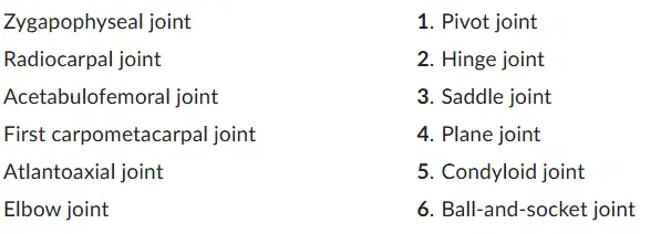
Match each synovial joint to its correct classification.
Zygapophyseal joint-1
Radiocarpal joint-3
Acetabulofemoral joint-4
First carpometacarpal joint-2
Atlantoaxial joint-6
Elbow joint-5
Zygapophyseal joint-3
Radiocarpal joint-6
Acetabulofemoral joint-2
First carpometacarpal joint-4
Atlantoaxial joint-1
Elbow joint-5
Zygapophyseal joint-1
Radiocarpal joint-6
Acetabulofemoral joint-3
First carpometacarpal joint-2
Atlantoaxial joint-4
Elbow joint-5
Zygapophyseal joint-4
Radiocarpal joint-5
Acetabulofemoral joint-6
First carpometacarpal joint-3
Atlantoaxial joint-1
Elbow joint-2
The shoulder joint is not very stable because the humerus head is much larger than the glenoid cavity.
A sprain involves stretching or tearing of tendons.

Match each type of fibrous joint to the correct description.
Gomphosis-3
Syndesmosis-2
Suture-1
Gomphosis-2
Syndesmosis-1
Suture-3
Gomphosis-3
Syndesmosis-1
Suture-2
Gomphosis-1
Syndesmosis-3
Suture-2
Whiplash is caused by hyperextension of the anterior longitudinal ligament.

Match each joint to the correct description.
Zygapophyseal joint-3
Atlantoaxial joint-1
Atlanto-occipital joint-2
Zygapophyseal joint-1
Atlantoaxial joint-3
Atlanto-occipital joint-2
Zygapophyseal joint-3
Atlantoaxial joint-2
Atlanto-occipital joint-1
Zygapophyseal joint-2
Atlantoaxial joint-3
Atlanto-occipital joint-1
Both shoulder separations and dislocations most often occur:
Posteriorly
Medially
Laterally
Anteriorly
In a synovial joint, the fibrous membrane of the articular capsule produces synovial fluid.
Tommy John surgery is surgical repair of the:
Fibular collateral ligament
Tibial collateral ligament
Radial collateral ligament
Ulnar collateral ligament
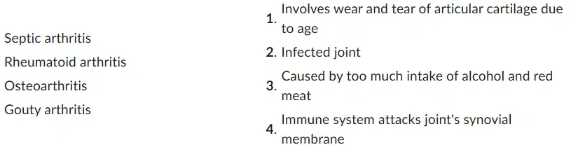
Match each arthritis type to the correct description.
Septic arthritis-1
Rheumatoid arthritis-3
Osteoarthritis-2
Gouty arthritis-4
Septic arthritis-2
Rheumatoid arthritis-4
Osteoarthritis-1
Gouty arthritis-3
Septic arthritis-3
Rheumatoid arthritis-1
Osteoarthritis-4
Gouty arthritis-2
Septic arthritis-4
Rheumatoid arthritis-3
Osteoarthritis-2
Gouty arthritis-1
Flexion is:
Anterior movement or increase of a joint angle
Posterior movement or increase of a joint angle
Posterior movement or decrease of a joint angle
Anterior movement or decrease of a joint angle
Ligaments are connective tissue bands that connect two bones, and tendons are connective tissue bands that connect a muscle to a bone.
The posterior cruciate ligament prevents the tibia from sliding too far forward.
In an eversion ankle sprain, the deltoid ligament can be torn.
In "student's elbow," the antecubital bursa becomes inflamed.

