
 Worksheets
Guided Notes: A and P/Skin, Musculoskeletal
Total questions: 65
Worksheet time: 34mins
Anika is learning about living things in her science class. Her teacher asks, "What are the basic units of life?" What should Anika answer?
cells
a type of tissue
a kind of organ
a chemical element
James is learning about cells in his biology class. His teacher asks him to describe the roles of the plasma membrane and cytoplasm in a cell. Which of the following best describes these parts?
The plasma membrane is the outer boundary of the cell that controls what enters and leaves the cell. The cytoplasm is the jelly-like substance within the cell that contains organelles.
The plasma membrane is the control center of the cell, and the cytoplasm is responsible for producing energy.
The plasma membrane stores genetic information, and the cytoplasm is the site of protein synthesis.
The plasma membrane is a rigid structure that supports the cell, and the cytoplasm is a storage area for waste.
William is learning about the parts of a cell in his biology class. He wants to know the roles of the nucleus, nuclear membrane, and nucleolus. Which of the following best describes their functions?
The nucleus contains the cell's genetic material (DNA) and controls cell activities. The nuclear membrane surrounds the nucleus, and the nucleolus is involved in producing ribosomes.
The nucleus is responsible for energy production, the nuclear membrane stores nutrients, and the nucleolus digests waste.
The nucleus helps in cell movement, the nuclear membrane produces proteins, and the nucleolus stores water.
The nucleus is involved in photosynthesis, the nuclear membrane transports oxygen, and the nucleolus breaks down toxins.
Aiden is learning about how his body gets energy to play soccer. His teacher explains that a part of his cells acts as the powerhouse, producing energy (ATP) through cellular respiration. Which part of the cell is Aiden's teacher describing?
Mitochondria are the powerhouses of the cell, producing energy (ATP) through cellular respiration.
Mitochondria are responsible for protein synthesis in the cell.
Mitochondria store genetic information and control cell activities.
Mitochondria are involved in the packaging and transport of proteins.
Abigail is learning about different parts of a cell in her biology class. She wants to know which part is responsible for protein synthesis. Which part of the cell should she focus on?
Ribosomes are responsible for protein synthesis in the cell.
Ribosomes store genetic information in the cell.
Ribosomes are responsible for energy production in the cell.
Ribosomes help in the transport of materials within the cell.
Nora is working in a laboratory and observes a network of membranes inside a cell that is involved in protein and lipid synthesis. What is the best description of this part of the cell?
The endoplasmic reticulum is a network of membranes involved in protein and lipid synthesis.
The endoplasmic reticulum is responsible for storing genetic information.
The endoplasmic reticulum is the site of cellular respiration and energy production.
The endoplasmic reticulum is a structure that packages and distributes proteins outside the cell.
Harper is working in a factory that processes and ships products. Which part of the factory is most similar to the Golgi Apparatus in a cell?
The Golgi apparatus modifies, sorts, and packages proteins and lipids for storage or transport out of the cell.
The Golgi apparatus generates energy for the cell through cellular respiration.
The Golgi apparatus is responsible for synthesizing DNA during cell division.
The Golgi apparatus controls the movement of substances into and out of the nucleus.
Oliver is cleaning up his room and uses a special box that contains tools to break down and dispose of old toys and trash. In a cell, which part acts like this special box by containing digestive enzymes to break down waste materials and cellular debris?
Lysosomes contain digestive enzymes to break down waste materials and cellular debris.
Lysosomes are responsible for producing energy in the form of ATP.
Lysosomes synthesize proteins for the cell.
Lysosomes store genetic information in the form of DNA.
Sophia is observing pond water under a microscope and notices some tiny organisms moving around. She wonders what structures help these organisms move. What part of the cell is responsible for this movement?
Cilia and flagella are hair-like structures that help with cell movement.
Cilia and flagella are responsible for protein synthesis in the cell.
Cilia and flagella store genetic information for the cell.
Cilia and flagella are the main site of cellular respiration.
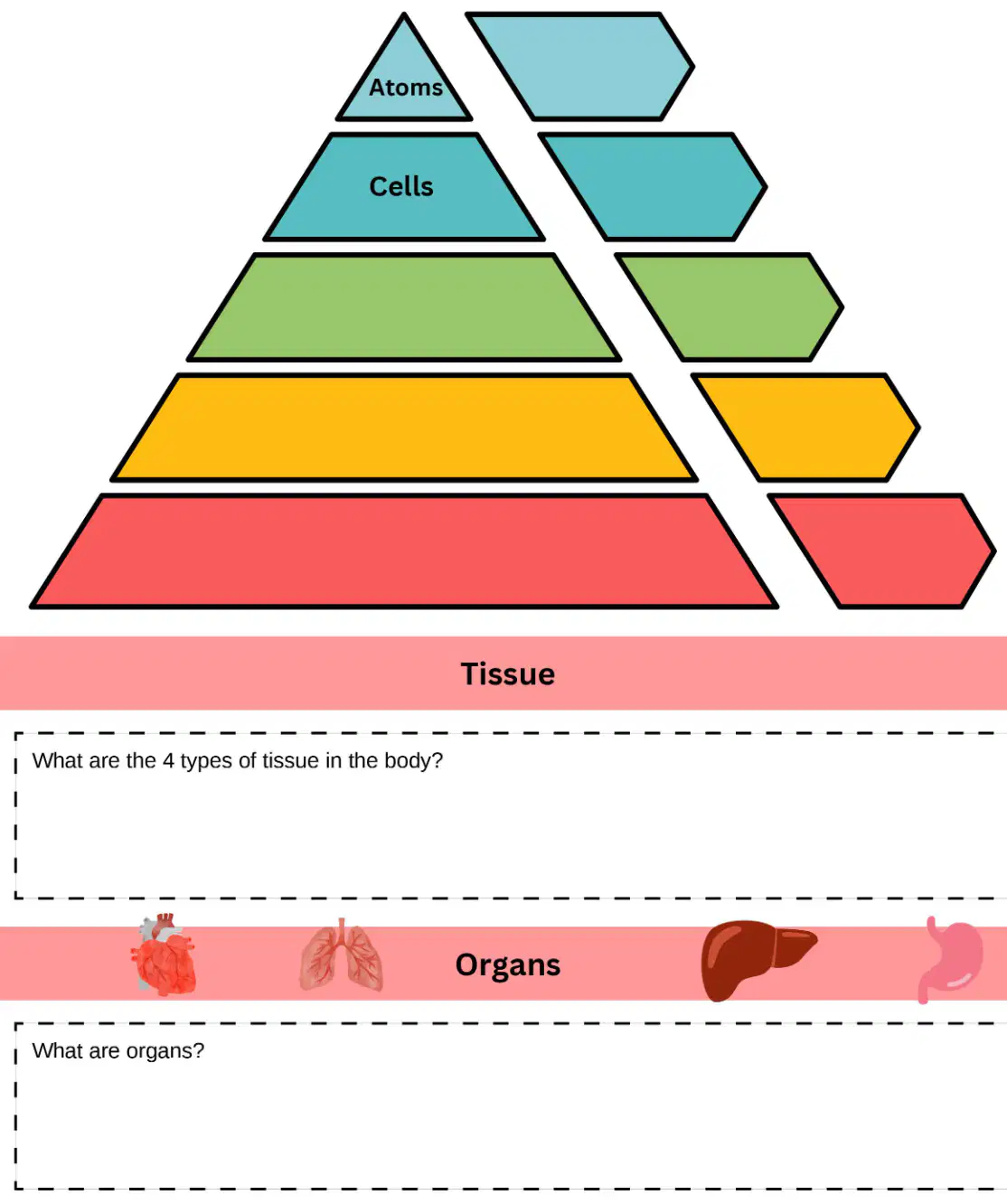
Aria is building a model to show how the human body is organized, starting from the smallest parts to the whole person. She begins with Atoms, then adds Cells, and needs to fill in the next three levels of increasing complexity. Complete Aria's pyramid: Atoms, Cells, _____, _____, _____.
Tissues, Organs, Organism
Organs, Tissues, Organism
Cells, Organs, Tissues
Tissues, Organism, Organs
Charlotte is studying for her biology exam and wants to remember the 4 main types of tissue in the body. Which of the following lists correctly identifies them?
Epithelial, connective, muscle, and nervous
Bone, blood, cartilage, and fat
Skin, bone, muscle, and nerve
Connective, skeletal, cardiac, and smooth
Grace is learning about the human body in her science class. Her teacher asks, "Can anyone tell me what organs are?" What should Grace answer?
groups of tissues that work together to perform specific functions
the smallest unit of life in the body
a type of cell found in animals
liquids that circulate in the body
Zoe is hiking on a hot day, and her body starts to sweat to cool her down and keep her internal temperature stable. What is the process called by which Zoe's body maintains a stable internal environment despite changes in external conditions?
Homeostasis is the process by which the body maintains a stable internal environment despite changes in external conditions.
Homeostasis is the process of breaking down food to release energy.
Homeostasis is the ability of an organism to move from one place to another.
Homeostasis is the process of converting oxygen into carbon dioxide.
Lily is learning about how her body protects itself from the environment. She wants to know which organs are involved and what their main functions are. Which of the following best describes the organs and function of the Integumentary System?
Organs: Skin, hair, nails. Function: Protects the body, regulates temperature, and provides sensory information.
Organs: Heart, blood vessels. Function: Pumps blood and transports nutrients.
Organs: Lungs, trachea. Function: Facilitates gas exchange and breathing.
Organs: Kidneys, bladder. Function: Removes waste and maintains fluid balance.
Avery is learning about how her body moves and stays upright. She wants to know which organs and functions are part of the Musculoskeletal System that help her run, jump, and play. Which of the following best describes the organs and function of the Musculoskeletal System?
Organs: Bones, muscles, joints. Function: Provides structure, support, movement, and protection for the body.
Organs: Heart, blood vessels. Function: Pumps blood and transports nutrients.
Organs: Lungs, trachea. Function: Facilitates gas exchange and breathing.
Organs: Kidneys, bladder. Function: Filters blood and removes waste.
Aria accidentally touches a hot stove and quickly pulls her hand away. Which body system is responsible for controlling this reaction and responding to the stimulus?
Organs: Brain, spinal cord, nerves. Function: Controls body activities and responds to internal and external stimuli.
Organs: Heart, blood vessels. Function: Pumps blood and transports nutrients.
Organs: Lungs, trachea. Function: Exchanges oxygen and carbon dioxide.
Organs: Stomach, intestines. Function: Digests food and absorbs nutrients.
Anika is studying for her biology exam and wants to understand how the body defends itself against infections and diseases. She comes across a system that includes organs like lymph nodes, lymph vessels, spleen, and thymus. Which of the following best describes the organs and function of this system?
Organs: Lymph nodes, lymph vessels, spleen, thymus. Function: Defends against infection and disease, returns tissue fluids to the bloodstream.
Organs: Heart, arteries, veins, capillaries. Function: Pumps blood and transports oxygen and nutrients.
Organs: Kidneys, ureters, bladder, urethra. Function: Removes waste from the blood and regulates water balance.
Organs: Stomach, intestines, liver, pancreas. Function: Breaks down food and absorbs nutrients.
Aria is learning about how her body works in science class. Her teacher asks her to identify which group of organs is responsible for exchanging oxygen and carbon dioxide between the blood and the environment. Which option should Aria choose?
Organs: Lungs, trachea, bronchi. Function: Exchanges oxygen and carbon dioxide between the blood and the environment.
Organs: Heart, arteries, veins. Function: Pumps blood throughout the body.
Organs: Stomach, intestines, liver. Function: Breaks down food and absorbs nutrients.
Organs: Kidneys, bladder, ureters. Function: Removes waste from the blood and regulates water balance.
Charlotte is preparing for her anatomy class demonstration. Her instructor asks her to stand in the anatomical position. Which of the following best describes how Charlotte should stand?
Standing upright, facing forward, arms at the sides with palms facing forward
Sitting with legs crossed and hands on knees
Lying flat on the back with arms at the sides
Standing with arms raised above the head
Noah is learning about the human body in his biology class. His teacher asks him to identify the correct definition for the directional term: Superior. Which option should Noah choose?
Above or toward the head end of the body; upper.
Below or toward the feet; lower.
Toward the front of the body; anterior.
Toward the back of the body; posterior.
James is learning about the human body in his biology class. His teacher asks him to identify the correct definition for the directional term: Inferior, as it relates to body position.
Below or toward the lower part of the body; lower.
Above or toward the upper part of the body; higher.
Toward the front of the body; anterior.
Toward the back of the body; posterior.
Daniel is studying anatomy and wants to describe the position of the chest relative to the spine. Which definition best matches the directional term: Anterior (Ventral)?
Toward the front of the body; in front of.
Toward the back of the body; behind.
Away from the midline of the body.
Toward the lower part of a structure or the body; below.
David is learning about anatomy in class. His teacher asks him to point to a part of the body that is medial. What does the term medial mean?
Toward the midline of the body.
Away from the midline of the body.
Toward the head or upper part of a structure.
Toward the back of the body.
During a soccer game, Maya injures the outer side of her knee. Which directional term best describes the location of her injury?
Away from the midline of the body.
Toward the midline of the body.
Above another part.
Closer to the point of attachment.
Aiden is learning about the human body in class. His teacher asks him: "If you are describing the location of the elbow in relation to the shoulder, which definition best fits the term 'proximal'?"
Closer to the point of attachment or origin.
Farther from the point of attachment or origin.
Toward the surface of the body.
Away from the midline of the body.
While examining a small cut on Maya's arm, the doctor explains that the wound is superficial. What does this mean?
Toward or at the body surface.
Away from the body surface; more internal.
Toward the midline of the body.
Away from the midline of the body.
During a radiology class, Ava is asked to identify a Sagittal plane:
A plane that divides the body into right and left parts.
A plane that divides the body into upper and lower parts.
A plane that divides the body into front and back parts.
A plane that runs diagonally across the body.
Emma is studying anatomy and is learning about the three main planes that describe sections of the body, which are also frequently used with various radiographic studies. She wants to know which describes a Transverse:
A plane that divides the body into superior (upper) and inferior (lower) parts.
A plane that divides the body into anterior (front) and posterior (back) parts.
A plane that divides the body into left and right halves.
A plane that runs diagonally between the horizontal and vertical planes.
During a science class, Ethan is learning about how the body protects its organs. The teacher asks: "Which organ is protected by the cranial cavity?"
Brain
Heart
Lungs
Liver
Ava is studying for her anatomy exam and wants to remember which organs are protected by the thoracic cavity. She writes down a list: Thoracic Cavity: __________
Heart, lungs, esophagus, trachea
Stomach, liver, pancreas, spleen
Kidneys, bladder, ureters, urethra
Brain, spinal cord, meninges
Oliver is studying for his anatomy exam and wants to know which organs are protected by the abdominal cavity. Which of the following lists includes organs found in the abdominal cavity?
Stomach, liver, intestines, spleen, pancreas, kidneys
Heart, lungs, trachea, esophagus
Brain, spinal cord, meninges
Bladder, reproductive organs, rectum
Mia is studying human anatomy and wants to know which organs are protected within the pelvic cavity. Which of the following lists the correct organs found in the pelvic cavity?
Bladder, reproductive organs, rectum
Heart, lungs, trachea
Stomach, liver, pancreas
Brain, spinal cord, eyes
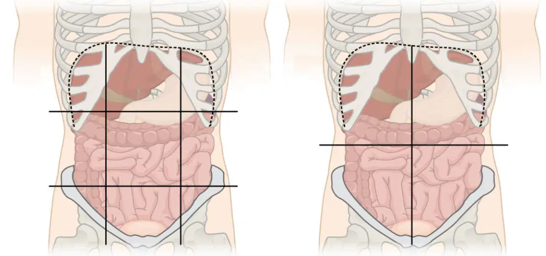
Grace is studying for her anatomy exam and needs to label the abdomen by listing the organs found in each quadrant. Can you help her by selecting the correct list of organs for each abdominal quadrant?
Right Upper Quadrant: Liver, gallbladder, part of pancreas, right kidney; Left Upper Quadrant: Stomach, spleen, left lobe of liver, left kidney, part of pancreas; Right Lower Quadrant: Appendix, right ovary and fallopian tube, right ureter; Left Lower Quadrant: Left ovary and fallopian tube, left ureter, part of intestines.
Right Upper Quadrant: Stomach, spleen, left kidney; Left Upper Quadrant: Liver, gallbladder, right kidney; Right Lower Quadrant: Left ovary, left ureter; Left Lower Quadrant: Appendix, right ovary, right ureter.
Right Upper Quadrant: Appendix, left ovary, left kidney; Left Upper Quadrant: Gallbladder, right ovary, right kidney; Right Lower Quadrant: Liver, spleen, pancreas; Left Lower Quadrant: Stomach, pancreas, intestines.
Right Upper Quadrant: Pancreas, spleen, left kidney; Left Upper Quadrant: Liver, appendix, right kidney; Right Lower Quadrant: Gallbladder, left ovary, left ureter; Left Lower Quadrant: Right ovary, right ureter, intestines.
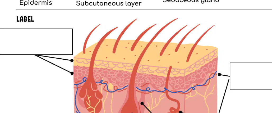
Emma is studying for her biology exam and is looking at an illustration of the human skin. She needs to label its parts and describe the function of each part. What is the part indicated by the top left blank?
Epidermis
Dermis
Sebaceous gland
Subcutaneous layer
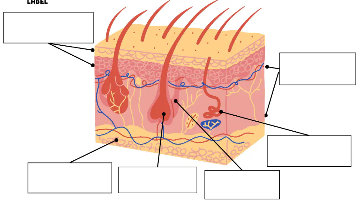
Grace is studying for her biology exam and is looking at an illustration of the human skin. She needs to label its parts and describe the function of each part. What is the part indicated by the bottom left blank?
Subcutaneous layer
Epidermis
Sebaceous gland
Hair Follicle
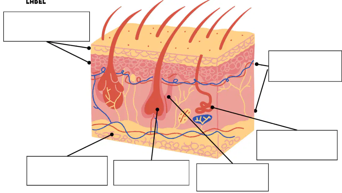
Luna is studying for her biology exam and is looking at an illustration of the human skin. She needs to label its parts and describe the function of each part. What is the part indicated by the bottom middle left blank?
Dermis
Epidermis
Sebaceous gland
Hair Follicle
Which of the following lists the essential functions performed by the skin and accessory structures?
Protection, sensation, temperature regulation, excretion, and vitamin D synthesis
Digestion, absorption, circulation, respiration, and excretion
Movement, support, reproduction, immunity, and sensation
Filtration, secretion, absorption, protection, and locomotion
For the disease 'Psoriasis', fill in the treatment.
Topical corticosteroids
Antibiotics
Insulin therapy
Antiviral drugs
For the disease 'Fungal Infections: Candida, Tinea corporis', fill in the etiology and signs/symptoms.
Etiology: Candida is caused by Candida species (yeast), often Candida albicans; Tinea corporis is caused by dermatophyte fungi (e.g., Trichophyton, Microsporum). Signs/symptoms: Candida—red, moist, itchy patches, often in skin folds; Tinea corporis—ring-shaped, scaly, itchy lesions with central clearing.
Etiology: Candida and Tinea corporis are both caused by bacteria; signs/symptoms include fever and cough.
Etiology: Candida is caused by a virus, and Tinea corporis by a parasite; signs/symptoms include joint pain and swelling.
Etiology: Both are caused by allergic reactions; signs/symptoms include hives and difficulty breathing.
For the disease 'Fungal Infections: Candida, Tinea corporis', fill in the treatment.
Topical antifungals such as clotrimazole or terbinafine
Oral antibiotics such as amoxicillin
Antiviral medications such as acyclovir
Corticosteroid creams only
For the disease 'Pediculosis', fill in the treatment.
Application of permethrin shampoo
Administration of insulin
Use of antiviral drugs
Surgical removal of lice
Describe this abnormal skin pigmentation: Albinism.
Albinism is a condition characterized by the absence of melanin pigment in the skin, hair, and eyes, resulting in very light skin and hair color.
Albinism is a condition where the skin becomes darker due to excess melanin production.
Albinism is a condition that causes red patches and scaling on the skin.
Albinism is a condition where the skin turns yellow due to liver problems.
Describe this abnormal skin pigmentation: Erythema.
Erythema is redness of the skin caused by increased blood flow to the capillaries, often due to inflammation or infection.
Erythema is a yellowing of the skin due to excess bilirubin.
Erythema is a bluish discoloration of the skin caused by lack of oxygen.
Erythema is a darkening of the skin due to increased melanin production.
Abnormal colors of the skin can indicate disease. Describe this abnormal skin pigmentation: Cyanosis.
Cyanosis is a bluish discoloration of the skin resulting from poor circulation or inadequate oxygenation of the blood.
Cyanosis is a yellowish discoloration of the skin caused by excess bilirubin in the blood.
Cyanosis is a reddish discoloration of the skin due to increased blood flow to the surface.
Cyanosis is a pale or white discoloration of the skin due to lack of blood flow.
Describe this abnormal skin pigmentation: Jaundice.
Jaundice is a yellowing of the skin and eyes caused by an excess of bilirubin in the blood, often due to liver disease.
Jaundice is a bluish discoloration of the skin caused by lack of oxygen in the blood.
Jaundice is a darkening of the skin due to increased melanin production.
Jaundice is a reddening of the skin caused by inflammation or infection.
Describe the following lesion: Macule.
A macule is a flat, distinct, discolored area of skin that is usually less than 1 centimeter wide and does not involve any change in the thickness or texture of the skin.
A macule is a raised, fluid-filled blister larger than 1 centimeter in diameter.
A macule is a thickened, rough area of skin with irregular borders and variable color.
A macule is a solid, elevated lesion greater than 1 centimeter in diameter.
Describe the following lesion: Papule.
A papule is a small, raised, solid pimple or swelling, often forming part of a rash and typically less than 1 centimeter in diameter.
A papule is a large, fluid-filled blister greater than 1 centimeter in diameter.
A papule is a flat, discolored area of skin that is not raised or depressed compared to the surrounding skin.
A papule is a pus-filled lesion that is usually associated with infection.
Describe the following lesion: Nodule.
A nodule is a larger, firm, raised lesion that is deeper than a papule and usually greater than 1 centimeter in diameter.
A nodule is a small, flat, non-palpable lesion less than 1 centimeter in diameter.
A nodule is a superficial, fluid-filled lesion less than 0.5 centimeters in diameter.
A nodule is a thin, flaky lesion that peels off easily from the skin surface.
Describe the following lesion: Tumor.
A tumor is a large, solid mass of tissue that may originate in the skin or underlying tissues and can be benign or malignant.
A tumor is a small, fluid-filled blister that only occurs on the surface of the skin.
A tumor is a flat, discolored patch that cannot be felt and is always benign.
A tumor is a scaly, crusted lesion that is always caused by a bacterial infection.
Describe the following lesion: Wheal.
A wheal is a raised, itchy area of skin that is usually a sign of an allergic reaction, often pale in the center with a red surrounding area.
A wheal is a flat, scaly patch of skin that is usually brown or black in color.
A wheal is a small, fluid-filled blister that is commonly caused by viral infections.
A wheal is a deep, pus-filled nodule that often results in scarring.
Describe the following lesion: Vesicle.
A vesicle is a small, fluid-filled blister on the skin, usually less than 1 centimeter in diameter.
A vesicle is a large, solid mass that forms deep within the skin.
A vesicle is a dry, scaly patch of skin caused by excessive keratin production.
A vesicle is a flat, discolored area of skin larger than 1 centimeter in diameter.
Describe the following lesion: Bulla.
A bulla is a large, fluid-filled blister on the skin, usually greater than 1 centimeter in diameter.
A bulla is a small, solid, raised lesion less than 0.5 centimeters in diameter.
A bulla is a flat, non-palpable discoloration of the skin less than 1 centimeter in diameter.
A bulla is a pus-filled lesion less than 1 centimeter in diameter.
Describe the following lesion: Pustule.
A pustule is a small, inflamed, pus-filled blister on the skin.
A pustule is a flat, non-palpable discoloration of the skin.
A pustule is a thickened, rough area of the skin with increased skin markings.
A pustule is a small, fluid-filled sac beneath the skin.
Describe the following lesion: Fissure.
A fissure is a linear crack or break in the skin extending into the dermis.
A fissure is a raised, fluid-filled lesion on the skin surface.
A fissure is a flat, non-palpable change in skin color.
A fissure is a small, solid, elevated lesion less than 1 cm in diameter.
Describe the following lesion: Ulcer.
An ulcer is an open sore or lesion in the skin or mucous membrane that extends into the deeper layers of the skin.
An ulcer is a raised, fluid-filled blister on the skin surface.
An ulcer is a solid, benign tumor found only in muscle tissue.
An ulcer is a patch of dry, scaly skin that does not break the surface.
The function of the skeletal system is:
to provide structure and support to the body
to digest food
to pump blood
to produce hormones
Hematopoiesis is defined as the formation of blood cells. Where does this process occur?
In the bone marrow
In the liver
In the lungs
In the kidneys
Fill in the blank: Long Bones - Description and Example: __________
Long bones are longer than they are wide and function as levers; examples include the femur and humerus.
Long bones are flat and protect internal organs; examples include the sternum and ribs.
Long bones are irregularly shaped and provide support; examples include the vertebrae and pelvis.
Long bones are short and cube-shaped; examples include the carpals and tarsals.
Fill in the blank: Short Bones - Description and Example: __________
Short bones are approximately equal in length, width, and thickness; examples include the carpals and tarsals.
Short bones are longer than they are wide; examples include the femur and humerus.
Short bones are flat and thin; examples include the sternum and ribs.
Short bones are irregularly shaped; examples include the vertebrae and pelvis.
Fill in the blank: Flat Bones - Description and Example: __________
Flat bones are thin, flattened, and usually curved; examples include the sternum and skull bones.
Flat bones are long and cylindrical; examples include the femur and tibia.
Flat bones are cube-shaped and mostly spongy bone; examples include the carpals and tarsals.
Flat bones are irregularly shaped and do not fit into other categories; examples include the vertebrae.
Fill in the blank: Irregular Bones - Description and Example: __________
Irregular bones have complex shapes; examples include the vertebrae and some facial bones.
Irregular bones are always flat and thin; examples include the ribs and sternum.
Irregular bones are long and cylindrical; examples include the femur and humerus.
Irregular bones are small and round; examples include the patella and carpals.
Fill in the blank: Sesamoid Bones - Description and Example: __________
Sesamoid bones are small and round, embedded in tendons; an example is the patella.
Sesamoid bones are long and cylindrical, found in the arms and legs; an example is the femur.
Sesamoid bones are flat and protect internal organs; an example is the scapula.
Sesamoid bones are irregularly shaped and form the vertebral column; an example is the vertebra.
Fill in the blank: Skeletal Muscle - Description: __________
Skeletal muscle is attached to bones and enables voluntary movement.
Skeletal muscle is found only in the heart and is involuntary.
Skeletal muscle lines the walls of internal organs and is involuntary.
Skeletal muscle is responsible for transmitting nerve impulses.
Fill in the blank: Smooth Muscle - Description: __________
Smooth muscle is found in the walls of internal organs and is involuntary.
Smooth muscle is attached to bones and is under voluntary control.
Smooth muscle is responsible for pumping blood throughout the body.
Smooth muscle is found only in the heart and is striated.
Fill in the blank: Cardiac Muscle - Description: __________
Cardiac muscle is found only in the heart and is responsible for pumping blood; it is involuntary.
Cardiac muscle is found in the digestive tract and is responsible for peristalsis; it is voluntary.
Cardiac muscle is found in the arms and legs and is responsible for movement; it is voluntary.
Cardiac muscle is found in the lungs and is responsible for gas exchange; it is involuntary.

