
 Worksheets
PRC-HEMA_2MT1
Total questions: 157
Worksheet time: 1hrs 19mins
Name
Class
Date
1.
Variation in the size of red blood cells.
a)
Anisocytosis
b)
Anisochromia
c)
Poikilocytosis
d)
Hypochromic cells
e)
Hyperchromic cells
2.
Variation in the color/central pallor (hemoglobin content) of red blood cells.
a)
Anisocytosis
b)
Anisochromia
c)
Poikilocytosis
d)
Hypochromic cells
e)
Hyperchromic cells
3.
With a central pallor greater than 1/3 of cell diameter.
a)
Anisocytosis
b)
Anisochromia
c)
Poikilocytosis
d)
Hypochromic cells
e)
Hyperchromic cells
4.
Usually microcytic also.
a)
Anisocytosis
b)
Anisochromia
c)
Poikilocytosis
d)
Hypochromic cells
e)
Hyperchromic cells
5.
Anulocyte (also known as Pessary cell or Ghost cell is an RBC with a thin rim of hemoglobin and a large, clear center. This may be observed in IDA.
a)
Macrocytic cell
b)
Microcytic cell
c)
Hypochromic cells
d)
Hyperchromic cells
6.
RBCs that lack a central pallor, usually caused by a shape change
a)
Macrocytic cell
b)
Microcytic cell
c)
Hypochromic cells
d)
Hyperchromic cells
7.
Occurs when there is a high MCHC
a)
Anisocytosis
b)
Anisochromia
c)
Poikilocytosis
d)
Hypochromic cells
e)
Hyperchromic cells
8.
Hereditary spherocytosis is the only disease in which MCHC is high or above the reference range.
a)
Anisocytosis
b)
Anisochromia
c)
Poikilocytosis
d)
Hypochromic cells
e)
Hyperchromic cells
9.
Larger than normal
a)
Macrocytes
b)
Microcytes
10.
Associated with impaired synthesis of DNA (megaloblastic anemia).
a)
Macrocytes
b)
Microcytes
11.
Smaller than normal.
a)
Macrocytes
b)
Microcytes
12.
Associated with defective formation of hemoglobin.
a)
Macrocytes
b)
Microcytes
13.
Usually, a microcytic cell will shift the curve to what direction?
a)
Shift curve to the right
b)
Shift curve to the left
c)
AOTA
d)
NOTA
14.
Ø Small, round dense RBC with no central pallor.
a)
Spherocyte
b)
Elliptocyte or Ovalocyte
c)
Stomatocyte
d)
Sickle Cell
15.
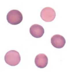
Identify the red blood cell morphology shown in the image.
a)
Spherocyte
b)
Elliptocyte
c)
Ovalocyte
d)
Stomatocyte
16.
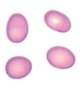
Identify the red blood cell morphology shown in the image.
a)
Spherocyte
b)
Elliptocyte
c)
Ovalocyte
d)
Stomatocyte
17.
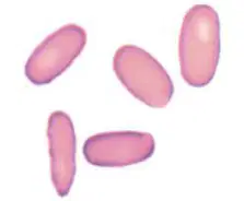
Identify the red blood cell morphology shown in the image.
a)
Spherocyte
b)
Elliptocyte
c)
Ovalocyte
d)
Stomatocyte
18.
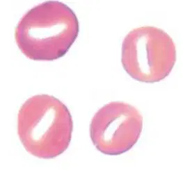
Identify the red blood cell morphology shown in the image.
a)
Spherocyte
b)
Elliptocyte
c)
Ovalocyte
d)
Stomatocyte
19.
Associated conditions: Hereditary spherocytosis, autoimmune hemolytic anemia, burns, ABO HDN, following transfusion of stored blood
a)
Spherocyte
b)
Elliptocyte or Ovalocyte
c)
Stomatocyte
d)
Sickle Cell
20.
Which of the following has a tendency to be wrongly reported if the feathered edge of a blood film is examined because the RBCs in this area lack central pallor?
a)
Spherocyte
b)
Elliptocyte or Ovalocyte
c)
Stomatocyte
d)
Sickle Cell
21.
Which of the following can result from natural RBC death (extravascular hemolysis)?
a)
Spherocyte
b)
Elliptocyte or Ovalocyte
c)
Stomatocyte
d)
Sickle Cell
22.
Associated conditions: Hereditary elliptocytosis or ovalocytosis, iron deficiency anemia, thalassemia major, myelophthisic anemias
a)
Spherocyte
b)
Elliptocyte or Ovalocyte
c)
Stomatocyte
d)
Sickle Cell
23.
Ø A.K.A. mouth cell.
a)
Spherocyte
b)
Elliptocyte or Ovalocyte
c)
Stomatocyte
d)
Sickle Cell
24.
Associated conditions: Rh deficiency syndrome, alcoholism, electrolyte imbalance, severe liver diseases, overhydrated heredit. stomatocytosis, dehydrated hereditary stomatocytosis (most common form)
a)
Spherocyte
b)
Elliptocyte or Ovalocyte
c)
Stomatocyte
d)
Sickle Cell
25.
Which of the following is an RBC with slit-like central pallor?
a)
Spherocyte
b)
Elliptocyte or Ovalocyte
c)
Stomatocyte
d)
Sickle Cell
26.
Ø Sickle or crescent-shaped RBCs.
a)
Spherocyte
b)
Elliptocyte or Ovalocyte
c)
Stomatocyte
d)
Sickle Cell
27.

Identify the red blood cell morphology shown in the image:
a)
Spherocyte
b)
Elliptocyte or Ovalocyte
c)
Stomatocyte
d)
Sickle Cell
28.
A.K.A. Drepanocyte, Meniscocyte
a)
Spherocyte
b)
Elliptocyte or Ovalocyte
c)
Stomatocyte
d)
Sickle Cell
29.
Associated conditions: Sickle cell anemia, hemoglobin SC disease, etc
a)
Spherocyte
b)
Elliptocyte or Ovalocyte
c)
Stomatocyte
d)
Sickle Cell
30.
Which of the following form of sickle cell is Crescent-shaped?
a)
Irreversible sickle cell
b)
Oat-shaped cell
31.
Which of the following form of sickle cell has long projections?
a)
Irreversible sickle cell
b)
Oat-shaped cell
32.
Which of the following form of sickle cell fragments when reoxygenated?
a)
Irreversible sickle cell
b)
Oat-shaped cell
33.
Which of the following form of sickle cell is reversible?
a)
Irreversible sickle cell
b)
Oat-shaped cell
34.
Which of the following form of sickle cell has less pronounced projections?
a)
Irreversible sickle cell
b)
Oat-shaped cell
35.
Which of the following form of sickle cell reverts to original biconcave shape when reoxygenated?
a)
Irreversible sickle cell
b)
Oat-shaped cell
36.
Hexagonal crystal of dense hemoglobin formed within the RBC membrane.
a)
Hb C crystal
b)
Hb SC crystal
c)
Codocyte
d)
Schizocyte
37.
Which of the following has a bar of gold appearance?
a)
Hb C crystal
b)
Hb SC crystal
c)
Codocyte
d)
Schizocyte
38.
Identify the red blood cell morphology shown in the image:
a)
Hb C crystal
b)
Hb SC crystal
c)
Codocyte
d)
Schizocyte
39.
Associated condition: Hb C disease
a)
Hb C crystal
b)
Hb SC crystal
c)
Codocyte
d)
Schizocyte
40.
Associated condition: Hb SC disease
a)
Hb C crystal
b)
Hb SC crystal
c)
Codocyte
d)
Schizocyte
41.
Fingerlike or quartzlike crystal of dense hemoglobin protruding from the RBC membrane
a)
Hb C crystal
b)
Hb SC crystal
c)
Codocyte
d)
Schizocyte
42.
Which of the following has a washington monument appearance?
a)
Hb C crystal
b)
Hb SC crystal
c)
Codocyte
d)
Schizocyte
43.
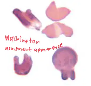
Identify the red blood cell morphology shown in the image:
a)
Hb C crystal
b)
Hb SC crystal
c)
Codocyte
d)
Schizocyte
44.
RBCs that show a centrally stained area with a thin outer rim of hemoglobin.
a)
Hb C crystal
b)
Hb SC crystal
c)
Codocyte
d)
Schizocyte
45.
A.K.A. Leptocytes, Platycyte, Greek Helmet cell, Mexican hat cell, Bull’s eye cell, Target cell
a)
Hb C crystal
b)
Hb SC crystal
c)
Codocyte
d)
Schizocyte
46.
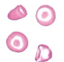
Identify the red blood cell morphology shown in the image:
a)
Hb C crystal
b)
Hb SC crystal
c)
Codocyte
d)
Schizocyte
47.
Associated conditions: Liver disease, some hemoglobinopathies, Thalassemia, LCAT deficiency
a)
Hb C crystal
b)
Hb SC crystal
c)
Codocyte
d)
Schizocyte
48.
Fragmented RBC caused by rupture in the peripheral circulation
a)
Hb C crystal
b)
Hb SC crystal
c)
Codocyte
d)
Schizocyte
49.
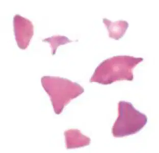
Identify the red blood cell morphology shown in the image:
a)
Hb C crystal
b)
Hb SC crystal
c)
Codocyte
d)
Schizocyte
50.
Associated conditions: Microangiopathic hemolytic anemia (along with microspherocytes), Macroangiopathic hemolytic anemia, extensive burns; patients with artificial heart valves, uremia.
a)
Hb C crystal
b)
Hb SC crystal
c)
Codocyte
d)
Schizocyte
51.
MAHA (Microangiopathic hemolytic anemias): Ø Group of disorders characterized by RBC fragmentation and thrombocytopenia. Ø Major MAHAs: TTP (Thrombotic thrombocytopenic purpura), HELLP syndrome (Hemolysis, elevated liver enzymes, low platelet count), HUS (Hemolytic uremic syndrome), DIC
a)
Hb C crystal
b)
Hb SC crystal
c)
Codocyte
d)
Schizocyte
52.
RBC fragment that is shaped like a helmet.
a)
Keratocyte
b)
Acanthocyte
c)
Echinocyte
d)
Dacryocyte
53.
Same conditions associated with schistocytes.
a)
Keratocyte
b)
Acanthocyte
c)
Echinocyte
d)
Dacryocyte
54.
Small, dense RBC with few irregularly spaced projections of different lengths.
a)
Keratocyte
b)
Acanthocyte
c)
Echinocyte
d)
Dacryocyte
55.
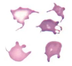
Identify the red blood cell morphology shown in the image:
a)
Keratocyte
b)
Acanthocyte
c)
Echinocyte
d)
Dacryocyte
56.
Associated conditions: Severe liver disease (Spur cell anemia), Neuroacanthocytosis (Abetalipoproteinemia or BassenKornzweig Syndrome, McLeod syndrome).
a)
Keratocyte
b)
Acanthocyte
c)
Echinocyte
d)
Dacryocyte
57.
RBC with blunt or pointed, short projections that are usually evenly spaced over the surface of the cell. (should be present in all fields of blood film).
a)
Keratocyte
b)
Acanthocyte
c)
Echinocyte
d)
Dacryocyte
58.
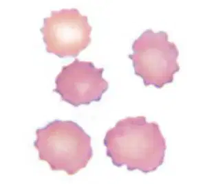
Identify the red blood cell morphology shown in the image:
a)
Keratocyte
b)
Acanthocyte
c)
Echinocyte
d)
Dacryocyte
59.
A.K.A. Burr cell
a)
Keratocyte
b)
Acanthocyte
c)
Echinocyte
d)
Dacryocyte
60.
Associated conditions: Uremia, Pyruvate Kinase deficiency
a)
Keratocyte
b)
Acanthocyte
c)
Echinocyte
d)
Dacryocyte
61.
When these cells are not evenly distributed in a blood film or not present in all fields, it is more likely to be a “drying artifact”. These artifacts are sometimes called “crenated RBCs”.
a)
Keratocyte
b)
Acanthocyte
c)
Echinocyte
d)
Dacryocyte
62.
RBC with a single pointed extension resembling a teardrop or pear.
a)
Keratocyte
b)
Acanthocyte
c)
Echinocyte
d)
Dacryocyte
63.
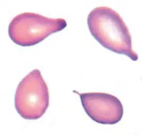
Identify the red blood cell morphology shown in the image:
a)
Keratocyte
b)
Acanthocyte
c)
Echinocyte
d)
Dacryocyte
64.
Associated conditions: Primary myelofibrosis, myelophthisic anemia, thalassemia, megaloblastic anemia.
a)
Keratocyte
b)
Acanthocyte
c)
Echinocyte
d)
Dacryocyte
65.
Which of the following inclusion bodies contains Precipitated RNA?
a)
Basophilic stippling
b)
Diffuse basophilia
c)
Howell-Jolly body
d)
Heinz body
66.
Associated with: Lead poisoning Thalassemia Hemoglobinopathy Megaloblastic anemia Myelodysplastic syndrome
a)
Basophilic stippling
b)
Diffuse basophilia
c)
Howell-Jolly body
d)
Heinz body
67.
Which of the following inclusion bodies contains RNA?
a)
Basophilic stippling
b)
Diffuse basophilia
c)
Howell-Jolly body
d)
Heinz body
68.
Associated with: Hemolytic anemia, post-treatment for iron, vitamin B12, or folate deficiency
a)
Basophilic stippling
b)
Diffuse basophilia
c)
Howell-Jolly body
d)
Heinz body
69.
Which of the following inclusion bodies contains DNA (Nuclear fragment)?
a)
Basophilic stippling
b)
Diffuse basophilia
c)
Howell-Jolly body
d)
Heinz body
70.
Associated with: Hyposplenism, Postsplenectomy, Megaloblastic anemia, Hemolytic anemia, Thalassemia, Myelodysplastic syndrome
a)
Basophilic stippling
b)
Diffuse basophilia
c)
Howell-Jolly body
d)
Heinz body
71.
Which of the following inclusion bodies contains Denatured Hgb?
a)
Basophilic stippling
b)
Diffuse basophilia
c)
Howell-Jolly body
d)
Heinz body
72.
Associated with: G6PD deficiency, Unstable Hgb, Oxidant drugs/chemicals
a)
Basophilic stippling
b)
Diffuse basophilia
c)
Howell-Jolly body
d)
Heinz body
73.
Which of the following inclusion bodies contains Iron?
a)
Pappenheimer bodies
b)
Cabot ring
c)
Hemoglobin H
74.
Associated with: Sideroblastic anemia, Hemoglobinopathies, Thalassemia, Megaloblastic anemia, Myelodysplastic syndrome, Hyposplenism, Postsplenectomy
a)
Pappenheimer bodies
b)
Cabot ring
c)
Hemoglobin H
75.
Which of the following inclusion bodies contains Mitotic spindle remnant?
a)
Pappenheimer bodies
b)
Cabot ring
c)
Hemoglobin H
76.
Associated with: Megaloblastic anemia, Myelodysplastic syndromes
a)
Pappenheimer bodies
b)
Cabot ring
c)
Hemoglobin H
77.
Associated with: Hb H disease
a)
Pappenheimer bodies
b)
Cabot ring
c)
Hemoglobin H
78.
Which of the following inclusion bodies contains Precipitated βglobin chains of hemoglobin?
a)
Pappenheimer bodies
b)
Cabot ring
c)
Hemoglobin H
79.
What does basophilic stippling contain?
a)
Precipitated RNA
b)
RNA only
c)
DNA (nuclear fragment)
d)
Denatured Hgb
80.
What does diffuse basophilia contain?
a)
Precipitated RNA
b)
RNA only
c)
DNA (nuclear fragment)
d)
Denatured Hgb
81.
What does howell-jolly body contain?
a)
Precipitated RNA
b)
RNA only
c)
DNA (nuclear fragment)
d)
Denatured Hgb
82.
What does heinz body contain?
a)
Precipitated RNA
b)
RNA only
c)
DNA (nuclear fragment)
d)
Denatured Hgb
83.
What does Pappenheimer bodies contain?
a)
Iron
b)
Mitotic spindle remnant
c)
Precipitated Beta globin chains of hemoglobin
84.
What does Cabot ring contain?
a)
Iron
b)
Mitotic spindle remnant
c)
Precipitated Beta globin chains of hemoglobin
85.
What does Hemoglobin H contain?
a)
Iron
b)
Mitotic spindle remnant
c)
Precipitated Beta globin chains of hemoglobin
86.
Ø Refers to the production of erythroid precursor cells that are defective
a)
Ineffective erythropoiesis
b)
Insufficient erythropoiesis
87.
Examples: Megaloblastic anemia, thalassemia, sideroblastic anemia.
a)
Ineffective erythropoiesis
b)
Insufficient erythropoiesis
88.
Refers to a decrease in the number of erythroid precursors in the bone marrow, resulting in decreased RBC production
a)
Ineffective erythropoiesis
b)
Insufficient erythropoiesis
89.
Examples: EPO deficiency, iron deficiency, aplastic anemia, acute pure red cell aplasia, infection.
a)
Ineffective erythropoiesis
b)
Insufficient erythropoiesis
90.
Ø Aplastic anemia
a)
Normal/Decreased Reticulocyte count:
b)
Increased Reticulocyte count:
91.
Ø Kidney disease
a)
Normal/Decreased Reticulocyte count:
b)
Increased Reticulocyte count:
92.
Ø Acute blood loss
a)
Normal/Decreased Reticulocyte count:
b)
Increased Reticulocyte count:
93.
PNH
a)
Normal/Decreased Reticulocyte count:
b)
Increased Reticulocyte count:
94.
PCH
a)
Normal/Decreased Reticulocyte count:
b)
Increased Reticulocyte count:
95.
SCD
a)
Normal/Decreased Reticulocyte count:
b)
Increased Reticulocyte count:
96.
Ø Enzyme deficiency
a)
Normal/Decreased Reticulocyte count:
b)
Increased Reticulocyte count:
97.
Ø Other hemolytic anemia
a)
Normal/Decreased Reticulocyte count:
b)
Increased Reticulocyte count:
98.
Rare but potentially deadly bone marrow failure syndrome
a)
Aplastic anemia
b)
Fanconi anemia
c)
Paroxysmal Nocturnal Hemoglobinuria
d)
Paroxysmal Cold Hemoglobinuria
e)
Anti-P autoantibody
99.
Characteristics: Ø Pancytopenia (markedly decreased number of all blood cells)
Reticulocytopenia (inability to produce effective reticulocytes)
Ø Bone marrow hypocellularity
Ø HSC depletion
a)
Aplastic anemia
b)
Fanconi anemia
c)
Paroxysmal Nocturnal Hemoglobinuria
d)
Paroxysmal Cold Hemoglobinuria
e)
Anti-P autoantibody
100.
An inherited associated disease of Aplastic anemia that is a member of Telomere biology disorders (TBD), a group of diseases caused by germline mutations that affect genomic stability, resulting in premature aging.
a)
Dyskeratosis congenita
b)
Schwachman-Bodian-Diamond syndrome
c)
Fanconi anemia
d)
Paroxysmal Nocturnal Hemoglobinuria
e)
Paroxysmal Cold Hemoglobinura
101.
What is the drug most frequently implicated in acquired aplastic anemia?
a)
Phenytoin
b)
Valproic acid
c)
Aminoglycoside
d)
Chloramphenicol
102.
An inherited associated disease of Aplastic anemia that clinically presents with a triad of abnormal skin presentation, dystrophic nails, and oral leukoplakia.
a)
Dyskeratosis congenita
b)
Schwachman-Bodian-Diamond syndrome
c)
Fanconi anemia
d)
Paroxysmal Nocturnal Hemoglobinuria
e)
Paroxysmal Cold Hemoglobinura
103.
Characterized by pancreatic insufficiency, cytopenia, skeletal abnormalities and predisposition for hematologic abnormalities
a)
Dyskeratosis congenita
b)
Schwachman-Bodian-Diamond syndrome
c)
Fanconi anemia
d)
Paroxysmal Nocturnal Hemoglobinuria
e)
Paroxysmal Cold Hemoglobinura
104.
The most common of the inherited bone marrow failure syndromes
a)
Dyskeratosis congenita
b)
Schwachman-Bodian-Diamond syndrome
c)
Fanconi anemia
d)
Paroxysmal Nocturnal Hemoglobinuria
e)
Paroxysmal Cold Hemoglobinura
105.
Physical presentations: skeletal abnormalities (thumb malformations, radial hypoplasia, microcephaly, hip dislocation, and scoliosis); skin pigmentation (hyperpigmentation, hypopigmentation, café-aulait lesions); short stature; and abnormalities of the eyes, kidneys, and genitals.
a)
Dyskeratosis congenita
b)
Schwachman-Bodian-Diamond syndrome
c)
Fanconi anemia
d)
Paroxysmal Nocturnal Hemoglobinuria
e)
Paroxysmal Cold Hemoglobinura
106.
Caused by the deficiency of CD55 (Decay-accelerating factor, DAF), and CD59 (Membrane inhibitor or reactive lysis, MIRL).
a)
Dyskeratosis congenita
b)
Schwachman-Bodian-Diamond syndrome
c)
Fanconi anemia
d)
Paroxysmal Nocturnal Hemoglobinuria
e)
Paroxysmal Cold Hemoglobinura
107.
A.k.a. Marchiafava-Micheli syndrome
a)
Dyskeratosis congenita
b)
Schwachman-Bodian-Diamond syndrome
c)
Fanconi anemia
d)
Paroxysmal Nocturnal Hemoglobinuria
e)
Paroxysmal Cold Hemoglobinura
108.
Acute form of cold-reactive hemolytic anemia.
a)
Dyskeratosis congenita
b)
Schwachman-Bodian-Diamond syndrome
c)
Fanconi anemia
d)
Paroxysmal Nocturnal Hemoglobinuria
e)
Paroxysmal Cold Hemoglobinura
109.
May be idiopathic or secondary
a)
Dyskeratosis congenita
b)
Schwachman-Bodian-Diamond syndrome
c)
Fanconi anemia
d)
Paroxysmal Nocturnal Hemoglobinuria
e)
Paroxysmal Cold Hemoglobinura
110.
Secondary form of this disease is most commonly seen in young children after a viral respiratory infection.
a)
Dyskeratosis congenita
b)
Schwachman-Bodian-Diamond syndrome
c)
Fanconi anemia
d)
Paroxysmal Nocturnal Hemoglobinuria
e)
Paroxysmal Cold Hemoglobinura
111.
A.k.a. Donath-Landsteiner antibody
a)
Anti-P autoantibody
b)
Anti-i autoantibody
c)
Anti-I autoantibody
d)
Anti-k autoantibody
112.
Anti-P autoantibody binds to the P antigen on RBCs at cold temperature leading to ____ as it react with complement C1 to C4.
a)
partial activation
b)
full activation
113.
Which type of activation of anti-P autoantibody only occurs on warming?
a)
partial activation
b)
full activation
114.
Ø Have normal MCV and MCHC.
a)
Normocytic, Normochromic
b)
Macrocytic, Normochromic
c)
Microcytic, Hypochromic
115.
Aplastic anemia
a)
Normocytic, Normochromic
b)
Macrocytic, Normochromic
c)
Microcytic, Hypochromic
116.
Fanconi anemia
a)
Normocytic, Normochromic
b)
Macrocytic, Normochromic
c)
Microcytic, Hypochromic
117.
PNH
a)
Normocytic, Normochromic
b)
Macrocytic, Normochromic
c)
Microcytic, Hypochromic
118.
PCH
a)
Normocytic, Normochromic
b)
Macrocytic, Normochromic
c)
Microcytic, Hypochromic
119.
Ø Have high MCV and normal MCHC.
a)
Normocytic, Normochromic
b)
Macrocytic, Normochromic
c)
Microcytic, Hypochromic
120.
Megaloblastic anemia
a)
Normocytic, Normochromic
b)
Macrocytic, Normochromic
c)
Microcytic, Hypochromic
121.
Nonmegaloblastic anemia
a)
Normocytic, Normochromic
b)
Macrocytic, Normochromic
c)
Microcytic, Hypochromic
122.
Ø Have low MCV and MCHC.
a)
Normocytic, Normochromic
b)
Macrocytic, Normochromic
c)
Microcytic, Hypochromic
123.
"CTAILS"
a)
Normocytic, Normochromic
b)
Macrocytic, Normochromic
c)
Microcytic, Hypochromic
124.
Sideroblastic anemia
a)
Normocytic, Normochromic
b)
Macrocytic, Normochromic
c)
Microcytic, Hypochromic
125.
Chronic blood loss
a)
Normocytic, Normochromic
b)
Macrocytic, Normochromic
c)
Microcytic, Hypochromic
126.
Lead poisoning
a)
Normocytic, Normochromic
b)
Macrocytic, Normochromic
c)
Microcytic, Hypochromic
127.
Iron Deficiency Anemia
a)
Normocytic, Normochromic
b)
Macrocytic, Normochromic
c)
Microcytic, Hypochromic
128.
Anemia of Chronic inflammation
a)
Normocytic, Normochromic
b)
Macrocytic, Normochromic
c)
Microcytic, Hypochromic
129.
Thalassemia
a)
Normocytic, Normochromic
b)
Macrocytic, Normochromic
c)
Microcytic, Hypochromic
130.
Characterized by impaired DNA synthesis caused by Vitamin B12 or folate deficiency
a)
Megaloblastic anemia
b)
Nonmegaloblastic anemia
131.
The MCV can be markedly increased (up to 150 fL), but 100- 115 fL is most common
a)
Megaloblastic anemia
b)
Nonmegaloblastic anemia
132.
Characterized by large RBCs and are typically related to membrane changes caused by disruption of the cholesterol-to-phospholipid ratio.
a)
Megaloblastic anemia
b)
Nonmegaloblastic anemia
133.
It is rare for the MCV to be >115 fL in
a)
Megaloblastic anemia
b)
Nonmegaloblastic anemia
134.
Ø Inadequate intake Ø Increased need Ø Renal dialysis
a)
Vitamin B12 deficiency
b)
Folate/folic acid deficiency (Vitamin B9 deficiency)
135.
Ø Increased need (pregnancy, lactation, growth). Ø Competition (infection with D. latum) Ø Lack of intrinsic factor (IF), aka Pernicious anemia
a)
Vitamin B12 deficiency
b)
Folate/folic acid deficiency (Vitamin B9 deficiency)
136.
has noticeable neurologic and neuropsychiatric symptoms.
a)
Vitamin B12 deficiency
b)
Folate/folic acid deficiency (Vitamin B9 deficiency)
137.
Characteristics:
ü Hypersegmented neutrophils (with >6 lobes)
ü Oval macrocytes
ü Presence of megaloblasts in the bone marrow
a)
Vitamin B12 deficiency
b)
Folate/folic acid deficiency (Vitamin B9 deficiency)
138.
The most common anemia among hospitalized patients
a)
Anemia of Chronic Inflammation
b)
Sideroblastic anemia
c)
Iron Deficiency Anemia (IDA)
139.
Associated with chronic infections
a)
Anemia of Chronic Inflammation
b)
Sideroblastic anemia
c)
Iron Deficiency Anemia (IDA)
140.
Central feature: Sideropenia in the face of abundant iron stores.
a)
Anemia of Chronic Inflammation
b)
Sideroblastic anemia
c)
Iron Deficiency Anemia (IDA)
141.
Develops when protoporphyrin production or the inclusion of iron into protoporphyrin is prevented.
a)
Anemia of Chronic Inflammation
b)
Sideroblastic anemia
c)
Iron Deficiency Anemia (IDA)
142.
Which of the following is a liver hormone that regulates iron absorption by inactivation of ferroportin?
a)
Hepcidin
b)
Lactoferrin
c)
Ferritin
143.
Which of the following competes with microorganisms for iron directly?
a)
Hepcidin
b)
Lactoferrin
c)
Ferritin
144.
Iron is trapped in mitochondria
a)
Anemia of Chronic Inflammation
b)
Sideroblastic anemia
c)
Iron Deficiency Anemia (IDA)
145.
Ø Characterized with a dimorphic peripheral blood picture.
a)
Anemia of Chronic Inflammation
b)
Sideroblastic anemia
c)
Iron Deficiency Anemia (IDA)
146.
The most common anemia
a)
Anemia of Chronic Inflammation
b)
Sideroblastic anemia
c)
Iron Deficiency Anemia (IDA)
147.
nucleated RBC precursor with cytoplasmic iron granules
a)
Sideroblast
b)
Siderocyte
148.
anucleated RBC with iron granules.
a)
Sideroblast
b)
Siderocyte
149.
(Stored iron)
a)
Hepcidin
b)
Lactoferrin
c)
Ferritin
150.
Only ferritin or stored iron is decreased.
a)
Storage Iron Depletion
b)
Transport Iron Depletion
c)
Functional Iron Depletion
151.
Ferritin and serum iron are decreased. ü Increased Total Iron Binding Capacity (TIBC). ü Hemoglobin level is normal
a)
Storage Iron Depletion
b)
Transport Iron Depletion
c)
Functional Iron Depletion
152.
Ferritin and serum iron are decreased. ü Increased Total Iron Binding Capacity (TIBC). ü Hemoglobin level is decreased
a)
Storage Iron Depletion
b)
Transport Iron Depletion
c)
Functional Iron Depletion
153.
Ferritin and serum iron in Transport Iron depletion
a)
Increased
b)
Decreased
c)
Normal
154.
Ferritin in storage iron depletion
a)
Increased
b)
Decreased
c)
Normal
155.
Hgb in functional iron depletion
a)
Increased
b)
Decreased
c)
Normal
156.
TIBC in Transport Iron depletion
a)
Increased
b)
Decreased
c)
Normal
157.
Hgb in Transport Iron depletion
a)
Increased
b)
Decreased
c)
Normal
100 %

