
 Worksheets
Chapter 3
Total questions: 98
Worksheet time: 49hrs 0mins
The major functions and structures of the skeletal system include:
Support, protection, movement, blood cell production, and mineral storage
Digestion, absorption, and excretion
Hormone production and regulation
Transmission of nerve impulses
By the end of the chapter, you should be able to: Identify three types of joints.
Fibrous, cartilaginous, and synovial joints
Hinge, ball-and-socket, and pivot joints
Elastic, rigid, and flexible joints
Rotary, sliding, and fixed joints
The axial and appendicular skeletons are differentiated by:
The axial skeleton forms the central axis of the body, while the appendicular skeleton consists of the limbs and girdles.
The axial skeleton includes the limbs, while the appendicular skeleton forms the skull and vertebral column.
Both skeletons consist only of the limbs and girdles.
The appendicular skeleton forms the central axis, while the axial skeleton consists of the limbs.
The medical specialists who treat disorders of the skeletal system are called:
Cardiologists
Orthopedists
Dermatologists
Neurologists
By the end of the chapter, you should be able to: Recognize, define, spell, and pronounce the primary terms related to the pathology and the diagnostic and treatment procedures of the skeletal system.
Recognize, define, spell, and pronounce the primary terms related to the pathology and the diagnostic and treatment procedures of the skeletal system.
List the bones of the skeletal system only.
Describe only the treatment procedures of the muscular system.
Identify only the spelling of skeletal system terms.
What is the framework of the body that supports and protects the internal organs?
Bones
Muscles
Skin
Blood
What structures work in conjunction with muscles, ligaments, and tendons to make possible the wide variety of body movements?
Joints
Veins
Nerves
Glands
Which mineral is required for normal nerve and muscle function and is stored in bones?
Calcium
Iron
Potassium
Zinc
What plays an important role in the formation of blood cells and is located within spongy bone?
Red bone marrow
Yellow bone marrow
Compact bone
Periosteum
Which of the following is NOT a function of bones?
Act as the framework of the body
Support and protect the internal organs
Produce hormones
Store calcium
Joints make possible the wide variety of body movements.
True
False
Red bone marrow: Where is red bone marrow located?
Within compact bone
Within spongy bone
In the skin
In the muscles
According to the information on the formation of bones, what does a newborn's skeleton begin as?
Hard bone
Fragile membranes and cartilage
Muscle tissue
Ligaments
What is the process called that starts turning membranes and cartilage into bone and continues through adolescence?
Ossification
Calcification
Myelination
Condensation
Which type of cells break down old or damaged bone after growth is complete?
Osteoblast cells
Osteoclast cells
Red blood cells
White blood cells
Which type of cells help rebuild the bone after growth is complete?
Osteoclast cells
Osteoblast cells
Platelet cells
Muscle cells
Ossification repairs minor damage from normal activity and also repairs bones after injuries, such as fractures.
True
False
What is the second hardest tissue in the human body?
Bone
Muscle
Cartilage
Skin
Which type of bone is also known as cortical bone?
Compact bone
Spongy bone
Cartilage
Red bone marrow
Which of the following is true about compact bone?
It is porous and light
It forms the protective outer layer of bones
It commonly contains red bone marrow
It is weaker than spongy bone
Spongy bone is also known as ______ bone.
cancellous
compact
medullary
cartilaginous
Which of the following bones are considered long bones?
A) Femur and humerus
B) Wrist and ankle
C) Skull and ribs
D) Vertebrae and pelvis
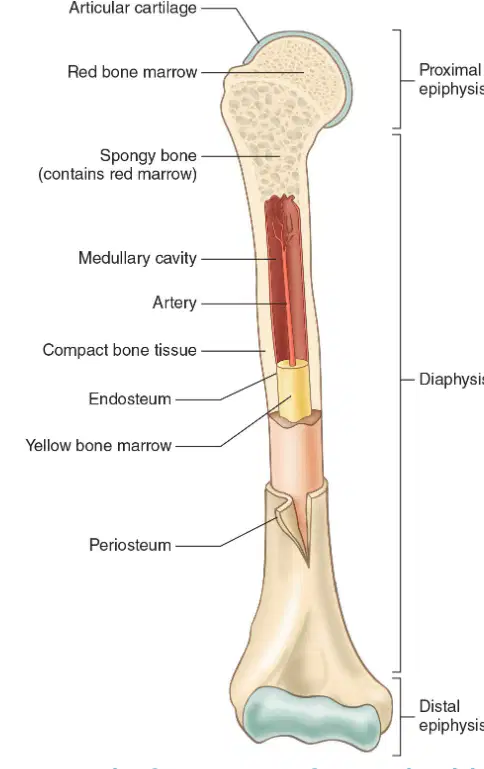
Which part of the long bone contains red bone marrow?
Spongy bone
Compact bone
Medullary cavity
Articular cartilage
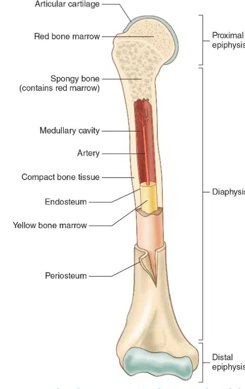
What is the function of the compact bone tissue in a long bone?
Forms the protective outer layer of bones
Produces red blood cells
Stores fat in the bone marrow
Connects muscles to bones
What is the central cavity located in the shaft of long bones and surrounded by compact bone called?
Medullary cavity
Epiphyseal plate
Periosteum
Articular cartilage
What type of tissue lines the medullary cavity?
Endosteum
Periosteum
Cartilage
Epithelium
Red bone marrow is a hematopoietic tissue.
True
False
Where is red bone marrow located?
Within the spongy bone
Inside the yellow bone marrow
On the surface of compact bone
In the periosteum
Which of the following does red bone marrow manufacture?
Red blood cells
Hemoglobin
White blood cells
Thrombocytes
All of the above
Yellow bone marrow functions as a fat storage area.
True
False
What type of tissue is described as smooth, rubbery, and blue-white, and acts as a shock absorber between bones?
Muscle
Cartilage
Ligament
Tendon
Cartilage makes up the flexible parts of the skeleton, such as the ______ and tip of the nose.
outer ear
shin bone
skull
elbow
Which of the following is NOT a function of articular cartilage?
Covers the surfaces of bones where they articulate
Makes smooth joint movement possible
Protects the bones from rubbing against each other
Produces red blood cells
The meniscus is a curved, fibrous cartilage found in some joints.
True
False
Give two examples of joints where the meniscus can be found.
Knee and temporomandibular joint of the jaw
Shoulder and elbow joint
Hip and ankle joint
Wrist and sacroiliac joint
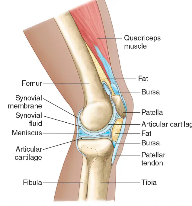
Label the following parts in the diagram of the knee:
Femur, Patella, Tibia, Fibula, Meniscus, Articular cartilage, Synovial membrane, Synovial fluid, Bursa, Fat, Patellar tendon, Quadriceps muscle (all are labeled in the diagram)
Femur, Humerus, Radius, Ulna, Meniscus, Articular cartilage, Synovial membrane, Synovial fluid, Bursa, Fat, Patellar tendon, Quadriceps muscle
Femur, Patella, Tibia, Fibula, Meniscus, Articular cartilage, Synovial membrane, Synovial fluid, Bursa, Fat, Achilles tendon, Biceps muscle
Femur, Patella, Tibia, Fibula, Meniscus, Articular cartilage, Synovial membrane, Synovial fluid, Bursa, Fat, Patellar tendon, Triceps muscle
What is the shaft of a long bone called?
Diaphysis
Epiphysis
Metaphysis
Periosteum
What are the wider ends of long bones such as the femurs of the legs called?
Epiphyses
Diaphyses
Metaphyses
Periosteum
The end of the bone located nearest to the midline of the body is called the ________.
Proximal epiphysis
Distal epiphysis
Diaphysis
Metaphysis
The end of the bone located farthest away from the midline of the body is called the ________.
Distal epiphysis
Proximal epiphysis
Medullary cavity
Diaphysis
What is an opening through which blood vessels, nerves, and ligaments pass called?
Foramen
Condyle
Tubercle
Fossa
Which of the following is an example of a foramen?
Mastoid process
Foramen magnum
Diaphysis
Epiphysis
A normal projection on the surface of a bone that most commonly serves as an attachment for a muscle or tendon is called a ________.
Process
Fossa
Foramen
Facet
Which of the following is an example of a process?
Foramen magnum
Mastoid process
Diaphysis
Epiphysis
Joints are also known as ________.
articulations
ligaments
cartilages
tendons
What is the place of union between two or more bones called?
Joint
Muscle
Ligament
Tendon
Joints are classified by construction or based on the degree of ________ they allow.
movement
pressure
temperature
color
Which type of joints hold the bones tightly together?
Fibrous joints
Cartilaginous joints
Synovial joints
Ball and socket joints
Fibrous joints consist of inflexible layers of ________ connective tissue.
dense
loose
elastic
reticular
In adults, fibrous joints are known as ________ and allow little or no movement.
sutures
synovial joints
cartilaginous joints
ligaments
In newborns and very young children, fibrous joints are called ________ or soft spots.
fontanelles
sutures
epiphyses
meninges
What type of joints allow only slight movement and consist of bones connected entirely by cartilage?
Cartilaginous joints
Fibrous joints
Synovial joints
Ball-and-socket joints
Where do the ribs connect to the sternum?
Breastbone
Collarbone
Shoulder blade
Pelvis
What is the function of the pubic symphysis during childbirth?
Allows some movement to facilitate childbirth
Produces hormones to induce labor
Prevents any movement of the pelvis
Connects the uterus to the pelvic wall
Where is the pubic symphysis located?
Between the pubic bones in the anterior (front) of the pelvis
At the base of the skull
Between the shoulder blades
At the back of the knee
Which of the following is NOT a function of cartilaginous joints?
A) Allow only slight movement
B) Consist of bones connected entirely by cartilage
C) Allow free movement
D) Are found in the pubic symphysis
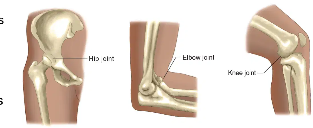
Which of the following is an example of a ball-and-socket joint?
Knee
Elbow
Hip
Wrist
Which type of synovial joint allows a wide range of movement in many directions?
Hinge joint
Ball-and-socket joint
Pivot joint
Saddle joint
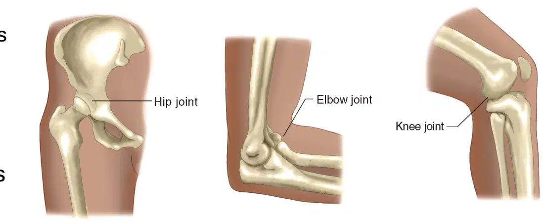
Fill in the blank: The elbow and knee are examples of ______ joints.
hinge
ball-and-socket
pivot
gliding
Which of the following is NOT a component of synovial joints?
Synovial capsule
Synovial membrane
Synovial fluid
Cartilage disc
Ball-and-socket joints allow movement in many directions.
True
False
Which of the following is a function of the synovial fluid in synovial joints?
Provides structure
Lubricates the joint
Connects bones
Produces blood cells
How many bones does a typical adult skeleton consist of?
100
206
300
80
The human skeleton is divided into which two main parts?
Axial and Appendicular
Cranial and Facial
Upper and Lower
Anterior and Posterior
What is the main function of the axial skeleton?
Protects major organs of the nervous, respiratory, and circulatory systems.
Facilitates movement of the limbs.
Stores minerals and fat.
Produces red blood cells.
The 80 bones of the head and body are organized into five parts. Name any two of these parts.
Bones of the skull and Rib cage
Femur and Tibia
Clavicle and Scapula
Pelvis and Patella
Which bone is NOT part of the axial skeleton?
Skull
Rib cage
Femur
Vertebral column
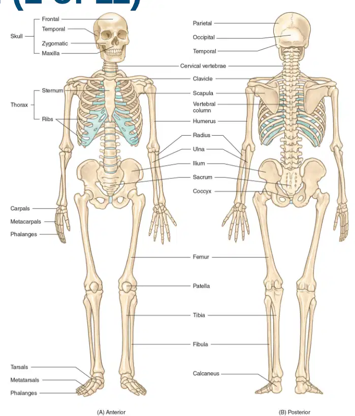
Refer to the diagram. Which bone is labeled as 'Clavicle' in the anterior view of the human skeleton?
The bone connecting the sternum to the shoulder (collarbone) is labeled as 'Clavicle'.
The bone forming the lower jaw is labeled as 'Clavicle'.
The bone forming the upper arm is labeled as 'Clavicle'.
The bone forming the thigh is labeled as 'Clavicle'.
The vertebral column is part of the axial skeleton.
True
False
What is the main function of the appendicular skeleton?
Protects the brain
Makes body movement possible
Produces blood cells
Stores minerals
The appendicular skeleton protects the organs of ________, excretion, and reproduction.
digestion
respiration
circulation
vision
How many bones does the appendicular skeleton consist of?
80
126
206
64
Which of the following are included in the upper extremities of the appendicular skeleton?
Hips, thighs, legs, ankles, and feet
Shoulders, arms, forearms, wrists, and hands
Skull and spine
Ribs and sternum
Lower extremities of the appendicular skeleton include: hips, thighs, legs, ankles, and ________.
feet
hands
shoulders
wrists
'Appendicular' means referring to an ________.
appendage
organ
cell
tissue
An appendage is anything that is attached to a major part of the body.
True
False
An extremity is the terminal end of a body part such as an arm or leg.
True
False
Which of the following are bones of the cranium?
Frontal, parietal, occipital, external auditory meatus, sphenoid, and ethmoid
Nasal, zygomatic, maxillary, palatine, lacrimal, inferior conchae, vomer, and mandible
Malleus, incus, and stapes
Radius, ulna, and humerus
What is the function of the bones of the cranium?
Enclose and protect the brain
Aid in digestion of food
Pump blood throughout the body
Facilitate movement of limbs
Which bones are known as the auditory ossicles?
Frontal, parietal, occipital
Malleus, incus, and stapes
Nasal, zygomatic, maxillary
Mandible, vomer, palatine
How many tiny bones are located in the middle ear?
Three
Two
Four
Five
Which of the following are bones of the face?
Frontal, parietal, occipital
Nasal, zygomatic, maxillary, palatine, lacrimal, inferior conchae, vomer, and mandible
Malleus, incus, and stapes
Sphenoid, ethmoid

Label the following parts on the anterior view of the adult human skull diagram:
Frontal bone (forehead), Parietal bone (upper side), Sphenoid bone (side near eye), Nasal bone (bridge of nose), Zygomatic bone (cheek), Maxillary bone (upper jaw), Mandible (lower jaw)
Temporal bone (forehead), Occipital bone (upper side), Ethmoid bone (side near eye), Lacrimal bone (bridge of nose), Palatine bone (cheek), Vomer bone (upper jaw), Hyoid bone (lower jaw)
Parietal bone (forehead), Frontal bone (upper side), Sphenoid bone (side near ear), Nasal bone (side of nose), Zygomatic bone (chin), Maxillary bone (lower jaw), Mandible (upper jaw)
Frontal bone (chin), Parietal bone (lower side), Sphenoid bone (side near mouth), Nasal bone (tip of nose), Zygomatic bone (forehead), Maxillary bone (cheek), Mandible (upper jaw)
What is the thoracic cavity also called?
Rib cage
Abdominal wall
Pelvic girdle
Cranial vault
What does the thoracic cavity consist of?
Ribs and sternum
Femur and tibia
Pelvis and sacrum
Scapula and clavicle
What is the function of the thoracic cavity?
It is a bony structure that protects the heart and lungs.
It produces digestive enzymes for the stomach.
It stores excess nutrients for the body.
It filters waste products from the blood.
How many pairs of ribs are there in the human body?
12 pairs
10 pairs
14 pairs
8 pairs
The first seven pairs of ribs are called true ribs and are attached anteriorly to the sternum.
True
False
Which ribs are called false ribs?
First seven pairs
Next three pairs
Last two pairs
Floating ribs are only attached posteriorly to the ______ but are not attached anteriorly.
vertebrae
sternum
clavicle
scapula
What is another name for the sternum?
Breast bone
Collar bone
Shin bone
Hip bone
Which part of the sternum forms the upper portion?
Manubrium
Body
Xiphoid process
Clavicle
The xiphoid process is cartilage that forms the ______ portion of the sternum.
lower
upper
middle
lateral
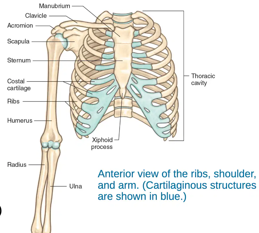
What is the common name for the clavicle?
Collarbone
Shinbone
Shoulder blade
Kneecap
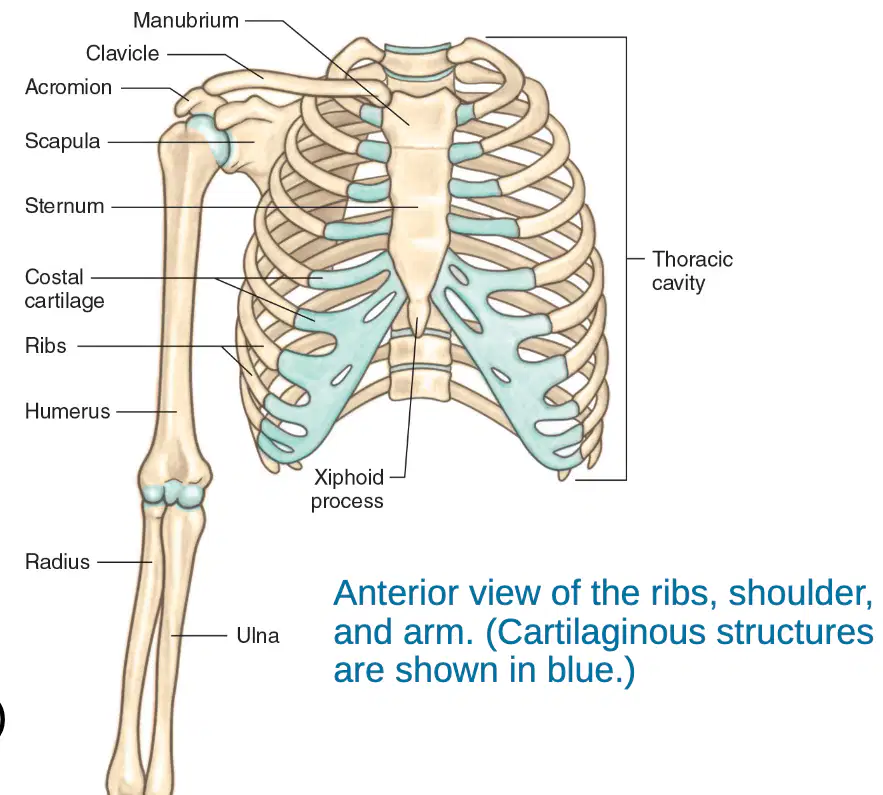
What is the bone of the upper arm called?
Humerus
Femur
Tibia
Radius

Which bone is known as the shoulder blade?
Scapula
Femur
Tibia
Clavicle

What is the extension of the scapula called?
Acromion
Olecranon
Coracoid process
Glenoid cavity
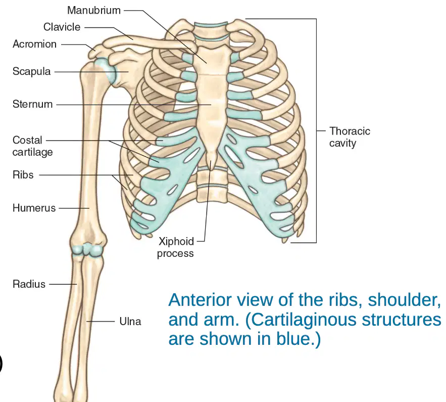
Which bone is the smaller, shorter forearm bone?
Radius
Ulna
Femur
Tibia

