Worksheets
The Skull
Total questions: 55
Worksheet time: 34mins
Which of the following statements is true about the ossification of cranial sutures?
All sutures close before birth
All sutures close during early childhood
Some sutures remain open through childhood and adolescence
Sutures never close
Explain why some cranial sutures remain patent (open) through childhood and adolescence, and discuss the significance of this process for skull development.
To allow for brain growth and skull expansion during development
To prevent the skull from becoming too heavy
To allow for easier movement of the head
To reduce the risk of head injuries
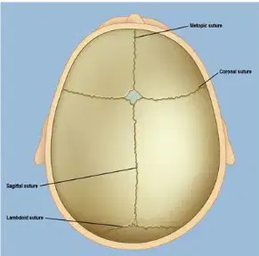
What are sutures in the context of the human skull?
Fibrous joints between skull bones
Spaces where multiple bones meet but do not fuse
Cartilage connecting the ribs to the sternum
Muscles that move the jaw
What happens to fontanelles over time after birth?
They close and ossify
They become larger
They turn into muscles
They remain open throughout life
What is the term for the condition in which a skull suture fuses prematurely, restricting skull growth in that direction?
Osteoporosis
Craniosynostosis
Scoliosis
Hydrocephalus
A child presents with abnormal skull shape and increased intracranial pressure. Which underlying process is most likely responsible for these symptoms?
Delayed tooth eruption
Premature fusion of a cranial suture (craniosynostosis)
Excessive calcium intake
Overexposure to sunlight
Explain how compensatory growth occurs in the skull when a suture fuses prematurely, and discuss the potential consequences.
Growth stops completely, leading to a smaller head size.
Growth occurs in other directions, leading to abnormal skull shapes and possibly increased intracranial pressure.
The skull becomes softer and more flexible.
The brain stops developing.
A student is asked to identify the part of the skull that forms the braincase. Which structure should they select?
Viscerocranium
Mandible
Neurocranium
Zygomatic bone
Which group of bones in the human skull is also known as the braincase?
Neurocranium
Viscerocranium
Auditory ossicles
Mandible
Explain the difference between the neurocranium and the viscerocranium in terms of their function and number of bones.
The neurocranium (braincase) protects the brain and has 8 bones, while the viscerocranium (facial skeleton) forms the face and has 14 bones.
The neurocranium forms the face and has 14 bones, while the viscerocranium protects the brain and has 8 bones.
Both the neurocranium and viscerocranium have 22 bones each.
The neurocranium and viscerocranium are the same and have no functional difference.
What is one of the main functions of the frontal bone?
Protects the frontal lobes of the brain
Supports the jaw
Forms the lower part of the nose
Houses the inner ear
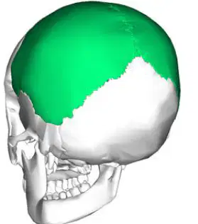
Which bone forms the sides and roof of the cranial vault?
Parietal bone
Temporal bone
Occipital bone
Frontal bone
Which structures does the occipital bone support?
Brainstem and cerebellum
Frontal lobe and parietal lobe
Temporal lobe and occipital lobe
Mandible and maxilla
Which nearby structure can be affected by pituitary tumors or sphenoid sinus infections?
Optic chiasm
Mandible
Zygomatic arch
Nasal septum
A patient presents with vision problems and a history of sphenoid sinus infection. Using your knowledge of the sphenoid bone, which structure is most likely affected and why?
Optic chiasm, because it is located near the sphenoid bone and can be impacted by infections or tumors in this area
Mandible, because it is directly attached to the sphenoid bone
Temporal bone, because it is the largest bone in the skull
Nasal septum, because it is the closest structure to the sphenoid bone
Where is the ethmoid bone located?
Between the orbits, forming part of the anterior cranial fossa, roof of nasal cavity, medial walls of orbit, and superior portion of nasal septum
At the base of the skull, forming the lower jaw
In the cheek, forming the zygomatic arch
At the back of the head, forming the occipital region
Why can a fracture of the ethmoid bone, especially the lamina papyracea, be dangerous?
It can lead to orbital complications and cerebrospinal fluid leaks.
It can cause paralysis of the facial muscles.
It can result in loss of taste.
It can cause permanent deafness.
What is the function of the alveolar process of the maxilla?
Forms the lower jaw
Houses the upper teeth
Supports the nasal septum
Forms the cheekbone
Which sinus is contained within the maxilla and is the largest paranasal sinus?
Frontal sinus
Sphenoidal sinus
Maxillary sinus
Ethmoidal sinus
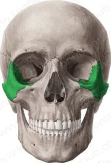
Which bones are highlighted in green in the diagram?
Zygomatic bones
Mandible bones
Nasal bones
Parietal bones
Which bones form the bridge of the nose?
Nasal bones
Zygomatic bones
Maxilla bones
Mandible bones
How many surfaces does each nasal bone have?
Two
Three
Four
One
Which of the following best describes the location of the lacrimal bones?
In the medial wall of each orbit
In the lower jaw
At the back of the skull
In the cheekbone
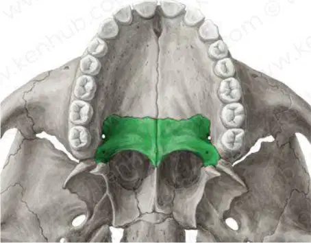
Which of the following best describes the shape and location of the palatine bones?
L-shaped paired bones tucked between the maxillae and the pterygoid process of sphenoid
U-shaped single bone at the base of the skull
Flat bone forming the forehead
Irregular bone forming the lower jaw
Which bone forms the inferior part of the nasal septum?
Vomer
Maxilla
Zygomatic
Frontal
What is the primary function of the vomer bone in the nasal cavity?
Divides left and right nasal airspaces
Supports the jaw
Forms the roof of the mouth
Protects the eyes
Which of the following is a component of the superior nasal septum?
Perpendicular plate of ethmoid
Mandible
Palatine bone
Inferior nasal concha
Which bone forms the posterior part of the hard palate?
Ethmoid bone
Vomer
Maxilla
Palatine bone
What is the main function of the zygomatic bones?
Divide the nasal cavity
Form the prominence of the cheeks and part of the lateral wall of the orbit
Support the lower teeth
Protect the brainstem
Which of the following bones contributes to the formation of the orbit, nasal cavity, and the roof of the mouth?
Occipital bone
Palatine bone
Parietal bone
Mandible
Which bone forms the posterior part of the hard palate and a portion of the floor of the nasal cavity?
Frontal bone
Vomer
Palatine bone
Maxilla
What is the main function of the vomer bone in the human skull?
Protects the brainstem
Forms part of the nasal septum
Supports the upper teeth
Forms the lower jaw
Which of the following bones is NOT a part of the orbit?
Maxilla
Ethmoid bone
Zygomatic bone
Mandible
Which bone forms the lower jaw and holds the lower teeth?
Palatine bone
Maxilla
Mandible
Zygomatic bone
What is the main function of cranial sutures in the human skull?
Support the nasal cavity
Produce cerebrospinal fluid
Allow movement between skull bones during growth
Anchor facial muscles
Which bone contributes to the formation of the medial wall of the orbit and contains air cells that form part of the paranasal sinuses?
Ethmoid bone
Occipital bone
Frontal bone
Temporal bone
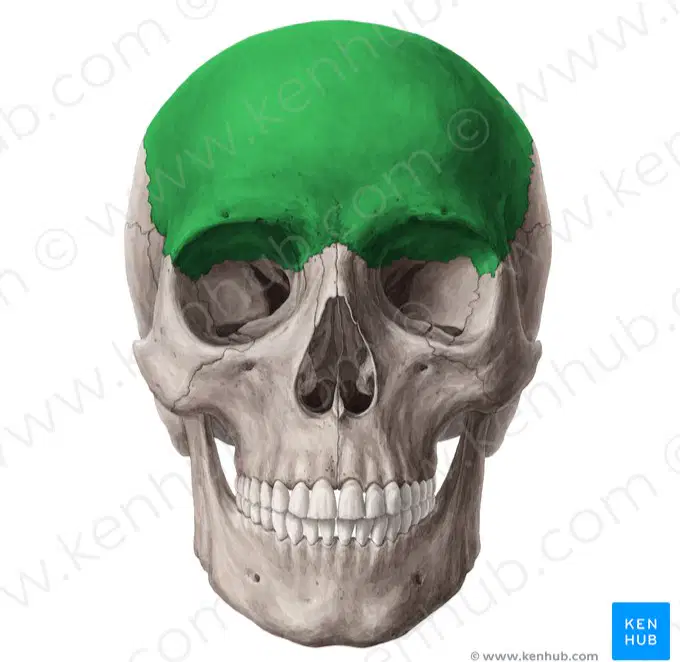
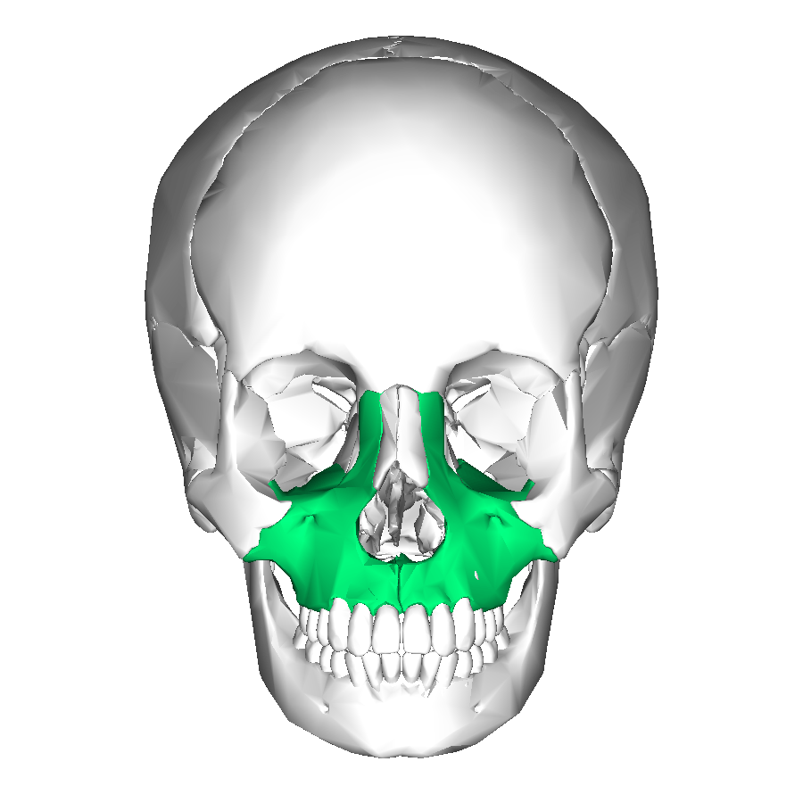
Name the highlighted bone
(a)
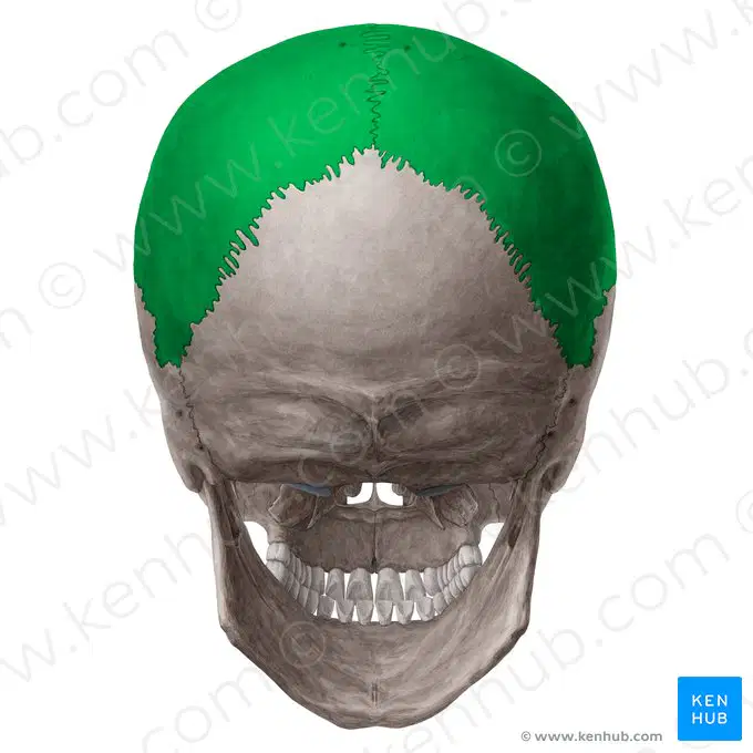
Name the highlighted bone
(a)
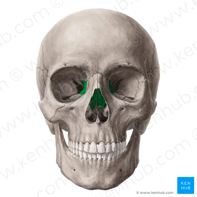
Name the highlighted bone
(a)

Name the highlighted bone
(a)
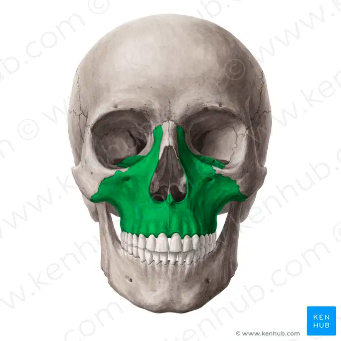
Name the highlighted bone
(a)
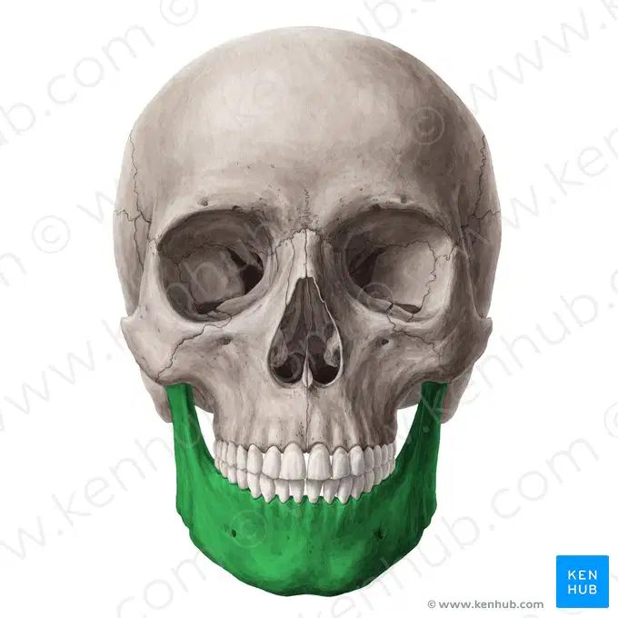
Name the highlighted bone
(a)
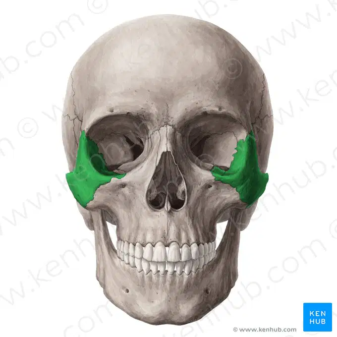
Name the highlighted bone
(a)
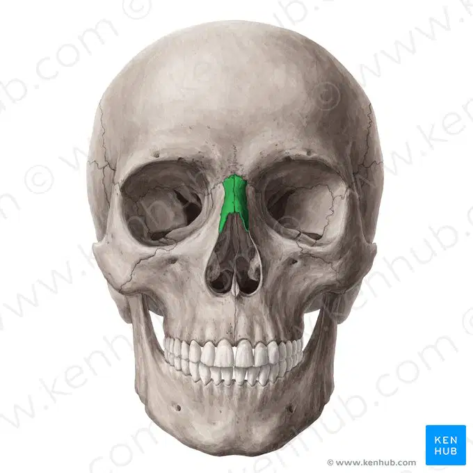
Name the highlighted bone
(a)
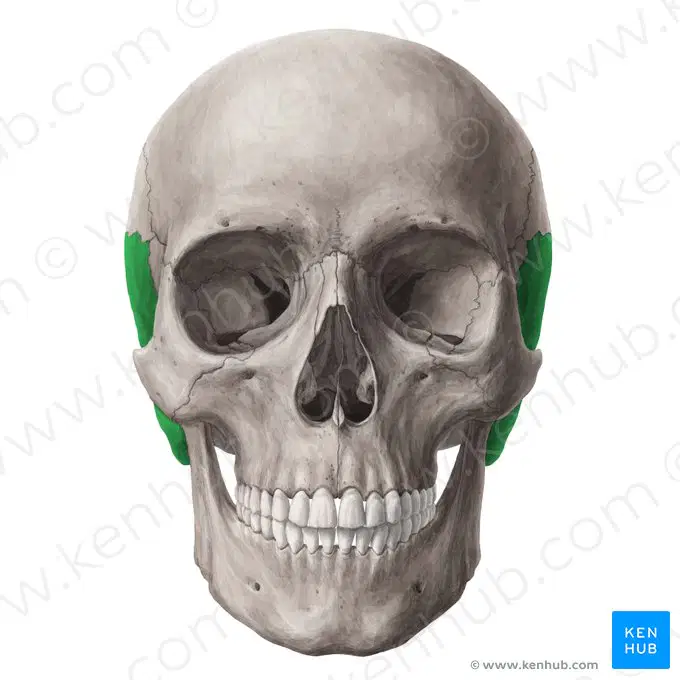
Name the highlighted bone
(a)


What bone is shown in red?
patella
pelvis