
 Worksheets
anatomy of foot ankle lower leg
Total questions: 50
Worksheet time: 41mins
Recall the counts: According to the instructional text, how many phalanges are in the human foot?
10
12
14
16
Which group correctly lists tarsal bones named in the section? Choose the option that includes only tarsal bones.
Calcaneus, Talus, Cuboid, Navicular, Cuneiforms
Metatarsals, Calcaneus, Phalanges
Phalanges, Navicular, Talus
Metatarsals, Cuneiforms, Phalanges
Fill in the blank with the exact term from the section: The heel bone of the foot is the (a) .
Match each category of foot bones to its description based on the diagram and list. Draw one line per pair.
Phalanges
Toe bones (proximal, middle, distal)
Metatarsals
Long bones of the midfoot that connect to toes
Tarsal bones
Seven bones of the hindfoot and midfoot including calcaneus and talus
Cuneiforms
Three specific tarsals: medial, intermediate, lateral
Reasoning with evidence: Explain how the arrangement of metatarsals and phalanges contributes to toe movement while maintaining foot stability, using the terminology provided (metatarsals, proximal/middle/distal phalanges, tarsal support).
BLANK: Name the thick fibrous tissue that supports the foot against downward force.
(a)
MATCH: Pair each labeled structure in the diagram with its role or location.
Spring ligament
Supports medial longitudinal arch and helps it recoil
Plantar fascia
Thick fibrous tissue resisting downward force under the foot
Transverse arch
Arch running across the midfoot from medial to lateral
Lateral longitudinal arch
Arch along the outer border of the foot from heel toward fifth metatarsal
Identify the bone that forms the lateral malleolus of the ankle. Choose the best answer.
Distal tibia
Distal fibula
Talus
Which structure is the medial malleolus a part of?
Fibula
Tibia
Talus
Calcaneus
Fill in the blank: The ankle mortise is formed by the distal tibia, distal fibula, and the (a) .
Using the labeled ankle diagram, match each term to its description.
Lateral malleolus
Distal projection on the outer side of the ankle
Medial malleolus
Distal projection on the inner side of the ankle
Talus
Bone seated within the ankle mortise
Fibula
Long bone of the lower leg located laterally
Explain why the ankle mortise view highlights a ‘clear space’ around the talus and what this indicates about joint alignment.
Refer to the diagram showing ankle joint movement. Which term describes moving the foot upward toward the shin at the ankle?
Plantarflexion
Dorsiflexion
Inversion
Eversion
In the ankle movement diagram, identify the motion represented by the foot pointing downward away from the shin.
(a)
Using the lateral ankle ligaments diagram, which ligament connects the fibula to the talus at the front of the ankle?
Anterior talofibular ligament (ATFL)
Calcaneofibular ligament (CFL)
Posterior talofibular ligament (PTFL)
Deltoid ligament
Match each lateral ankle ligament to its primary bony attachment pair as shown in the labeled lateral view.
Anterior talofibular ligament (ATFL)
Fibula ↔ Talus (anterior)
Calcaneofibular ligament (CFL)
Fibula ↔ Calcaneus
Posterior talofibular ligament (PTFL)
Fibula ↔ Talus (posterior)
According to the medial ankle diagram, the deltoid ligament is composed of three parts. Which list correctly names them?
Anterior tibiotalar, tibionavicular, tibiocalcaneal
Posterior tibiotalar, tibionavicular, tibiocalcaneal
Posterior talofibular, calcaneofibular, anterior talofibular
Plantar fascia, spring ligament, Achilles tendon
Refer to the described X-ray image of two lower legs: Identify which bone is the main weight-bearing bone in the lower leg.
Fibula
Tibia
Talus
Calcaneus
Complete the sentence with a precise term: The interosseous membrane between the tibia and fibula is also called the (a) .
Using the labeled illustration of the tibia and fibula, match each structure to its correct description.
Anterior tibiofibular ligament
Ligament connecting tibia and fibula at the front of the distal leg
Posterior tibiofibular ligament
Ligament connecting tibia and fibula at the back of the distal leg
Interosseous membrane
Broad connective tissue spanning between the shafts of tibia and fibula
Proximal tibiofibular joint
Articulation between tibia and fibula near the knee
Recall: According to the overview diagram, where do all muscles that act on the foot and ankle originate?
In the lower leg
In the foot bones
In the pelvis
In the toes
Match each muscle to its primary action. Use the labeled calf and lower-leg diagrams for reference.
Gastrocnemius
Plantar flexion
Posterior Tibialis
Inversion
Tibialis Anterior
Dorsiflexion
Peroneals
Eversion
Skill/Concept: Which insertion site is correctly paired with its muscle?
Gastrocnemius — calcaneus
Posterior Tibialis — base of 1st metatarsal
Tibialis Anterior — navicular
Peroneals — femoral condyles
Which bone forms the heel of the foot?
Talus
Calcaneus
Navicular
Cuboid
What is the main function of the plantar fascia?
To provide shock absorption
To support the arch of the foot
To facilitate foot inversion
To enhance toe flexion

A
Calcaneus
Talus
Cuboid
Intermediate Cuneiform
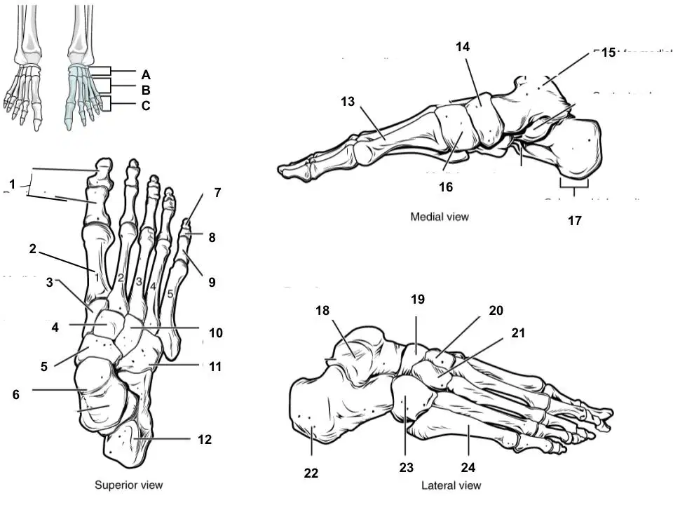
Which structure of the foot is labeled #5?
(a)
Which structure of the foot is labeled #6?
(a)
Which structure of the foot is labeled #11?
(a)
Which structure of the foot is labeled #12?
(a)
Which structure of the foot is labeled #14?
(a)
Which structure of the foot is labeled #15?
(a)
Which structure of the foot is labeled #17?
(a)
Which structure of the foot is labeled #18?
(a)
Which structure of the foot is labeled #19?
(a)
Which structure of the foot is labeled #23?
(a)
Name of muscle: Tibialis _________________
longus
anterior
brevis
mangus
What actions does the tibialis anterior perform?
dorsiflexion and plantarflexion
dorsiflexion and eversion
dorsiflexion and inversion
plantarflexion and inversion

What is #1
tibialis anterior
tibialis posterior
soleus
gastrocnemius
What is #3
tibialis anterior
tibialis posterior
fibularis longus
gastrocnemius
What is #5
soleus
tibialis posterior
fibularis longus
gastrocnemius

Identify the lateral ankle ligament 1.
ATF Ligament
PTF Ligament
CF Ligament
Deltoid Ligament
Identify the lateral ankle ligament 2.
ATF Ligament
PTF Ligament
CF Ligament
Deltoid Ligament
Identify the lateral ankle ligament 3.
ATF Ligament
PTF Ligament
CF Ligament
Deltoid Ligament

Which ligament is represented by the letter A?
Anterior Talofibular Ligament (ATFL)
Posterior Talofibular Ligament (PTFL)
Calcaneofibular Ligament (CFL)
Deltoid Ligament

Which ligament is represented by the letter A?
Anterior Talofibular Ligament (ATFL)
Posterior Talofibular Ligament (PTFL)
Calcaneofibular Ligament (CFL)
Deltoid Ligament

Which ligament is represented by the letter C?
Anterior Talofibular Ligament (ATFL)
Posterior Talofibular Ligament (PTFL)
Calcaneofibular Ligament (CFL)
Deltoid Ligament
Which ligament is represented by the letter B?
Anterior Talofibular Ligament (ATFL)
Posterior Talofibular Ligament (PTFL)
Calcaneofibular Ligament (CFL)
Deltoid Ligament

Which structure is shown
spring ligament
plantar fascia
syndesmosis
deltoid ligament
Which ligament supports the medial ankle
deltoid
anterior talofibular ligament
calcaneofibular ligament
posterior talofibular ligament

