
 Worksheets
How it works?
Total questions: 73
Worksheet time: 37mins
What type of images does a CT scanner provide?
Low-resolution anatomical images
High-resolution cross-sectional anatomical images
3D animated images
Only bone images
The table moves through a circular opening in the CT scanner called the ________, while an x-ray tube emits x-rays as it spins 360 degrees inside the gantry.
gantry
aperture
collimator
detector
What does a detector array measure in a CT scanner?
The amount of x-rays that pass through the anatomic part
The speed of the table
The temperature inside the scanner
The color of the images
Computed tomography (CT) scanning is an extremely common imaging modality in modern medicine.
True
False
With advancements in technology, CT scanning is rapidly replacing many diagnostic radiographic procedures.
True
False
What does CT in CT scan stand for?
Computerized Tomography
Computer Aided Tomography
Computed Telemetry
Central Tomography
Who invented the CT scan?
Wilhelm Röntgen
Sir Godfrey Hounsfield
Marie Curie
Alexander Fleming
In which year was the CT scan invented?
1960
1972
1985
1955
A CT scan is mainly used for:
Cooking
Medical imaging and diagnosis
Painting
Construction
Instead of sending out a single X-ray through the body as with ordinary x-ray, what does a CT scan do?
Several beams are sent at the same time from different angles.
It uses only sound waves to create images.
It only measures the temperature of the body.
It produces images using magnetic fields only.
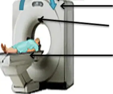
Which of the following is NOT a part of the CT scan machine?
Rotating X-ray source
Motorized platform
Image reconstruction unit
Stethoscope
What is the typical structure of a CT scanner?
Small handheld device
Large, box like machine with a hole, or short tunnel in the center
Flat table
Open frame
In a CT scanner, there is ______ X ray source but a large number of detectors.
one
two
four
ten
What is the function of the examination table in a CT scanner?
To rotate the X-ray source
To slide into and out of the tunnel, allowing the patient to lie on it during examination
To display images
To detect X-rays
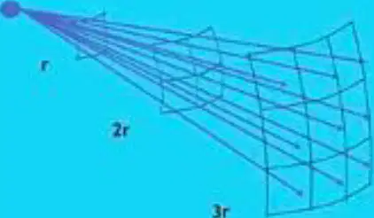
According to the information provided, how do X-rays travel?
In curved lines
In straight lines
In circles
In zig-zag lines
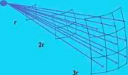
What happens to the number of X-rays per unit area as the distance from the source increases?
It increases
It stays the same
It decreases
It doubles
Fill in the blank: The number of X-rays per unit area is inversely proportional to the ______ of the distance from the source.
square
cube
sum
product
According to the table, if the distance from the X-ray source is tripled (3r), what is the number of X-rays per unit area?
N/9
N/3
N/6
N/12
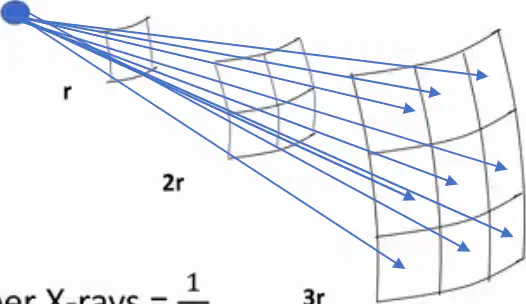
The inverse square law as it applies to X-rays can be explained using the example provided in the image.
The intensity of X-rays decreases proportionally to the square of the distance from the source.
The intensity of X-rays increases as the distance from the source increases.
The intensity of X-rays remains constant regardless of distance from the source.
The intensity of X-rays is not affected by distance from the source.
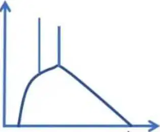
What primarily determines the input X-ray spectrum before the X-rays pass through the patient?
The patient's weight
The kVp (max Energy) and pre-patient filtration
The room temperature
The type of detector used
About __% of the X-rays are typically stopped inside the patient.
99
10
50
75
There are many FEWER photons in the exit spectrum as most have been attenuated by the patient.
True
False
Since the lower energy photons are preferentially attenuated, the average energy is ______ in the remnant spectrum.
Lower
Higher
The same
Unchanged
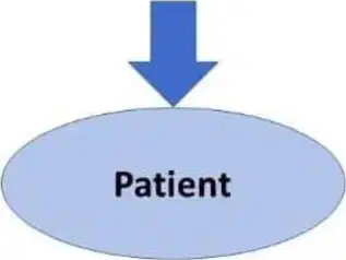
What happens to the number of photons after passing through the patient?
The number of photons decreases.
The number of photons increases.
The number of photons remains the same.
The number of photons doubles.
When travelling through the body of the patient, which interactions of x-rays with matter take place?
A) Photoelectric and Compton interactions
B) Nuclear fusion
C) Beta decay
D) Gamma emission
This process means x-rays are knocking out electrons from the matter, ______ (removing electrons) and releasing energetic electrons near the body's DNA (which can cause damage). Fill in the blank.
ionizing
polarizing
oxidizing
neutralizing
It is important to consider carefully the patient radiation dose in each case because:
Excessive radiation can be harmful to the patient.
It always improves image quality.
It reduces the need for medical staff.
It guarantees a faster recovery.
Preparation for CT exam: Any metal objects should be removed from the body before starting the scanning. Metal can interfere with the CT scan results. These items include ________, glasses, and dentures.
jewellery
shoes
socks
scarf
What is the usual position of the patient on the CT examination table?
Sitting upright
Lying flat on back
Standing
Kneeling
Why may straps and pillows be used during a medical imaging exam?
To make the patient comfortable
To help maintain the correct position and to hold still during the exam
To reduce the cost of the exam
To speed up the process
Movement during a medical imaging exam can result in ________ pictures.
blurry
clear
colorful
sharp
What is the purpose of using contrast material in X-ray imaging?
To make the patient feel better
To help internal structures show up more clearly on the X-ray images
To reduce radiation exposure
To shorten the exam time
Fill in the blank: Next, the table will move quickly through the scanner to determine the correct _______ position for the scan.
starting
ending
middle
final
Fill in the blank: Then, the table will move _______ through the machine as the actual CT scanning is performed.
slowly
quickly
backwards
randomly
Depending on the type of CT scan, the machine may make several passes.
True
False
The patient may be asked to hold their breath during the scanning.
True
False
What is the ring called in which the X-ray tube and detectors are located opposite to each other?
gantry
collimator
anode
rotor
With CT scanning, numerous X-ray beams and a set of electronic X-ray detectors rotate around the patient, measuring the amount of radiation being absorbed through the body. True or False: The X-ray tube and detectors are located on the same side of the ring.
True
False
What is the function of the detectors in a CT scanner?
To measure the amount of radiation being absorbed through the body.
To generate X-rays for imaging.
To move the patient through the scanner.
To reconstruct the final image from raw data.

Refer to the diagram showing the X-ray tube and detectors arranged in a ring around the patient. What is the purpose of this arrangement in CT scanning?
To allow the X-ray tube and detectors to rotate around the patient and measure the amount of radiation absorbed through the body from different angles.
To increase the brightness of the X-ray image for better visualization.
To reduce the amount of radiation required for a single scan by half.
To focus the X-rays on a single point for higher resolution images.
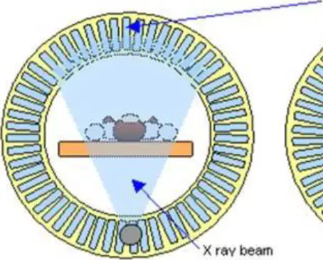
At the same time, the examination table is moving through the scanner, so that the X-ray beam follows a ______ path. (Fill in the blank)
spiral
linear
zigzag
circular
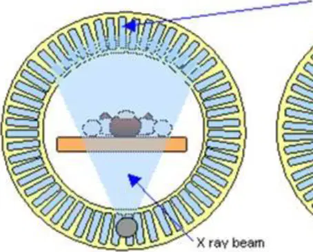
Refer to the diagram labeled 'CT scanner with the X ray source and detectors shown in three positions.' Which of the following statements is correct?
The X-ray source and detectors remain stationary while the patient moves.
The X-ray source and detectors rotate around the patient while the examination table moves through the scanner.
The patient rotates while the X-ray source and detectors remain stationary.
The X-ray beam does not move during the scan.

According to the passage, what is a CT scan image compared to?
A loaf of bread
A slice of bread
A cake
A pizza
Each 'slice' in a CT scan shows an image from the _______ to the center part of the body.
outer skin
muscle layer
bone marrow
internal organs
A CT scan produces multiple images or 'slices' because:
it takes images from different angles to create detailed cross-sectional views of the body.
it uses only a single X-ray image for diagnosis.
it is designed to show only the outer surface of organs.
it cannot differentiate between different tissues.
What is digital geometry processing used for?
To generate a three-dimensional volume of the inside of the object from a series of two-dimensional radiographic images taken around a single axis of rotation.
To create two-dimensional images from three-dimensional objects.
To process sound waves for medical imaging.
To monitor patient heart rate.
Fill in the blank: The computer workstation that processes the imaging information is located in a separate _______ room, where the technologist operates the scanner and monitors the examination.
control
waiting
patient
storage
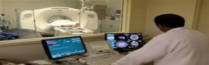
What is shown in the image labeled 'CT Control room'?
A technologist operating a computer workstation in a separate control room, monitoring a CT scanner and the examination.
A patient lying inside an MRI scanner.
A doctor performing surgery in an operating room.
A nurse administering medication in a hospital ward.
After the examination is completed, the patient must wait until the ________ verifies that the images are of high enough quality for accurate interpretation.
technologist
receptionist
janitor
patient
The CT examination is usually completed within 30 minutes. The portion requiring intravenous contrast injection usually lasts only ________ to ________ seconds. Fill in the blanks.
10, 30
30, 60
60, 90
5, 15
What does the variable 'I' represent in the equation I=I0e−μx ?
The intensity after radiation absorption
The initial intensity before absorption
The absorption coefficient of the material
The thickness of the material
What does the variable ' I0 ' represent in the equation ' I=I0e−μx '?
The original intensity of the incident radiation
The thickness of the material
The absorption coefficient of the material
The final intensity after passing through the material
What does the variable 'μ' represent in the equation I = I₀e^(-μx)? I=I0e−μx
The attenuation coefficient (cm⁻¹)
The initial intensity
The thickness of the material
The transmitted intensity
What does the variable 'X' represent in the equation I = I0e−μx ?
The thickness
The initial intensity
The absorption coefficient
The transmitted intensity
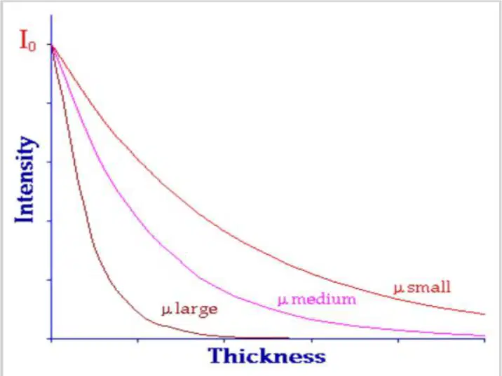
Based on the graph, which attenuation coefficient (μ) results in the fastest decrease in intensity as thickness increases?
μ large
μ medium
μ small
What did Hounsfield envision for dividing a slice in CT imaging?
Dividing a slice into a matrix of 2-dimensional boxes
Dividing a slice into a matrix of 3-dimensional rectangular boxes (voxels) of material (tissue)
Dividing a slice into a single large box
Dividing a slice into circular regions
In CT imaging, the Z direction of the voxels corresponds to the _________?
slice thickness
matrix size
pixel spacing
field of view
The X and Y voxel dimensions in CT imaging depend on:
The size of the scan circle and the size of the matrix
The color of the tissue
The speed of the scan
The temperature of the room
If a scan circle is divided into a matrix of 250 rows × 250 columns, each voxel is ______ mm.
1 × 1 mm
0.5 × 0.5 mm
2 × 2 mm
5 × 5 mm
If a 512 × 512 matrix is used in CT imaging, each voxel is approximately:
1 × 1 mm
0.5 × 0.5 mm
2 × 2 mm
5 × 5 mm

What are the small boxes of tissue in a scanned slice called, as envisioned by Hounsfield?
Pixels
Voxels
Cells
Grids

Each voxel in the reconstruction matrix has an attenuation coefficient denoted by _____?
μ (mu)
λ (lambda)
σ (sigma)
θ (theta)

X-ray transmission measurements (N_i) can be expressed as the sum of ______ values occurring in voxels along the path of the ray for N_i.
Density
Attenuation
Volume
Intensity
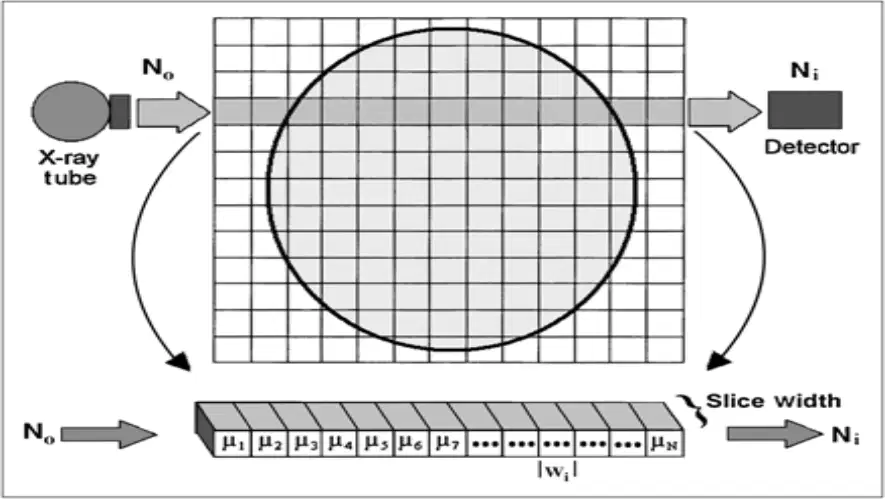
The diagram illustrates the relationship between the X-ray tube, the matrix of voxels, and the detector in the reconstruction matrix. What does this relationship show?
The X-ray tube emits beams that pass through the matrix of voxels and are detected by the detector to create an image.
The detector emits X-rays that are absorbed by the matrix of voxels and then received by the X-ray tube.
The matrix of voxels generates X-rays that are detected by both the X-ray tube and the detector.
The X-ray tube and detector are unrelated to the matrix of voxels in image reconstruction.
To carry out reconstruction, what does N_i represent in the context of x-ray imaging?
The x-ray intensity entering the subject
The transmitted x-ray intensity measured by the detector
The attenuation value of a voxel
The sum of all voxel values
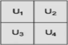
What does U_1 represent in the context of the reconstruction process?
U_1 represents the attenuation value in the first voxel along the path of the ray.
U_1 represents the total attenuation along the entire ray path.
U_1 represents the source intensity before entering the object.
U_1 represents the detector response after the ray exits the object.
According to the passage, the derived measurement X_i can be related to which of the following?
The product of the attenuation values in the voxels
The sum of the attenuation values in the voxels along the path of the ray
The difference between the entering and exiting x-ray intensities
The average of the x-ray intensities
Fill in the blank: The formula for the derived measurement X_i is X_i = ____ + u_2 + u_3 + u_4 + ... + u_n.
u_1
X_1
n_1
v_1
What are CT numbers calculated from?
The linear attenuation coefficient values for each tissue individual voxel
The density of water
The atomic number of materials
The energy of x-ray photons
What is the reference material for CT numbers and what value is it assigned?
Water is the reference material for CT numbers and it is assigned a value of zero.
Air is the reference material for CT numbers and it is assigned a value of 1000.
Bone is the reference material for CT numbers and it is assigned a value of zero.
Fat is the reference material for CT numbers and it is assigned a value of 100.
Tissues or materials with attenuation (density) greater than water will have ______ CT numbers. Those that are less dense will have ______ CT numbers.
positive; negative
negative; positive
zero; positive
positive; zero
CT numbers are determined by which of the following?
The density of the tissues or materials
The color of the tissues
The shape of the tissues
The temperature of the tissues
X-ray attenuation depends on both the ______ and ______ of materials and the energy of the x-ray photons.
density; atomic number (Z)
temperature; color
mass; volume
conductivity; melting point

