
 Worksheets
Muscle Types Quiz
Total questions: 88
Worksheet time: 44mins
Which of the following is NOT one of the three main types of muscles classified in the human body?
Skeletal muscle
Cardiac muscle
Smooth muscle
Connective muscle
Which type of muscle is primarily found in the heart?
Skeletal muscle
Cardiac muscle
Smooth muscle
Voluntary muscle
If a patient has a disorder affecting the muscles in their digestive tract, which type of muscle is most likely involved?
Skeletal muscle
Cardiac muscle
Smooth muscle
Striated muscle
Which type of muscle is found in the heart?
Cardiac muscle
Skeletal muscle
Smooth muscle
Voluntary muscle
What is a unique structural feature of cardiac muscle fibers?
They show both longitudinal and transverse striations
They are completely smooth
They lack any striations
They are multinucleated like skeletal muscle
Which type of nerves supply the cardiac muscles?
Autonomic (involuntary) nerves
Somatic (voluntary) nerves
Sensory nerves only
Cranial nerves only
What is the primary function of cardiac muscles?
Responsible for cardiac contractility, contracting spontaneously and rhythmically
Responsible for voluntary movement of limbs
Responsible for digestion in the stomach
Responsible for secretion of hormones
Cardiac muscle contraction is considered involuntary because:
It is controlled by autonomic nerves, ensuring the heart beats without conscious effort, which is vital for continuous blood circulation.
It only contracts when we sleep, ensuring rest for the heart.
It is not connected to any nerves, so it does not contract at all.
It is controlled by voluntary nerves, allowing conscious control of heart rate.
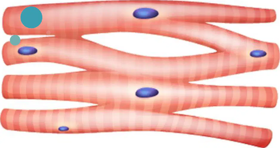
Which type of muscle is shown in the diagram labeled "Cardiac muscle"?
Cardiac muscle
Skeletal muscle
Smooth muscle
Voluntary muscle
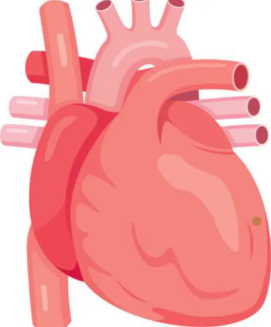
Where in the human body is cardiac muscle primarily found?
Heart
Lungs
Stomach
Brain
Compare the structure of cardiac muscle to skeletal muscle. What is a key difference in their function?
Cardiac muscle contracts involuntarily, while skeletal muscle contracts voluntarily.
Cardiac muscle is found in the arms, while skeletal muscle is found in the heart.
Cardiac muscle is responsible for digestion, while skeletal muscle is responsible for breathing.
Cardiac muscle is smooth, while skeletal muscle is striated.
Which type of muscle is found in the visceral wall?
Smooth muscle
Skeletal muscle
Cardiac muscle
Connective tissue
What is a key characteristic of smooth muscle?
They are non-striated (smooth)
They are striated
They are voluntary
They are attached to bones
Which type of nerves supply smooth muscle?
Autonomic (involuntary) nerves
Somatic (voluntary) nerves
Sensory nerves
Cranial nerves
What is the primary function of smooth muscle in the body?
Responsible for visceral movements
Responsible for voluntary movements
Responsible for pumping blood
Responsible for producing hormones
Smooth muscle is considered involuntary because:
It is controlled by autonomic nerves and is responsible for movements in internal organs without conscious control.
It is attached to bones and moves the skeleton.
It is found only in the heart and pumps blood.
It is under conscious control and helps in walking.
Which of the following factors can influence the contraction of certain muscle fibers?
Local stretch and hormones
Blood pressure and temperature
Oxygen concentration and light
Sound and humidity
How may muscle fibers be arranged in the body?
Only circularly
Only longitudinally
Circularly or longitudinally
Randomly
What is the function of circular muscle arrangement in blood vessels?
To increase the lumen size
To narrow the lumen
To aid in peristalsis
To produce hormones
Which muscle arrangement is primarily responsible for peristalsis in the intestine?
Circular
Oblique
Longitudinal
Radial
A patient is experiencing difficulty with peristalsis in their intestines. Which muscle arrangement is most likely affected?
Circular, because it narrows the lumen
Longitudinal, because it is responsible for peristalsis
Oblique, because it aids in digestion
Radial, because it controls blood flow
In which system do contractions lead to the proper mixing of food?
Digestive system
Urogenital system
Respiratory system
Nervous system
What is the primary effect of contractions in blood vessels?
They narrow the lumen
They mix blood with nutrients
They speed up blood flow
They increase the size of the lumen
The function of contractions differs between the digestive system and the urogenital system in which of the following ways?
In both systems, contractions only mix contents.
In the digestive system, contractions mix food, while in the urogenital system, contractions are slow and sustained to expel content.
Both systems use contractions to narrow the lumen.
In the digestive system, contractions are slow and sustained, while in the urogenital system, they mix food.
Which type of muscle tissue is shown in the image associated with the digestive tract?
Smooth muscle tissue
Skeletal muscle tissue
Cardiac muscle tissue
Nervous tissue
What is the primary function of smooth muscle tissue in the digestive system?
Voluntary movement of limbs
Pumping blood through the heart
Involuntary contraction to move food along the digestive tract
Transmitting nerve impulses
Smooth muscle tissue in the walls of arteries is important because:
It allows voluntary control of blood flow.
It helps arteries contract and relax to regulate blood pressure and flow.
It transmits electrical impulses to the heart.
It stores nutrients for the body.
Which type of muscles are attached to the skeleton?
Skeletal muscles
Cardiac muscles
Smooth muscles
Involuntary muscles
What characteristic feature do skeletal muscles show under a microscope?
They show longitudinal and transverse striations
They appear smooth and unstriated
They have intercalated discs
They are multinucleated without striations
Which type of nerves supply skeletal muscles?
Voluntary nerves
Involuntary nerves
Autonomic nerves
Sensory nerves
What is the primary function of skeletal muscles?
Responsible for voluntary motor activity
Responsible for involuntary movements
Responsible for hormone secretion
Responsible for digestion
Skeletal muscles are essential for voluntary motor activity because:
They are attached to the skeleton and controlled by voluntary nerves, allowing conscious movement. Their striated structure enables rapid and forceful contractions needed for movement.
They contract automatically and are controlled by the autonomic nervous system.
Their structure allows secretion of hormones into the bloodstream.
They line the digestive tract and move food through peristalsis.
What are the two types of muscle attachments called?
Origin and insertion
Flexor and extensor
Ligament and tendon
Agonist and antagonist
Which muscle attachment is associated with a lesser range of movement?
Insertion
Origin
Flexor
Extensor
A muscle's insertion point is best described as:
The attachment with lesser range of movement
The attachment with greater range of movement
The attachment closest to the heart
The attachment that never moves
Given a diagram of a muscle with two attachment points, how would you determine which is the origin and which is the insertion?
The origin is the attachment with greater movement, and the insertion is the one with lesser movement.
The origin is always at the top, and the insertion is always at the bottom.
The origin is the attachment with lesser movement, and the insertion is the one with greater movement.
The origin and insertion are interchangeable terms.
Which of the following is a characteristic of all skeletal muscles?
They have fleshy, reddish, contractile portions.
They are always white and noncontractile.
They do not have any heads or bellies.
They are only found in the heart.
What term is used to describe the fleshy, contractile portion of a skeletal muscle?
Muscle belly
Ligament
Cartilage
Bone marrow
Some skeletal muscles are fleshy throughout, but what is true about others?
They have white noncontractile portions.
They are always attached to bones by cartilage.
They contain only tendons and no muscle fibers.
They are never contractile.
Based on the diagram, which part of the skeletal muscle is labeled as the "muscle belly"?
The central, fleshy part between the tendons
The white, noncontractile ends
The outermost layer of the muscle
The bone attached to the muscle
Some skeletal muscles have white noncontractile portions while others are fleshy throughout because:
White noncontractile portions are tendons that connect muscle to bone, while fleshy portions are responsible for contraction.
White noncontractile portions store energy, while fleshy portions store fat.
White noncontractile portions are responsible for muscle contraction, while fleshy portions provide support.
All muscles are fleshy throughout; white portions are a sign of disease.
What are tendons primarily composed of?
Collagen bundles
Muscle fibers
Fatty tissues
Nerve cells
Where are tendons commonly present in the body?
At the attachments of the muscles
Inside the bone marrow
In the skin layers
Surrounding the heart
Which of the following best describes the function of tendons in the muscular system?
They connect muscles to bones
They produce red blood cells
They store energy for muscles
They transmit nerve impulses
What is an aponeurosis?
A thin, broad sheet of collagen tissues representing the attachment of flat muscles.
A thick, round band of muscle fibers.
A type of nerve tissue connecting bones.
A gland that secretes hormones.
Which of the following best describes the function of an aponeurosis?
It attaches flat muscles to the structures they move.
It transmits nerve impulses to muscles.
It stores fat for energy.
It produces red blood cells.
Based on the diagram, where is the aponeurosis located in relation to the skull?
It is located on the top of the skull, connecting the frontalis and occipitalis muscles.
It is found inside the jawbone.
It is located at the base of the skull.
It is present within the nasal cavity.
What is a raphe in anatomical terms?
Inter-digitations of the tendinous end of flat muscles
A type of bone joint
A nerve bundle
A blood vessel
Which of the following is an example of a raphe?
Linea alba
Femoral artery
Tibial nerve
Patella
Why might the structure of a raphe be important for the function of flat muscles?
It allows for strong attachment and distribution of force across the muscle
It increases the flexibility of the muscle
It provides a pathway for nerves
It stores energy for muscle contraction
What is the sheath that encloses a group of muscle fibers in skeletal muscle called?
Epimysium
Perimysium
Endomysium
Sarcolemma
Which of the following best describes the possible directions of muscle fibers in skeletal muscle?
Parallel and oblique
Circular and spiral
Transverse and longitudinal
Radial and concentric
Which of the following best describes parallel muscles?
The fibers run parallel to each other
The fibers run perpendicular to each other
The fibers form a circular pattern
The fibers are arranged in a pennate pattern
Which muscle is an example of a parallel muscle with tendinous bands?
Rectus abdominis muscle
Biceps brachii muscle
Supinator muscle
Deltoid muscle
What is the main structural difference between a parallel muscle and a wrapping muscle?
Parallel muscles have fibers running straight, while wrapping muscles have fibers that wrap around structures
Parallel muscles are only found in the legs, while wrapping muscles are only found in the arms
Wrapping muscles have tendinous intersections, while parallel muscles do not
Parallel muscles are circular, while wrapping muscles are triangular
If a muscle has fibers that run parallel and contains tendinous intersections, which type of muscle is it?
Parallel muscle with tendinous bands
Wrapping muscle
Circular muscle
Pennate muscle
The arrangement of muscle fibers in a wrapping muscle differs from that in a simple parallel muscle in which of the following ways?
Wrapping muscles have fibers that spiral or wrap around a structure, allowing for rotational movement, while parallel muscles have straight fibers that contract in a single direction, producing linear movement.
Wrapping muscles and parallel muscles have identical fiber arrangements and functions.
Wrapping muscles have circular fibers, while parallel muscles have triangular fibers, both producing rotational movement.
Wrapping muscles have fibers that run perpendicular to the bone, while parallel muscles have fibers that run parallel to the bone, both producing the same type of movement.
Which type of muscle fiber arrangement is described as fibers running oblique to the long axis of the muscle, similar to a feather?
Pennate type
Parallel type
Circular type
Fusiform type
Which of the following is an example of a unipennate muscle?
Extensor digitorum muscle
Deltoid muscle
Rectus femoris muscle
Biceps brachii muscle
What distinguishes a multipennate muscle from unipennate and bipennate muscles?
It has multiple tendons with fibers arranged in several directions.
It has fibers running parallel to the tendon.
It has only one row of muscle fibers.
It forms a circular pattern around an opening.
If a muscle has fibers attaching obliquely to both sides of a central tendon, what type of muscle is it?
Bipennate
Unipennate
Multipennate
Circumpennate
Analyze the structural differences between unipennate, bipennate, and multipennate muscles and explain how these differences might affect muscle function.
Unipennate muscles have fibers on one side of the tendon, bipennate on both sides, and multipennate have multiple rows with central tendons, allowing for greater force generation in multipennate muscles.
All three types have the same structure and function.
Unipennate muscles are circular, bipennate are parallel, and multipennate are fusiform.
Only unipennate muscles can generate high force.
What does "unipennate" mean in relation to muscle structure?
The muscle has a single tendon on one side and the fibers run toward it.
The muscle has tendons on both sides and fibers run parallel to them.
The muscle fibers run in a circular pattern around a tendon.
The muscle has multiple tendons on both sides and fibers run in different directions.
Which of the following best describes the arrangement of fibers in a unipennate muscle?
Fibers run toward a single tendon on one side.
Fibers run parallel to the tendon on both sides.
Fibers run in a spiral around the tendon.
Fibers are arranged in a circular pattern.
What does the term "bipennate" mean in relation to muscle structure?
The muscle has a single tendon in the center and fibers run toward it from both sides.
The muscle has multiple tendons on one side only.
The muscle fibers run parallel to each other without a central tendon.
The muscle has no tendons and fibers run in a circular pattern.
Which of the following best describes the arrangement of fibers in a bipennate muscle?
Fibers run toward a single central tendon from both sides.
Fibers run in a spiral around the tendon.
Fibers are arranged in a parallel fashion along the tendon.
Fibers attach to multiple tendons on one side only.
A student is asked to identify a muscle type where fibers run toward a single tendon in the center from both sides. Which muscle type should they choose?
Bipennate
Unipennate
Multipennate
Fusiform
What does the term "multipennate" mean in relation to muscle structure?
The muscle is formed of several units of bipennate
The muscle is formed of parallel fibers only
The muscle is formed of a single unit of unipennate
The muscle is formed of circular fibers
Which of the following best describes the structure of a multipennate muscle?
Several bipennate units forming a single muscle
Only parallel muscle fibers
A single unipennate unit
Muscle fibers arranged in a circular pattern
If a muscle is described as multipennate, what can you infer about its internal structure?
It contains multiple bipennate arrangements within the muscle
It is made up of only one type of muscle fiber
It has no tendons
It is always smaller than unipennate muscles
When a muscle contracts, by how much does it typically shorten compared to its original length?
By about 1/3 or 1/2 of its original length
By about 1/4 of its original length
By about 3/4 of its original length
It does not shorten at all
What factor determines the strength of movement allowed by a muscle?
The number of its fibers
The color of the muscle
The length of the muscle
The temperature of the muscle
If a muscle has more fibers, what can you infer about its strength of movement?
The strength of movement will be greater
The strength of movement will be less
The strength of movement will not change
The muscle will not be able to contract
Which type of muscle is stronger?
Pennate muscles
Muscles with parallel fibers
Cardiac muscles
Smooth muscles
Why are pennate muscles considered stronger than muscles with parallel fibers?
Because their fiber arrangement allows for greater force production
Because they are longer in length
Because they contract faster
Because they have more blood vessels
Given the information that pennate muscles are stronger than muscles with parallel fibers, which type of muscle would be more suitable for activities requiring high force output?
Pennate muscles
Muscles with parallel fibers
Cardiac muscles
Smooth muscles
Which of the following is NOT a category used to classify muscles?
According to the shape
According to their action
According to the number of joints acting upon
According to their color
Muscles can be classified according to which of the following criteria?
Shape, action, and number of joints acting upon
Size, weight, and length
Color, density, and flexibility
Temperature, location, and age
If a muscle is classified based on the number of joints it acts upon, which category does this represent?
According to the shape
According to their action
According to the number of joints acting upon
According to their location
A student is asked to classify a muscle based on how it moves the body. Which classification category should they use?
According to the shape
According to their action
According to the number of joints acting upon
According to their length
Which of the following is an example of a strap muscle?
Rectus abdominis
Pronator quadratus
Biceps brachii
Deltoid
What is the primary shape of the pronator quadratus muscle?
Quadrate
Strap
Fusiform
Triangular
Compare the structural arrangement of strap muscles and quadrate muscles. Which statement is correct?
Strap muscles have parallel fibers, while quadrate muscles are square-shaped.
Strap muscles are square-shaped, while quadrate muscles have parallel fibers.
Both strap and quadrate muscles have triangular fibers.
Quadrate muscles are always found in the abdomen.
What is the shape of a fusiform muscle?
Spindle-shaped with a central belly and tendons at both ends
Flat and broad with no central belly
Circular with fibers radiating from a central point
Triangular with a wide base and narrow apex
Which of the following is an example of a fusiform muscle?
Digastric muscle
Deltoid muscle
Rectus abdominis
Pectoralis major
The structure of a fusiform muscle compared to a flat muscle is characterized by which of the following fiber arrangements?
Fibers running parallel to the length, allowing for greater range of motion and speed.
Fibers arranged in a circular pattern, providing stability.
Fibers running perpendicular to the length, increasing strength.
Random fiber arrangement, allowing for flexibility.
Which type of muscles are shown in the title of the provided material?
Circular muscles
Skeletal muscles
Cardiac muscles
Smooth muscles
The functional significance of circular muscles in the human body, such as the orbicularis oculi, is that they:
help in voluntary movement of limbs.
allow for the opening and closing of body passages, such as the eyelids.
are responsible for pumping blood throughout the body.
provide structural support to bones.

