
 Worksheets
Filtration PP Recap
Total questions: 47
Worksheet time: 24mins
Which statement best defines x-ray filtration in radiographic imaging?
Insertion of absorbing materials to eliminate undesirable low-energy x-ray photons from the primary beam
Amplification of all x-ray photons to increase image brightness
Reflection of x-ray photons away from the patient to reduce dose
Alignment of the x-ray tube to center the primary beam
Filtration primarily removes which portion of the x-ray beam?
High-energy photons
Low-energy photons
Mid-energy photons only
All photons equally
According to the concept of filtration, shaping the emission spectrum creates a more useful x-ray beam by favoring which property?
Lower frequency/longer wavelength
Higher frequency/shorter wavelength
Constant frequency/constant wavelength
Variable frequency/longer wavelength

Based on the graph comparing unfiltered and filtered spectra, which effect does filtration have on photon intensity at low energies?
It increases low-energy photon intensity above the unfiltered curve.
It leaves low-energy photon intensity unchanged.
It decreases low-energy photon intensity relative to the unfiltered beam.
It eliminates high-energy photons only.

A radiographer adds filtration to the x-ray tube. Which outcome most directly explains why the resulting beam is considered more useful for imaging?
More low-energy photons improve skin contrast.
Removal of low-energy photons reduces non-diagnostic exposure while preserving higher-energy photons.
All photons are amplified equally, improving detail.
The peak energy of photons is lowered to reduce scatter.
Which statement best describes the primary purpose of filtration in X-ray imaging?
To soften the beam by adding low-energy photons
To remove low-energy or soft photons and harden the beam
To decrease the average beam energy for better contrast
To increase patient dose for higher image quality
Filtration has which effect on the average X-ray beam energy?
Decreases it
Does not change it
Increases it
Randomly fluctuates it
Approximately how much photon energy is needed for X-rays to pass through soft tissues like muscles and organs?
5 to 10 keV
15 to 20 keV
30 to 40 keV
60 to 80 keV
Why are low-energy photons considered undesirable in patient imaging?
They improve image receptor exposure
They cannot penetrate the part and only contribute to patient dose
They enhance spatial resolution
They reduce motion blur
As filtration increases, what happens to image receptor (IR) exposure?
IR exposure increases
IR exposure decreases
IR exposure remains unchanged
IR exposure fluctuates unpredictably
Which material is identified as the main material used for X-ray beam filtration?
Lead
Copper
Aluminum
Tungsten
What does aluminum equivalency (Al/Eq) specifically measure in radiographic filtration?
The percentage of scatter radiation removed
How thick the aluminum layer is used for filtration
The peak kilovoltage of the X-ray beam
The number of photons produced per second
The Half-Value Layer (HVL) is defined as the thickness of aluminum needed to do which of the following to an X-ray beam?
Double the beam intensity
Reduce the beam’s intensity by half
Eliminate all low-energy photons
Increase beam energy by 25%
Which outcome is a direct purpose of using HVL as stated in the material?
To increase patient dose for clearer images
To verify that the beam is strong enough to create a clear image
To calibrate image receptor sensitivity
To measure tube current (mA) accuracy
According to the description of HVL, which statement best explains how it supports patient safety?
HVL adds more low-energy rays to enhance contrast
HVL removes lower-energy rays that increase dose without improving the image
HVL reduces the need for any filtration
HVL shortens exposure time regardless of technique
HVL is measured in which units and indicates what property of the X-ray beam?
Millimeters of aluminum; the beam’s strength/quality
Centimeters of lead; the beam’s size
Kilovolts; the beam’s intensity
Milliamperes; the beam’s exposure time
Which reason is given for why monitoring HVL is important for quality control of X-ray machines?
It confirms the beam is homogeneous in energy
It provides an easy way to check X-ray output and ensure the machine is working correctly
It guarantees zero radiation scatter
It replaces the need for protective shielding
Which statement reflects the heterogenous nature of the X-ray beam as related to HVL?
All rays have the same energy, so a single HVL is unnecessary
The beam contains different energy levels, so HVL indicates overall beam quality
Energy levels do not affect filtration needs
HVL only applies when using lead filters
Which term refers to the combined effect of inherent and added filtration in an X‑ray system?
Compound filtration
Compensating filtration
Total filtration
Partial filtration
A radiographer places a specially shaped filter to even out exposure across a body part with varying thickness. Which type of filtration is being applied?
Inherent filtration
Added filtration
Compound filtration
Compensating filtration
Which statement best defines added filtration in radiographic imaging?
Filtering materials inside the X-ray tube target
Any filtering material located outside the X-ray tube and housing but before the image receptor
Software processing that smooths the image after exposure
Protective shielding placed behind the image receptor
What is a major source of added filtration and approximately how much aluminum equivalency does it provide due to the silver backing on its mirror?
The grid; about 0.2 mm Al/Eq
The collimator; about 1 mm Al/Eq
The cassette; about 2 mm Al/Eq
The anode; about 0.5 mm Al/Eq
Why might extra sheets of aluminum or copper be added to the beam path in addition to the collimator?
To increase low-energy X-rays and raise patient dose
To remove low-energy X-rays, reduce patient exposure, and improve image quality
To focus the X-ray beam for higher spatial resolution only
To compensate for inadequate tube current at the anode
In the context of added filtration, what does the abbreviation Al/Eq most directly refer to?
Average lumen/equilibration of the collimator
Aluminum equivalency of a filtering component
Alignment equipment used in collimation
Alternating current equipment for X-ray production
Which statement best describes a K-edge filter in X-ray imaging?
A single metal layer used to uniformly attenuate all X-ray energies
A combination of two or more different metal layers that shape the X-ray beam and reduce unnecessary patient radiation
A device that increases low-energy photons to improve contrast
A filter placed only at the image receptor to sharpen images
In a properly arranged K-edge filter stack, where should the higher atomic number metal (such as copper) be placed?
Nearest to the patient to absorb secondary photons
Nearest to the X-ray tube to absorb low-energy photons first
Between two aluminum layers to balance beam hardening
At the collimator exit to shape the field edges
What is the primary role of the second metal layer (e.g., aluminum) in a K-edge filter after X-rays pass through the first layer?
To generate high-energy photons
To absorb secondary photons produced by the first layer, making the beam cleaner and more uniform
To reflect unused photons back to the tube
To convert X-rays into visible light
Which pair of materials is commonly combined to exploit their K-edge properties in compound filtration, forming a Thoraeus filter?
Lead and tin
Copper and aluminum
Tungsten and molybdenum
Silver and iodine
Approximately what are the K-edge energies for copper and aluminum used in K-edge filtration?
Copper: 1.5 keV; Aluminum: 8 keV
Copper: 8 keV; Aluminum: 1.5 keV
Copper: 44 keV; Aluminum: 25 keV
Copper: 25 keV; Aluminum: 44 keV
Using K-edge filters in clinical imaging can reduce the patient’s entrance surface exposure (ESE) by what approximate range?
2%–10%
12%–20%
25%–44%
55%–70%
Why does stacking different metals with specific K-edge energies improve image quality and patient safety?
It amplifies low-energy photons that create sharper images
It removes weaker X-rays that do not contribute to image formation and would increase patient dose, while producing a more uniform beam
It randomizes photon energies to reduce artifacts
It blocks all high-energy photons to prevent overexposure
What is the primary purpose of compensation filtration in radiography?
To increase overall X-ray intensity for all body parts
To even out radiographic density when imaging parts with uneven thickness or density
To sharpen image edges by reducing scatter only
To shorten exposure time regardless of patient size
Which filter is most appropriate to equalize exposure across a body part where one side is thicker than the other, such as the foot or thoracic spine?
Boomerang filter
Grid filter
Wedge filter
Notch filter
In a chest X-ray intended to balance exposure between the lungs and the mediastinum, which compensating filter should be used?
Wedge filter
Trough filter
Neutral density filter
Edge enhancement filter
Which statement best describes how compensating filters improve image quality?
They block all scatter radiation, increasing contrast only.
They add an absorber to reduce X-ray intensity over thinner regions, producing a more evenly exposed image.
They increase tube current to penetrate thicker anatomy.
They compress tissue to make thickness uniform.
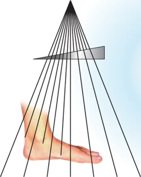
A diagram shows an X-ray beam passing through an angled plate over a foot, with more attenuation on the thinner toes and less on the thicker heel. Which filter does this diagram most likely illustrate?
Trough filter
Wedge filter
Boomerang filter
Compensating grid
Which scenario most strongly justifies the use of a trough filter rather than a wedge filter?
Imaging an ankle with uniform thickness
AP projection of the thoracic spine where one side is thicker
Chest radiography where the center is denser than the lateral regions
Any extremity with metallic implants
Which statement best defines total filtration in an X‑ray system?
The thickness of aluminum added to the beam only
The sum of inherent and added filtration
Any filter that changes the X‑ray beam, including compensating filters
Only the glass envelope and oil surrounding the tube
Which type of filters are NOT counted as part of total filtration?
Inherent filters
Added aluminum sheets
Compound (K‑edge) or compensating filters
Aluminum equivalent (Al/Eq) standards
A facility increases aluminum filtration from 2.5 mm Al/Eq to 4.0 mm Al/Eq for routine exams above 70 kVp. Based on the guidance, what is the most likely consequence?
Improved image quality due to higher contrast
Reduced patient dose with no impact on image quality
Reduced image quality because more than 3 mm Al can weaken the beam too much
No change, because filtration does not affect image quality
For X‑ray machines operating above 70 kVp, what is the minimum total filtration required by safety standards?
1.0 mm Al/Eq
2.0 mm Al/Eq
2.5 mm Al/Eq
3.0 mm Al/Eq
What is the primary goal of filtration in an X-ray tube?
To increase patient dose for better image quality
To remove low-energy X-ray photons that do not help form the image
To eliminate all high-energy photons from the beam
To reduce exposure time regardless of image quality
Which statement best describes a consequence of filtration on the X-ray beam?
Only low-energy photons are removed, with no effect on useful X-rays
Some higher-energy photons may also be filtered out, resulting in a small loss of useful X-rays
Filtration increases the number of photons reaching the image receptor
Filtration has no effect on the composition of the X-ray beam
After filtration, what adjustment is often needed to maintain image quality?
Decrease in kVp or mAs
Slight increase in exposure technique (mAs or kVp)
Switch to non-filtered imaging
Increase in exposure time only
Why does filtration reduce patient dose despite a minor increase in technique settings?
Because it completely blocks all photons
Because it removes low-energy photons that contribute to dose but not image formation
Because it lowers kVp significantly
Because it shortens the exposure time to near zero
Which outcome is an overall benefit of filtration according to the summary?
Significant loss of image quality with no change in dose
Greatly reduced patient exposure with only a minor increase in technique
No change in patient exposure but a major increase in technique
A heterogeneous beam with a shorter contrast scale
Which change in beam characteristics is most directly associated with filtration’s removal of low-energy photons?
Creation of a more homogeneous beam and an extended contrast scale
Increase in scatter and a shorter contrast scale
Randomization of photon energies with no effect on contrast
Reduction of beam intensity without any change in energy distribution

