
 Worksheets
Interactions with Matter PP Breakdown
Total questions: 73
Worksheet time: 37mins
Which statement best explains why understanding x‑ray photon interaction matters in clinical imaging?
It minimizes harm to the patient and produces better quality images.
It allows images to be captured without any radiation.
It removes the need for image processing and shielding.
It ensures all x‑rays are absorbed by the detector.
According to the introduction, which TWO goals are directly linked to knowledge of x‑ray photon interaction?
Minimizing patient harm and producing better quality images
Increasing exposure time and reducing tube current
Maximizing scatter and lowering spatial resolution
Eliminating the need for technologist positioning
Which of the following is listed as one of the five ways x‑rays interact with matter?
Compton scattering
Beta decay
Thermal conduction
Nuclear fusion
Which option correctly lists three interactions that occur within the energy range used in diagnostic radiography?
Classical interactions, Compton scattering, and Photoelectric effect
Pair production, Photodisintegration, and Compton scattering
Photoelectric effect, Pair production, and Beta emission
Classical interactions, Photodisintegration, and Nuclear fission
Which interaction type is NOT typically within the diagnostic radiography energy range?
Pair production
Compton scattering
Photoelectric effect
Classical interactions
In the list provided, how many distinct x‑ray interaction types with matter are named?
Five
Three
Four
Six
A student claims that understanding x‑ray photon interaction is only about improving image sharpness. Based on the introduction, what important aspect does this claim ignore?
Minimizing harm to the patient
Calibrating the x‑ray tube filament
Eliminating all scatter radiation
Using contrast media in every exam
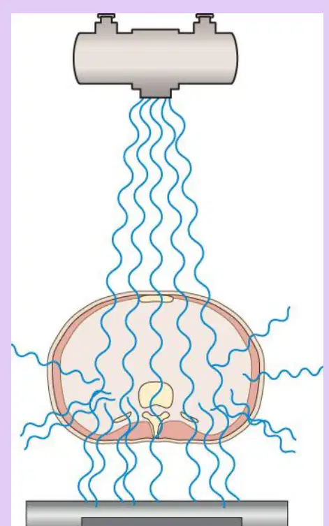
Refer to the schematic showing an x‑ray beam passing through a patient toward a detector. What concept does this visual support from the introduction?
That knowledge of photon interactions helps both reduce patient harm and improve image quality
That all photons are reflected by the patient and never reach the detector
That detectors work only when the beam is turned off
That image quality is unrelated to how photons interact with matter
Which statement best defines classical (coherent or Thomson) scattering in x‑ray interactions?
A low‑energy x‑ray photon changes direction after interacting with an orbital electron, with negligible energy transfer to the patient
A high‑energy x‑ray photon ejects an inner‑shell electron, creating a photoelectron and characteristic cascade
A mid‑energy x‑ray photon transfers part of its energy to an outer electron and continues with reduced energy
Multiple x‑ray photons combine to form a single higher‑energy photon inside tissue
According to the description of classical scattering, what primarily causes the photon to scatter?
Absorption by the nucleus followed by pair production
Interaction of a low‑energy incident x‑ray photon with an orbital electron, exciting the atom
Bremsstrahlung deceleration near the nucleus
Annihilation of a positron with an electron
In coherent (Thomson) scattering, how does patient dose compare to other interactions?
It is typically the highest contributor to patient dose because energy is deposited locally
It is moderate because part of the photon energy is transferred to an outer‑shell electron
It is very little to none because the energy is released as a scatter photon in a new direction
It is unpredictable because the photon energy is converted to mass
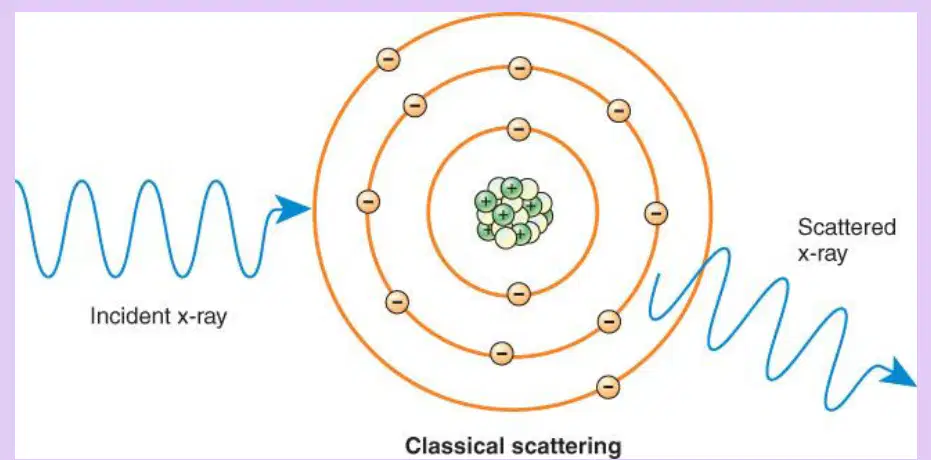
Refer to the diagram of classical scattering (wave enters atom and exits as a redirected wave). Which statement about the scattered photon is accurate?
It leaves with essentially the same energy as the incident photon but with a changed direction
It leaves with double the energy due to constructive interference inside the atom
It leaves with zero energy after complete absorption by the atom
It leaves only after ejecting an inner‑shell electron
Recall the diagnostic energy range in which the incident x-ray photon commonly causes photoelectric interactions.
2–12 kVp
20–120 kVp
120–320 kVp
0.2–1.2 MeV
During a photoelectric interaction, which electron does the incident x‑ray photon primarily interact with to cause ejection?
An outer‑shell valence electron
A free electron in the patient’s skin
An inner‑shell electron of a tissue atom
A neutron in the nucleus
What happens to the energy of the incident x‑ray photon at the moment the inner‑shell electron is ejected in a photoelectric interaction?
It is partially scattered and partially absorbed
It is totally absorbed by the inner‑shell electron
It is converted entirely into visible light
It is transferred to the nucleus as binding energy
The ejected electron produced by a photoelectric interaction is specifically called a:
Compton electron
Auger electron
Photoelectron
Pair‑production electron
Choose the correct expression for the kinetic energy of the photoelectron produced in a photoelectric interaction.
Kinetic energy = binding energy + incident photon energy
Kinetic energy = incident photon energy − binding energy of the electron’s original shell
Kinetic energy = incident photon energy × binding energy
Kinetic energy = incident photon energy only, because binding energy is negligible
Which statement best explains why photoelectric interactions contribute to patient dose?
They produce only high‑energy scatter that exits the patient
They completely absorb the incident photon’s energy in tissue and create an energetic photoelectron
They only deflect the photon without energy transfer
They occur exclusively in air rather than in tissue
Strategic thinking: A 60 keV x‑ray photon ejects an inner‑shell electron that had a binding energy of 40 keV. Based on the relationship provided, what is the photoelectron’s kinetic energy, and what does this imply for tissue dose?
20 keV; the photoelectron can further interact and increase patient dose
40 keV; no additional interactions will occur
60 keV; all energy escapes as scatter, lowering dose
0 keV; the electron remains bound so dose is minimal
Skill/Concept: After ejection, what can the new photoelectron do before being captured by another atom, and why is this important?
It loses all energy instantly, which reduces image contrast
It has enough kinetic energy to undergo additional interactions elsewhere, adding to patient dose
It annihilates with a positron, producing two 511 keV photons
It returns immediately to its original shell, eliminating dose
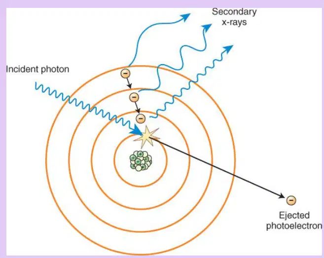
Visual reasoning: Refer to the diagram showing concentric electron shells, an incident photon striking an inner-shell electron, and an arrow showing an ejected particle leaving the atom. What does the long arrow pointing outward represent?
A secondary x‑ray returning to the nucleus
An ejected photoelectron carrying kinetic energy
An incident photon being reflected
A neutron emitted from the nucleus
When a photoelectric interaction ejects an inner-shell electron, what immediate atomic condition results?
The atom becomes ionized and unstable due to an inner-shell vacancy
The atom gains stability because outer electrons move outward
The atom emits a high-energy gamma ray that leaves the body
The atom becomes negatively charged but remains stable
Which sequence best explains how secondary x-ray photons are produced after a photoelectric event?
Compton scatter causes outer-shell excitation, releasing bremsstrahlung photons
A characteristic cascade fills an inner-shell vacancy, emitting characteristic radiation
Pair production creates an electron-positron pair that annihilates into photons
Coherent scatter aligns electron spins, releasing microwave radiation
According to the material, what is true about the energy and fate of secondary photons produced by characteristic cascades in tissue?
They are high energy and readily exit the body, reducing dose
They are low energy and are absorbed in tissue, increasing patient dose
They have variable energy and always improve image contrast without dose impact
They are neutral particles that do not interact with matter
Which statement best defines characteristic radiation in the context of photoelectric interactions?
Radiation produced when electrons decelerate near a nucleus
Radiation emitted when an electron transitions to fill an inner-shell vacancy
Radiation resulting from photon energy being redirected without ionization
Radiation produced only by external filtration of the x-ray beam
Which interaction contributes the most to patient dose in diagnostic imaging, according to the content?
Compton scattering
Coherent scattering
Photoelectric absorption
Pair production
What professional responsibility is emphasized for the radiographer regarding exposure parameters?
Maximize absorption to guarantee image formation regardless of dose
Select technical factors that balance image quality and patient dose
Eliminate all absorption to prevent any patient dose
Use the highest possible kVp to avoid photoelectric interactions
Complete the statement: In the context of these slides, "Photoelectric = _____."
partial transmission
complete absorption
coherent scattering
dose neutrality
A patient’s tissues undergo many low-energy characteristic emissions after inner-shell vacancies are filled. Based on this, what is the most likely outcome if all other factors remain the same?
Patient dose decreases because photons escape the body
Patient dose increases because these photons are absorbed in tissue
Image quality degrades because no absorption occurs
Scatter to the detector increases without affecting dose
Recall the first event in a photoelectric interaction. Which statement best describes what initially happens?
The incident photon is completely absorbed by an inner-shell electron
An outer-shell electron is ejected by partial photon absorption
The atom emits a secondary photon that strikes the incident photon
The nucleus absorbs the photon and releases a neutron
In a photoelectric interaction, the ejected electron has a specific name. What is it called?
Auger electron
Photoelectron
Compton electron
Valence electron
After an inner-shell vacancy is created during a photoelectric event, what happens next inside the atom?
The nucleus captures an electron, stopping all emissions
An outer electron drops down to fill the vacancy
The atom immediately ionizes a neighboring atom
The incident photon reappears with lower energy
The drop of an outer electron into an inner-shell vacancy releases what?
A bremsstrahlung x-ray
A secondary (characteristic) photon
A positron
A gamma cascade
Which statement best explains why characteristic photons from photoelectric interactions typically add to patient dose rather than image formation?
They are high energy and escape the body to the detector
They are scattered forward and increase detector signal
They are low energy and are absorbed in nearby tissues
They convert to heat before leaving the atom
Which factor increases the probability of a photoelectric interaction occurring in tissue?
Lower incident photon energy relative to inner-shell binding energy
Lower atomic number of the tissue atoms
Higher detector sensitivity to x-rays
Greater source-to-image distance
Consider two tissues: A with atomic number Z=7 and B with atomic number Z=20, exposed to the same x-ray beam. Based on photoelectric principles, which tissue has the higher chance of photoelectric absorption?
Tissue A (Z=7), because lower Z increases inner-shell binding
Tissue B (Z=20), because higher atomic number increases photoelectric absorption probability
Both have equal probability since photon energy is identical
Neither, because photoelectric effect is independent of atomic number
Which question directly assesses whether a photoelectric interaction can occur for a given photon?
Is the photon’s energy sufficient to eject an inner-shell electron?
Will the photon scatter at 180 degrees from the atom?
Does the atom have more neutrons than protons?
Is the photon traveling perpendicular to the detector?
Which energy range best characterizes the incident x-ray photon that typically undergoes Compton scattering in tissue?
Below 10 keV
20–40 keV moderate-energy
60–100 keV high-energy
Above 200 keV very high-energy
In Compton scattering, with which electron does the incident photon primarily interact to cause ionization?
A K-shell inner electron
An outer- or middle-shell orbital electron
A nucleus-bound proton
A free electron outside the atom
During Compton scattering, what typically happens to the incident photon as it ejects an electron from its shell?
It gains energy and continues straight
It gives up up to one third of its energy and is deflected
It is completely absorbed without deflection
It splits into two photons of equal energy
What is the ejected electron called after a Compton interaction, and what is a key consequence of its departure from the atom?
Photoelectron; decreases patient dose
Auger electron; stabilizes the atom
Compton (secondary) electron; can interact with adjacent atoms and add to patient dose
Beta particle; exits without further interactions
Which statement best describes the effect of Compton scatter photons on imaging?
They improve image sharpness by traveling directly to the receptor through the anatomy
They add scatter to the image because they travel in a new direction before reaching the receptor
They are always absorbed in tissue and never reach the receptor
They remove artifacts by neutralizing background radiation
Select the most accurate sequence of events for a Compton interaction.
Photon absorption by nucleus → pair production → annihilation
Incident moderate-energy photon hits inner-shell electron → electron captured → photon continues unchanged
Incident moderate-energy photon interacts with outer/middle-shell electron → electron ejected → photon deflected with reduced energy
Bremsstrahlung emission → characteristic radiation → photoelectric effect
A radiography setup uses predominantly 30 keV photons. Predict the dominant interaction with soft tissue and its primary patient-dose contributor based on this section.
Photoelectric effect dominates; dose from characteristic x-rays
Compton scattering dominates; dose from Compton (secondary) electrons
Coherent scatter dominates; negligible dose from electrons
Pair production dominates; dose from positron annihilation
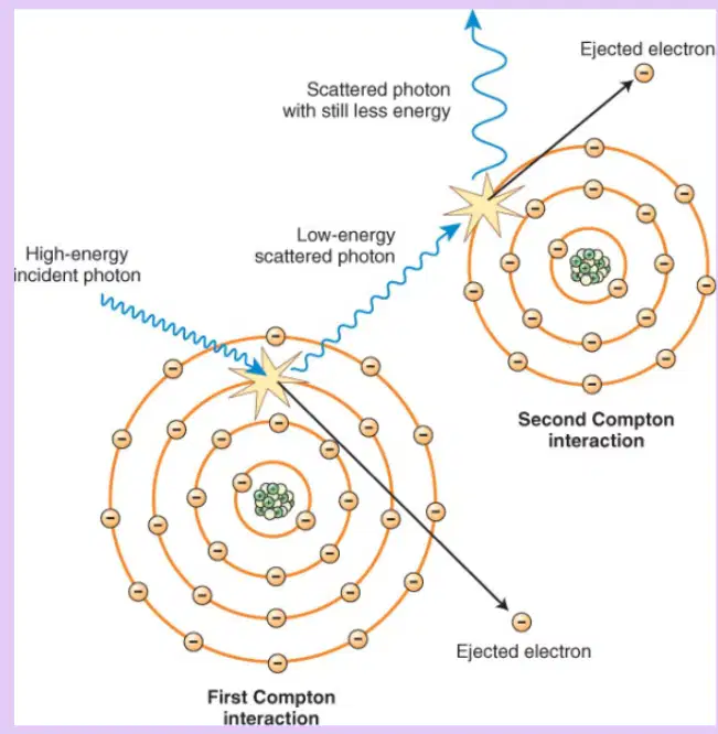
Refer to the diagram showing a high-energy incident photon striking an orbital electron and producing an ejected electron and a scattered photon. Which labeled particle is most likely to undergo further interactions in adjacent atoms, contributing to patient dose?
The scattered photon with higher remaining energy
The nucleus at the center of the atom
The ejected electron (Compton electron) leaving the atom
The inner-shell vacancy created after ejection
Which statement best describes how Compton scatter affects image quality in diagnostic imaging?
It produces uniform brightening by adding primary photons to the receptor
It strikes the image receptor in unintended locations, creating image fog
It increases subject contrast by selectively absorbing high-energy photons
It removes all scattered radiation before it reaches the detector
In general diagnostic imaging, Compton scatter is characterized as one of the most prevalent interactions between x‑ray photons and the human body. What is the most appropriate implication for radiographic practice?
Its frequency is low, so scatter control is optional
Its prevalence requires minimizing scatter production and exposure whenever possible
It only occurs in high‑atomic‑number tissues, so soft tissues are unaffected
It is negligible at all photon energies used clinically
According to the material, the probability of a Compton interaction depends primarily on which factor?
Atomic number of the irradiated atoms
Mass density of the patient alone
Energy of the incident photon
Thickness of the image receptor
Which statement about Compton scatter photons and occupational exposure is correct?
They lose nearly all energy and cannot leave the patient, so exposure to staff is rare
They retain about two thirds of their energy and can exit the patient, potentially exposing the radiographer
They are completely absorbed by lead aprons before leaving the patient
They only travel backward to the x‑ray tube and never toward staff
When should shielding be used during x‑ray procedures to limit occupational dose from Compton scatter?
Only when the exposure time exceeds 1 second
During any procedure where staff may be near the patient and x‑ray tube during exposure
Only in rooms without fixed barriers
Only for pediatric patients due to higher scatter
Which option best identifies the major source of occupational exposure to radiographers in diagnostic imaging?
Leakage radiation from the x‑ray tube housing
Primary beam transmission through the table
Background environmental radiation in the department
Compton scattering from the patient
A technologist increases kVp for a thicker patient. Based on the material, which change most directly influences the likelihood of Compton interactions?
Raising photon energy, which affects Compton probability
Changing the atomic number of tissue, which controls Compton probability
Altering grid ratio, which determines if Compton occurs
Adjusting SID, which eliminates Compton interactions
Which practice most directly addresses image fog created by Compton scatter?
Maximizing scattered radiation to boost receptor exposure
Minimizing Compton scatter through technique and shielding
Using tissues with higher atomic number to prevent Compton events
Removing the anti‑scatter grid to reduce attenuation
Which condition must be met for pair production to occur during an x-ray interaction with matter?
The photon energy is less than the binding energy of K-shell electrons
The incident x-ray photon has energy greater than 1.02 MeV and interacts with the nucleus
The photon scatters off an outer-shell electron with any energy
The photon is absorbed entirely by a single orbital electron
In pair production, what immediate products are created when a very-high-energy photon interacts with the nucleus?
Two electrons with opposite spin
A positron and an electron
Two lower-energy x-ray photons
A neutron and a proton
During pair production, why is the threshold energy approximately 1.02 MeV?
It equals the binding energy of the innermost electron shell
It is the energy needed to overcome nuclear repulsion
It corresponds to the combined rest-mass energy of an electron and a positron
It is the minimum to create two Compton-scattered photons
After pair production, each created particle must have what minimum energy to exist before further interactions?
0.25 MeV each
0.51 MeV each
1.02 MeV each
Varies with atomic number
What ultimately happens to the positron produced in pair production according to the instructional text?
It remains bound to the nucleus as a positronium state indefinitely
It quickly captures an electron to become a neutral atom
It travels until it strikes an electron, causing an annihilation event
It emits characteristic radiation and returns to the nucleus
What is produced during the annihilation event that follows the meeting of a positron with an electron?
One photon with energy 1.02 MeV
Two x-ray photons as the particle mass energy is converted
Bremsstrahlung radiation with a continuous spectrum
A single gamma ray with variable energy
Which statement best distinguishes pair production from interactions relevant to general radiography?
Pair production dominates image formation at standard diagnostic energies
Pair production does not occur in general radiography because photon energies are too low
Pair production primarily involves outer-shell electrons rather than the nucleus
Pair production requires high atomic number contrast media to proceed
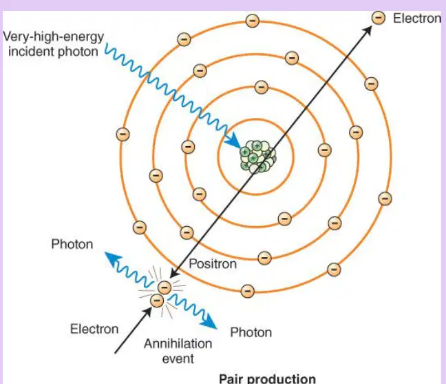
According to the diagram of pair production, where does the initial interaction of the very-high-energy photon occur?
With an orbital electron in the K-shell
With the nucleus of the tissue atom, leading to creation of a positron and an electron
With the crystal lattice of the detector
With a free electron outside the atom
Which statement best defines photodisintegration in atomic interactions?
Absorption of low-energy photons by outer-shell electrons causing heat
Scattering of moderate-energy photons by orbital electrons without ionization
Interaction in which extremely high-energy photons strike the nucleus and make it unstable
Electron capture by the nucleus leading to characteristic radiation
In photodisintegration, how does the atom regain stability after the nucleus becomes unstable?
By emitting a bremsstrahlung photon from the electron cloud
By ejecting a nuclear particle such as a proton or neutron
By filling an inner-shell vacancy with an outer-shell electron
By spontaneously reducing photon energy through elastic scattering
Which scenario most likely involves photodisintegration rather than other photon interactions?
A diagnostic x-ray photon of typical radiography energy interacting with an outer-shell electron
An extremely high-energy photon directly interacting with the atomic nucleus and causing a particle to be expelled
A moderate-energy photon changing direction after colliding with an electron
An incident photon being completely absorbed in the photoelectric effect
What is the relevance of photodisintegration to routine radiography practice?
It is the dominant interaction that forms radiographic contrast
It occasionally occurs and slightly increases patient dose
It does not occur in radiography due to the energy levels used
It is only observed when using contrast media
Which statement best defines attenuation in diagnostic imaging?
Complete transmission of x-ray photons through tissue without interaction
Increase in photon number as they pass through tissue due to scattering
Photon loss as x-ray photons interact with tissue and are absorbed or scattered
Conversion of x-ray photons into visible light within the image receptor
Differential absorption most directly explains which outcome in an x-ray image?
Uniform brightness because all tissues absorb photons equally
Image contrast because different tissues absorb different amounts of radiation
Motion blur because tissues move during exposure
Magnification of structures due to beam divergence
According to the content, which tissues are most likely to appear white on an x‑ray image?
Soft tissue and fat due to low atomic number and low absorption
Lung tissue because it scatters most photons
Bone and contrast media due to high atomic number and greater photon absorption
Air-filled regions because they transmit no photons
A technologist wants to increase visualization of soft tissue detail. Based on differential absorption, which change would most likely increase contrast between soft tissue and bone?
Select anatomy with similar densities so attenuation is equal
Use a contrast agent to raise the atomic number in the soft tissue region
Decrease photon interactions so all photons reach the receptor
Eliminate scattering so no photons are absorbed
Which statement correctly contrasts transmission and absorption?
Transmission refers to photons that are attenuated by the body and stopped before the receptor; absorption refers to photons that reach the receptor
Transmission refers to x-ray photons that pass through the body to the image receptor; absorption refers to photons attenuated by the body that do not reach the receptor
Both transmission and absorption describe photons that become scatter radiation
Transmission is caused by photoelectric interactions, while absorption is due to Compton scatter only
Which pairing correctly matches radiographic term with example and property?
Radiolucent—bone; readily absorbs x-rays
Radiopaque—air; much lower probability of absorption
Radiolucent—air; less dense with lower probability of absorption
Radiopaque—soft tissue; transmits most photons
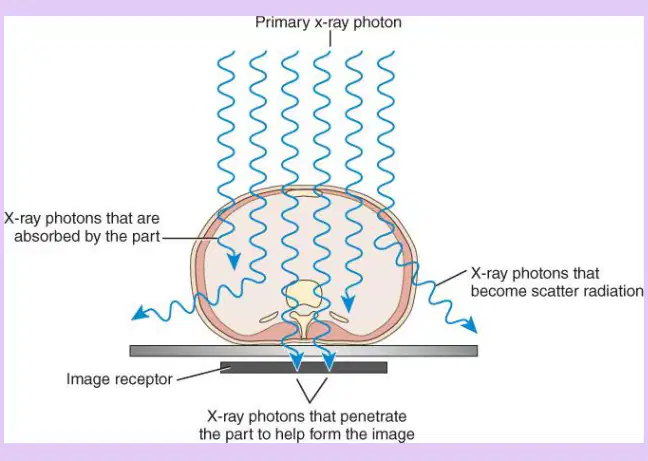
Refer to the diagram showing incident x-ray photons, absorbed photons, scattered photons, and photons reaching the image receptor. Which path of photons is primarily responsible for forming useful image signal?
Photons that are absorbed by the part and never reach the receptor
Photons that become scatter radiation and deviate from the primary beam
Photons that penetrate the part and reach the image receptor
Photons reflected off the skin surface back to the source
A region appears gray to black on an x-ray image. Based on attenuation principles, what is the most likely explanation?
The region contains lower density tissues that absorb fewer photons, allowing more to reach the receptor
The region contains high atomic number material that absorbs more photons, decreasing receptor exposure
The region contains tissues that do not transmit or scatter any photons
The region has equal attenuation to surrounding bone

