
 Worksheets
Cardiovascular Exam
Total questions: 121
Worksheet time: 1hrs 1mins
Which statement best explains why the muscle wall of the left ventricle is thicker than that of the right ventricle?
It pumps blood only to the lungs, which are nearby.
It must generate higher pressure to pump blood to the entire body.
It receives oxygen directly from the aorta, making it thicker.
It stores extra blood before it enters the atria.
Pulmonary circulation is defined as the movement of blood from the heart to the lungs for oxygenation and then back to the heart. Which pathway does this describe?
Right ventricle → pulmonary arteries → lungs → left atrium
Left ventricle → aorta → body tissues → right atrium
Left atrium → coronary arteries → heart muscle → right ventricle
Right atrium → vena cavae → body → left ventricle
Cardiac circulation primarily serves which tissue with nutrients and oxygen?
Lung tissue
Brain tissue
Heart muscle
Skeletal muscle
In cardiac circulation, venous blood from the heart muscle gathers in the coronary sinus and drains where?
Into the left atrium through the mitral valve
Directly through separate openings into the right atrium
Into the superior vena cava
Into the pulmonary veins
Systemic circulation is best described as blood leaving the heart through the aorta to deliver oxygen and nutrients to what?
Only the lungs
All other parts of the human body
Only the heart muscle
Only the brain and kidneys
Which valve controls blood flow from the right atrium to the right ventricle?
Mitral (bicuspid) valve
Tricuspid valve
Aortic valve
Pulmonary valve
Which pair correctly matches the valve with the chambers it separates?
Mitral valve: right atrium and right ventricle
Tricuspid valve: left atrium and left ventricle
Aortic valve: left ventricle and aorta
Pulmonary valve: left ventricle and pulmonary artery
Heart valves have "leaflets" called what?
Chordae tendineae
Papillary muscles
Cusps
Septae
The mitral valve has how many cusps, giving rise to its other name?
One cusp, called unicuspid
Two cusps, called bicuspid
Three cusps, called tricuspid
Four cusps, called quadricuspid
The tricuspid valve typically has how many cusps?
Two
Three
Four
Five
Cord-like tissues that hold atrioventricular valves in place are called what?
Chordae tendineae
Purkinje fibers
Coronary sinuses
Papillae
These cords are anchored within the ventricles by which muscles?
Smooth muscles
Papillary muscles
Intercostal muscles
Skeletal muscles
Which arteries supply the heart muscle itself with the freshest blood from the aorta?
Carotid arteries
Pulmonary arteries
Right and left coronary arteries
Renal arteries
Where is the main coronary artery located relative to the aortic valve?
Far from the aortic valve in the descending aorta
At the location of the aortic valve as the first blood vessel off the aorta
Behind the pulmonary valve in the pulmonary trunk
Within the right atrium next to the coronary sinus
Which statement correctly contrasts pulmonary and systemic circulation?
Pulmonary circulation delivers blood to body tissues; systemic circulation delivers blood to the lungs.
Pulmonary circulation oxygenates blood in the lungs; systemic circulation distributes oxygenated blood to body tissues.
Both pulmonary and systemic circulation only serve the heart muscle.
Systemic circulation returns blood to the lungs for oxygenation; pulmonary circulation carries it to the aorta.
Identify the correct route for venous return from the heart muscle described in the material.
Cardiac veins → pulmonary veins → right atrium
Cardiac veins → coronary sinus → right atrium
Cardiac veins → superior vena cava → right atrium
Cardiac veins → inferior vena cava → right atrium
Which task of the heart is emphasized in the lesson overview?
To store oxygen in the ventricles
To pump blood through distinct circulatory systems
To produce hormones for digestion
To filter waste from blood like kidneys
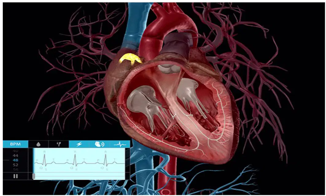
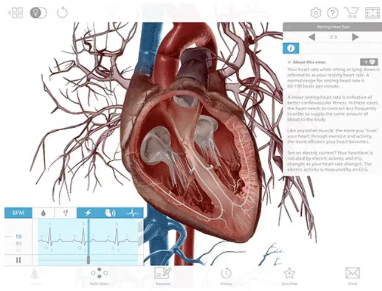
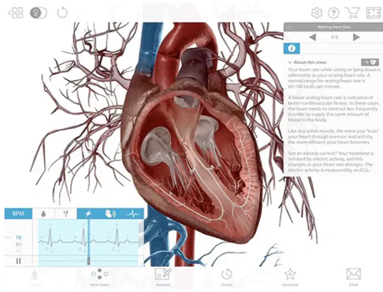
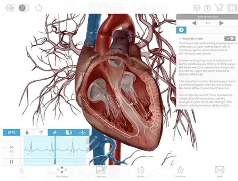
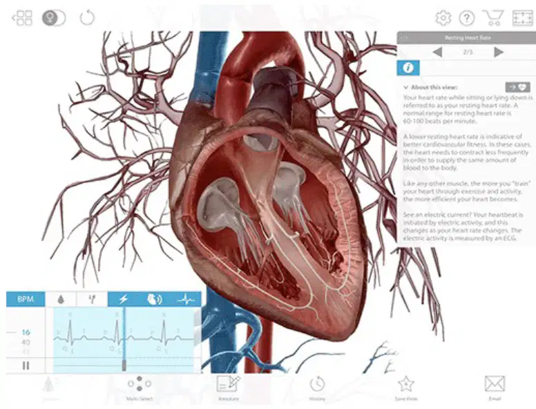
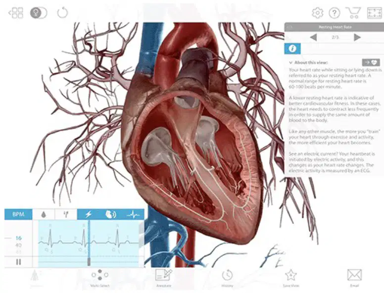
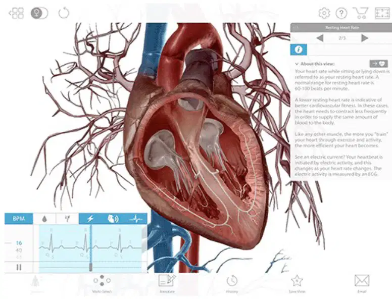
In the image of the heart with highlighted internal structures and an ECG panel, what tool is mentioned as the medical diagnostic device for electrical activity?
EEG (electroencephalogram)
ECG (electrocardiogram) often called an EKG
EMG (electromyogram)
Echo (echocardiogram)
In heart anatomy, what is meant by saying the coronary arteries are end arteries? Choose the best definition.
They form many large collateral loops that can bypass blockages easily.
They terminate in capillary beds without significant alternate routes, so blockage can deprive tissue of blood.
They drain directly into the coronary sinus rather than into veins.
They only supply the atria and not the ventricles.
Which statement best distinguishes a Right Dominant Heart from a Left Dominant Heart?
Right dominant heart: posterior interventricular artery arises from the right coronary artery; left dominant heart: posterior interventricular artery arises from the circumflex artery.
Right dominant heart: SA node supplied only by the left coronary; left dominant heart: SA node supplied only by the right coronary.
Right dominant heart: coronary sinus drains to the left atrium; left dominant heart: coronary sinus drains to the right atrium.
Right dominant heart: ventricles contract during diastole; left dominant heart: atria contract during systole.
Which coronary artery branch most commonly supplies the sinoatrial (SA) node, the heart’s pacemaker?
A branch of the right coronary artery dedicated to the SA node
The great cardiac vein
The coronary sinus
The circumflex vein of Marshall
What is the primary function of the coronary sinus?
To deliver oxygenated blood to the left ventricle
To collect deoxygenated blood from coronary veins and return it to the right atrium
To initiate the heartbeat as the pacemaker
To open and close the atrioventricular valves
Where does the coronary sinus open into the heart?
Into the left ventricle near the aortic valve
Into the right atrium near the opening of the inferior vena cava and the right atrioventricular orifice
Into the pulmonary trunk at the bifurcation
Into the left atrium within the left atrioventricular groove
Which statement correctly describes systole compared to diastole?
Systole is the heart’s relaxation phase; diastole is contraction.
Systole is the contraction phase generating higher pressure; diastole is the relaxation phase with lower pressure.
Both systole and diastole are relaxation phases with equal pressures.
Systole only occurs in the atria, while diastole only occurs in the ventricles.
Normal blood pressure is about 120 mmHg during systole and about 80 mmHg during diastole. What best explains why systolic pressure is higher?
Systole reflects ventricular contraction propelling blood forcefully into arteries.
Diastole reflects ventricular contraction being stronger than systole.
Systole occurs when the valves are closed, trapping blood.
Diastole occurs only in the atria, increasing pressure there.
Which description best defines the sinoatrial (SA) node?
A venous reservoir that drains the heart
The pacemaker region that initiates the heartbeat
The valve guarding the coronary sinus opening
A muscular ridge that separates the ventricles
What are Purkinje fibers primarily responsible for in cardiac conduction?
Collecting venous blood from the myocardium
Delivering electrical impulses rapidly throughout the ventricular walls for coordinated contraction
Regulating coronary artery diameter to control flow
Producing lymphatic fluid for the pericardial cavity
What is the main function of the atrioventricular (AV) node?
To initiate atrial depolarization faster than the SA node
To delay and relay electrical impulses from the atria to the ventricles
To open the pulmonary semilunar valve
To drain blood into the coronary sinus
Which statement about the pericardium is most accurate?
The heart is covered by the visceral pericardium, and the cardiac cavity is lined by the parietal pericardium; a small amount of fluid separates them to reduce friction.
The heart is covered by the parietal pericardium only, with no fluid present.
The pericardium is a vein that drains into the coronary sinus.
The pericardium produces electrical impulses for the SA node.
Which structure helps guard the opening of the coronary sinus into the right atrium?
Chordae tendineae
Papillary muscle
Thebesian valve (endocardial fold)
Ligamentum arteriosum
Which veins are tributaries to the coronary sinus according to the description?
Great cardiac vein, small and middle cardiac veins, and inferior vein of the left ventricle
Superior vena cava, pulmonary veins, and azygos vein
Internal thoracic vein, cephalic vein, and basilic vein
Portal vein, splenic vein, and superior mesenteric vein
Which layer directly lines the inside chambers of the heart and is continuous with the valves?
Pericardium
Epicardium
Myocardium
Endocardium
What is the tough, protective sac that surrounds the heart?
Pericardium
Endocardium
Interatrial septum
Coronary sinus
The epicardium is best described as which of the following?
Outer surface layer of the heart
Inner lining of the heart chambers
Muscular middle layer of the heart
Fibrous sac outside the heart
Which structure separates the right and left atria?
Interventricular septum
Interatrial septum
Coronary sinus
Tricuspid valve
Which structure separates the right and left ventricles?
Interatrial septum
Interventricular septum
Bicuspid (mitral) valve
Pulmonary valve
Which statement best describes the coronary arteries?
They carry oxygen-poor blood from the heart to the lungs
They supply oxygen and nutrients to the heart muscle
They drain deoxygenated blood into the coronary sinus
They form the valves between atria and ventricles
Which vessel collects most venous blood from the heart muscle before it enters the right atrium?
Pulmonary vein
Aorta
Coronary sinus
Superior vena cava
Where does blood go immediately after leaving the coronary sinus?
Left atrium
Right atrium
Left ventricle
Pulmonary artery
Which valve is located between the right atrium and right ventricle?
Bicuspid (mitral) valve
Aortic valve
Tricuspid valve
Pulmonary valve
Which valve is located between the left atrium and left ventricle?
Tricuspid valve
Pulmonary valve
Bicuspid (mitral) valve
Coronary valve
Which statement correctly contrasts the left and right sides of the heart in a double-pump model?
Right side pumps to the body; left side pumps to the lungs
Left side pumps oxygenated blood to the body; right side pumps deoxygenated blood to the lungs
Both sides pump deoxygenated blood to the body
Both sides pump oxygenated blood to the lungs
Which pair correctly names the upper receiving chambers of the heart?
Left and right ventricles
Left and right atria
Aorta and pulmonary trunk
Coronary sinus and superior vena cava
Which pair correctly names the lower pumping chambers of the heart?
Left and right atria
Left and right ventricles
Aorta and vena cava
Pulmonary veins
Which option best explains where blood entering the right atrium typically comes from?
Pulmonary veins carrying oxygen-rich blood
Systemic veins such as the superior and inferior vena cava
Coronary arteries bringing oxygen
Aorta delivering high-pressure blood
Which option best explains where blood entering the left atrium typically comes from?
Pulmonary veins returning oxygen-rich blood from the lungs
Systemic veins carrying deoxygenated blood
Coronary sinus draining the myocardium
Aorta feeding the chambers
Which coronary vessels are arteries rather than veins?
Great cardiac, middle cardiac, small cardiac
Left coronary, right coronary, circumflex
Coronary sinus, anterior cardiac veins, small cardiac
Superior vena cava, inferior vena cava, coronary sinus
Which statement best matches the function of the interatrial and intraventricular septa?
They allow mixing of oxygenated and deoxygenated blood
They separate the chambers to keep right and left heart flows distinct
They act as valves to prevent backflow
They are part of the pericardial sac
Which structure acts as the heart’s natural pacemaker by initiating the electrical impulse that starts each heartbeat?
Atrio-Ventricular (AV) node
Bundle of His
Sino-Atrial (SA) node
Purkinje fibers
What is the primary role of the Atrio-Ventricular (AV) node in the heart’s conduction system?
It speeds up the impulse so the ventricles contract before the atria.
It delays the impulse to allow the atria to finish contracting before the ventricles.
It generates the first impulse that sets the heart rate.
It distributes impulses through the ventricular walls for coordinated contraction.
Which pathway conducts the impulse from the AV node down the interventricular septum toward the ventricles?
Purkinje fibers
Bundle of His
SA node
Papillary muscles
Which structures rapidly spread the electrical signal throughout the ventricular walls to produce a coordinated ventricular contraction?
Sino-Atrial node
Aortic semilunar valve
Purkinje fibers
Cordae tendinea
Which statement best defines an ECG for a patient undergoing the test?
A painful procedure that uses needles to measure heart muscle movement
A noninvasive method that measures the heart’s electrical activity during the cardiac cycle
A scan that measures the heart’s blood flow using ultrasound
A surgical test that directly inspects the heart’s chambers
What useful information can doctors determine from an ECG?
Whether the heart’s valves are physically torn
Whether a patient’s heart rate is normal or abnormal and if parts of the heart are getting overworked
Exactly how much oxygen is in each blood cell
The total amount of blood pumped in a day
What is another common abbreviation for an ECG and why does it exist?
EHG, from the English term electro-heart graph
EKG, from the Dutch term elektrokardiogram used by Einthoven
ECX, from the Latin term electrocardius
EKC, from the German term elektrocardium

How is an ECG administered to measure heart activity without entering the body?
By placing a stethoscope on the chest and recording sounds
By using skin electrodes connected by wires to a machine that plots voltage over time
By injecting dye into the bloodstream to trace pathways
By using a thermometer to track heat changes in the heart
In a standard 12-lead ECG, how are the leads obtained?
Using 12 separate machines placed around the room
Using 12 electrodes on the chest only
Using 10 electrodes that provide 12 different electrical views of the heart
Using 8 electrodes that create 10 views of the lungs
Where are the electrodes placed for a typical 12-lead ECG?
Eight on the back and two on the neck
Six on the chest and one on each limb
Ten on the abdomen
Four on the chest and six on the scalp
Which waves make up one round of the cardiac cycle on an ECG?
P wave, QRS complex, and T wave
A wave, B complex, and C wave
Q wave, R wave, and S wave only
Delta wave and gamma wave
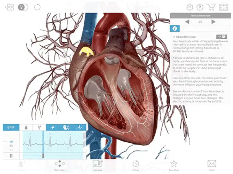
What does the P wave on an ECG represent?
Electrical activity of ventricular contraction
Electrical signal that begins at the SA node and passes to the AV node, leading to atrial stimulation
Recovery period between contraction and relaxation
Return to baseline after ventricular depolarization
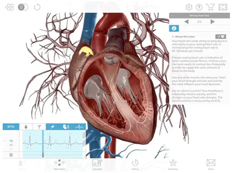
What immediate effect does the electrical signal traveling from the SA node to the AV node have on the heart’s chambers?
It relaxes the ventricles, stopping blood flow
It stimulates the atria to contract, pushing blood into the ventricles
It opens the heart valves permanently
It causes the aorta to constrict
Which statement correctly describes the PR interval on an ECG?
The time between the end of the QRS complex and the start of the T wave
The time between the start of the P wave and the start (first deflection) of the QRS complex
The time from the peak of the R wave to the end of the T wave
The duration of the T wave alone
What does the QRS complex primarily reflect?
Electrical signals leading to ventricular contraction
Atrial relaxation
The heart’s recovery to baseline
Movement of blood into the atria
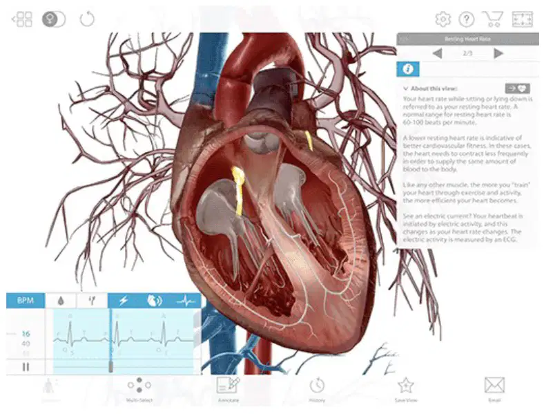
Which pathway correctly traces the electrical signal after it reaches the AV node?
Directly to the aorta and pulmonary artery
To the atrioventricular bundle (bundle of His), down bundle fibers to Purkinje fibers
Back to the SA node for recycling
Straight to the ventricles without specialized pathways
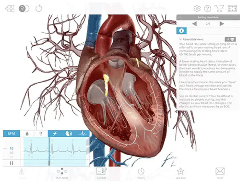
What mechanical action in the heart is stimulated by the conduction through the Purkinje fibers?
Atrial relaxation and valve closure
Ventricular contraction pushing blood into the pulmonary artery and aorta
Coronary artery dilation only
Complete stop of heart movement
Which description correctly defines the ST interval (ST segment)?
Time between the P wave and the start of the QRS complex
Time between the end of the QRS complex and the start of the T wave, representing the period of zero potential between ventricular depolarization and repolarization
Time from the beginning of the T wave to the next P wave
Time of atrial repolarization clearly visible on the ECG
What does the T wave represent on an ECG?
Ventricular depolarization leading to contraction
Atrial depolarization only
Electrical activity returning to baseline—ventricular repolarization
Mechanical relaxation of the atria
If an ECG shows an unusually wide QRS complex, which process is most likely affected based on what the QRS represents?
Atrial depolarization is delayed
Ventricular depolarization and conduction through bundle branches are altered
Ventricular repolarization is faster than normal
The SA node stopped initiating signals
Why does the electrical signal on an ECG appear slightly before the heart’s muscle movement?
Because the ECG records mechanical motion first
Because electrical activation triggers contraction, so the electrical event precedes the physical movement
Because the ECG records blood pressure changes, which lag behind motion
Because electrodes cause a delay in sensing
During a 12-lead ECG, how many different views of the heart’s electrical activity are produced, and with how many electrodes?
12 views using 12 electrodes
10 views using 10 electrodes
12 views using 10 electrodes
8 views using 6 electrodes
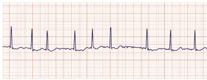
Which heart condition is identified on an ECG by the absence of a normal P wave with QRS complexes occurring at irregular intervals?
Atrial fibrillation
Atrial flutter
Ventricular fibrillation
Myocardial infarction

On an ECG, a sawtooth pattern of the P wave most closely indicates which condition?
Atrial flutter
Atrial fibrillation
ST elevation
Ventricular fibrillation
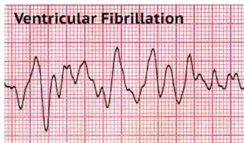
Which statement best describes ventricular fibrillation as seen on an ECG?
Chaotic, rapid baseline with no organized QRS complexes
Regular rhythm with missing P waves
Normal P-QRS-T pattern with slight ST depression
Sawtooth P waves preceding each QRS
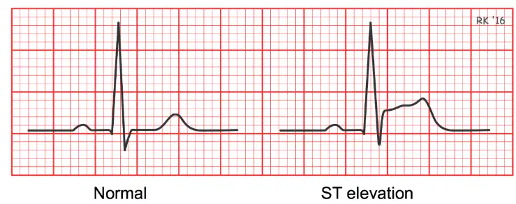
Elevation of the ST segment on certain leads can be a sign of which condition?
Myocardial infarction (heart attack)
Atrial fibrillation
Atrial flutter
Sinus bradycardia
Which best explains why ECGs are important in cardiology?
They assess the heart’s electrical conduction to diagnose and monitor conditions
They directly measure the amount of oxygen in the blood
They visualize coronary arteries without imaging
They replace the need for medications
Which symptom is most likely to lead a clinician to order an ECG?
Chest pain or shortness of breath
Vision changes only
Skin rash without other symptoms
Joint stiffness after exercise
Which statement about atrial fibrillation outcomes is supported by the material?
Untreated AFib increases risk of death and stroke
AFib always causes ST elevation
AFib eliminates QRS complexes
AFib is the same as atrial flutter
Which waveform component corresponds to the ventricles relaxing after repolarization?
T wave phase following QRS
P wave before QRS
PR interval only
Sawtooth P wave
Which heart condition most directly results from blocked coronary arteries preventing adequate blood flow to heart muscle?
Myocardial infarction
Atrial flutter
Ventricular fibrillation always
Sinus arrhythmia
Which best describes the role of ECG in monitoring treatments like pacemakers and medications?
ECG tracks electrical effects of treatments on heart rhythm
ECG measures blood sugar levels directly
ECG replaces physical exams entirely
ECG is only used during surgery
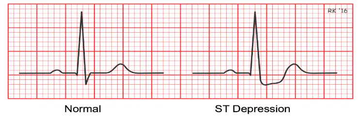
If an ECG shows ST depression on multiple leads, which process is most likely occurring in the heart tissue?
Ischemia causing reduced oxygen to tissue
Hyperoxygenation of myocardium
Complete atrioventricular block
Immediate ventricular fibrillation
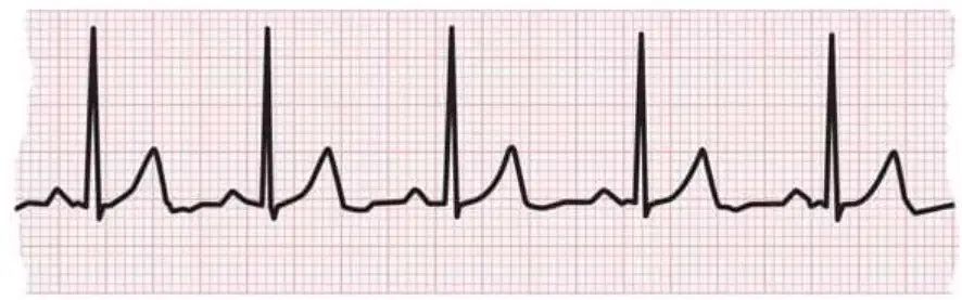
Which statement correctly compares atrial fibrillation and atrial flutter?
AFib lacks normal P waves; flutter shows sawtooth P waves
AFib and flutter show identical patterns
Flutter lacks QRS complexes; AFib does not
AFib always shows ST elevation; flutter shows ST depression
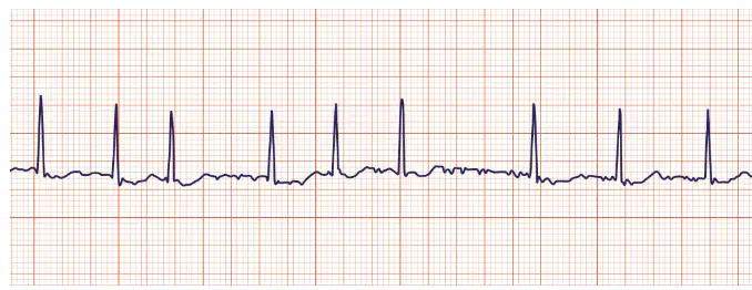
Which ECG feature remains present during atrial fibrillation even though P waves are absent?
QRS complexes at irregular intervals
Consistent sawtooth P waves
Elevated ST segments on all leads
Wide, regular T waves only
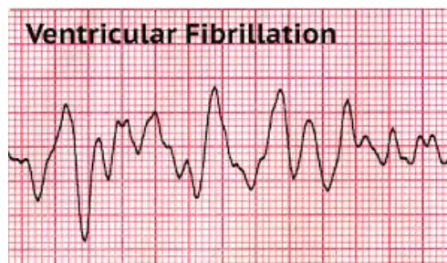
What is a likely immediate effect of ventricular fibrillation on the patient’s ability to function?
Severe inability to pump blood effectively
Mild increase in heart rate only
Improved cardiac output
No change unless ST elevation occurs
Which of the following is NOT listed as a heart condition ECGs can help identify or monitor?
Blocked arteries
Heart failure
High blood pressure only
Arrhythmias
In future technology, ECG might noninvasively detect episodes of which metabolic condition, especially relevant for people with diabetes?
Hypoglycemia (low blood sugar)
Hypercalcemia (high calcium)
Hypernatremia (high sodium)
Ketonemia (high ketones)
Which structure acts as the heart’s primary pacemaker by initiating rhythmic electrical impulses?
Sinoatrial (SA) node
Atrioventricular (AV) node
Bundle of His
Purkinje fibers
If the heart’s primary pacemaker becomes damaged, what can happen to keep the heartbeat going?
Other specialized cardiac cells can generate pacemaker activity at a slower rate
The heart immediately stops beating with no backup mechanism
Skeletal muscle cells take over as the pacemaker
Only the brain can replace the pacemaker function via motor neurons
What is the main advantage of gap junctions between cardiac muscle cells?
They allow rapid electrical coupling so the heart contracts as a coordinated unit
They store extra calcium for stronger contractions
They form thick connective tissue that prevents overstretching
They slow electrical signals to save energy
In typical skeletal muscle, the resting potential is about −70 mV and the threshold is about −55 mV. Cardiac pacemaker cells do not have a true resting potential. What best describes this difference?
Pacemaker cells slowly depolarize on their own toward threshold
Pacemaker cells remain fixed at −70 mV without change
Pacemaker cells require external nerves to reach threshold
Pacemaker cells hyperpolarize to −90 mV before every beat
The plateau phase of the cardiac action potential depends on high levels of which ion in the sarcoplasmic reticulum and extracellular fluid?
Calcium (Ca2+)
Sodium (Na+)
Potassium (K+)
Chloride (Cl−)
Why is the plateau phase critical for proper heart function?
It prolongs contraction, allowing time for coordinated pumping of blood
It makes contractions shorter so the heart can beat faster
It stops calcium entry to prevent any contraction
It only helps the atria while the ventricles rest
Compared with skeletal muscle, which has a total refractory period of about 1 millisecond, what is true about the refractory period of cardiac muscle?
It is much longer, preventing tetany and ensuring rhythmic beats
It is identical, allowing sustained tetanic contractions
It is shorter, enabling the heart to tetanize
It varies randomly from beat to beat without a pattern
Which term best describes the procedure of puncturing a vein to draw blood or deliver medication?
Arteriotomy
Venipuncture
Phlebotomy training
Capillary sampling
Which vessel type typically carries blood away from the heart?
Veins
Arteries
Venules
Capillaries
Which vessel type typically carries blood toward the heart?
Arteries
Arterioles
Capillaries
Veins
Where are capillaries primarily located, and what is their main function?
Inside the heart; to generate blood pressure
Between arteries and veins in body tissues; to allow exchange of gases and nutrients
In the lungs only; to store oxygen
Under the skin; to regulate body temperature
Which statement correctly differentiates arteries from veins?
Arteries have valves; veins do not
Veins have thicker muscular walls than arteries
Arteries carry blood away from the heart; veins carry blood toward the heart
Both arteries and veins always carry oxygenated blood
Which layer of an artery contains smooth muscle that adjusts vessel diameter?
Tunica intima
Tunica media
Tunica externa
Endocardium
Which outer protective layer of an artery is composed mostly of connective tissue?
Tunica externa
Tunica media
Tunica intima
Myocardium
Which inner layer of an artery provides a smooth lining to reduce friction as blood flows?
Tunica media
Tunica externa
Tunica intima
Endothelium of the myocardium
Which condition involves a balloon-like bulge in a weakened arterial wall that can rupture?
Aneurism
Hemorrhage
Varicose vein
Ischemia
Which term refers to active bleeding due to broken blood vessels?
Aneurism
Hemorrhage
Phlebitis
DVT
Which condition describes enlarged, twisted superficial veins often in the legs due to valve failure?
Varicose veins
Arteriosclerosis
Atherosclerosis
Ischemia
Which term describes hardening and loss of elasticity of arterial walls, often with age?
Aneurism
Arteriosclerosis
Phlebitis
Hypotension
Which condition is specifically the build-up of fatty plaques within arteries that can narrow the lumen?
Atherosclerosis
Arteriosclerosis
Phlebitis
TIA
What is ischemia?
Excess blood flow to tissues causing swelling
Inadequate blood supply to tissues leading to oxygen shortage
Formation of clots in deep veins of the legs
Inflammation of arteries due to infection
Transient Ischemic Attack (TIA) is best described as which event?
A brief, reversible reduction of blood flow to the brain causing short-lived neurological symptoms
Permanent blockage of a coronary artery causing heart attack
Chronic high blood pressure with no symptoms
A rupture of the aorta resulting in immediate hemorrhage
Which phrase matches cerebrovascular incident (stroke)?
Temporary muscle spasm in leg arteries
Disorder of veins returning blood to the heart
Loss of brain function due to interrupted blood flow
Widening of capillaries in the skin
Hypertension is defined as which condition?
Abnormally low blood pressure
Abnormally high blood pressure
Normal blood pressure with occasional spikes
Blocked lymphatic drainage
Hypotension is defined as which condition?
Abnormally high blood pressure
Abnormally low blood pressure
Normal blood pressure after exercise
Clot formation in the deep veins
Deep vein thrombosis (DVT) primarily involves which problem?
Plaque build-up in coronary arteries
Clot formation in deep veins, often in the legs
Swelling of arterioles in the hands
Rupture of cerebral arteries
Peripheral Vascular Disease (PVD) most commonly means which situation?
A disease of vessels outside the heart and brain causing poor limb circulation
Inflammation of the heart valves only
A disorder limited to pulmonary arteries
An infection of capillaries in the skin
Intermittent claudication is best described as which symptom pattern?
Sudden chest pain during sleep
Leg pain during activity that improves with rest due to reduced blood flow
Persistent abdominal cramps unrelated to activity
Headache triggered by high altitude
Phlebitis refers to which condition?
Inflammation of a vein
Inflammation of an artery
Hardening of capillaries
Enlargement of the aorta
Which arteries first branch from the aorta to supply the heart itself?
Renal arteries
Carotid arteries
Coronary arteries
Subclavian arteries
Which sequence correctly names the three major branches of the aortic arch to reach the head and upper limbs?
Coronary, renal, femoral
Brachiocephalic trunk, left common carotid, left subclavian
Right subclavian, left coronary, celiac trunk
Inferior vena cava, superior vena cava, pulmonary trunk
Which arteries branching from the abdominal aorta primarily supply the stomach, spleen, and liver?
Celiac trunk and its branches
Common iliac arteries
Pulmonary arteries
Carotid arteries
Which arteries supply the kidneys directly from the abdominal aorta?
Femoral arteries
Renal arteries
Mesenteric arteries
Subclavian arteries
Starting at the common iliac arteries, which vessel directly continues down the thigh to reach the knee region?
Brachial artery
Femoral artery
Radial artery
Carotid artery
Which veins return blood from abdominal organs to the inferior vena cava primarily through the hepatic portal system and then the hepatic veins?
Pulmonary veins
Hepatic veins after portal circulation
Jugular veins
Great saphenous veins

