
 Worksheets
Embryology I
Total questions: 142
Worksheet time: 2hrs 21mins
Choose the three correct statements!
In the fifth month of prenatal development, the total number of female germ cells in the ovary is approx. 7 million.
At birth, the total number of female germ cells in the ovary is significantly lower than in the fifth month of prenatal development.
During childhood, most oocytes become atretic; fewer than 500 will be ovulated.
In the fifth month of prenatal development, the total number of female germ cells in the ovary is approx. 2 million.
At puberty, the total number of female germ cells in the ovary is approx. 400.
Choose the only correct statement!
The process of spermatogenesis and spermiogenesis takes approx. 74 days.
The process of spermatogenesis and spermiogenesis takes approx. 4 days.
The process of spermatogenesis takes approx. 36 days, the spermiogenesis takes approx. 64 days.
The process of spermatogenesis and spermiogenesis takes approx. 100 day.
The process of spermatogenesis and spermiogenesis shows individual variation.
Choose the only correct statement!
The viability of sperm cell is 4-5 days.
The viability of sperm cell is 1-2 days.
The viability of sperm cell is 1-2 weeks.
The viability of sperm cell is 15 days.
The viability of sperm cell is 4 weeks.
Choose the only correct statement!
The viability of oocyte is 24 hours.
The viability of oocyte is 2 hours.
The viability of oocyte is 4 hours.
The viability of oocyte is 10 days.
The viability of oocyte is 30-40 hours.
Choose the two correct statements!
The average ejaculate of semen contains 40-100 million/mL.
The average ejaculate of semen has a volume of about 3 mL.
The average ejaculate of semen has a volume of about 1 mL.
The average ejaculate of semen contains 100 000/mL.
The average ejaculate of semen contains 1-2 million/mL.
Choose the three correct statements!
The semen contains prostaglandins.
The semen contains fructose.
In the semen, the fructose is the source of energy for the sperm cells.
In the semen, the ATP is the source of energy for the sperm cells.
In the semen, the ATP and the fructose are the source of energy for the sperm cells.
Choose the four correct statements!
Gametogenesis is the process whereby the germ cells are converted into male and female gametes.
During the second week of embryonic period, the primordial germ cells are formed in the epiblast, then move through the primitive streak and migrate to the wall of the yolk sac.
Gametogenesis includes the reduction of number of chromosomes and cytodifferentiation.
During the fourth week of embryonic period, the primordial germ cells migrate from the yolk to the developing gonads.
During the gametogenesis the primordial germ cell are formed in the wall of the yolk sac.
Choose the only correct statement!
Oogenesis is the process whereby the oogonia are converted into mature oocytes.
Oogenesis is the process whereby the primordial germ cells develop into oogonia.
The oogenesis begins after birth.
Oogenesis is the process whereby the primary oocyte and the first polar body are formed.
Oogenesis begins at the puberty.
Choose the two correct statements!
Spermatogenesis is the process whereby the spermatogonia are converted into spermatids.
Spermatogenesis begins at the puberty, the Type A dark spermatogonium divide to form the Type A pale spermatogonia.
Spermatogenesis begins at birth, the Type A dark spermatogonium divides to form the primary spermatocyte.
Spermatogenesis begins before birth, the Type A pale spermatogonium divides to form the primary spermatocyte.
Spermatogenesis begins at the puberty, the Type A pale spermatogonium divide to form the spermatid.
Choose the cells of spermatogenesis in sequence! Only one correct answer!
Type A dark spermatogonium, Type A pale spermatogonium, Type B spermatogonium, primary spermatocyte, secondary spermatocyte, spermatid
Type A dark spermatogonium, Type A pale spermatogonium, Type B spermatogonium, spermatid, primary spermatocyte, secondary spermatocyte
Spermatid, Type A dark spermatogonium, Type A pale spermatogonium, Type B spermatogonium, primary spermatocyte, secondary spermatocyte
Type A pale spermatogonium, primary spermatogonium, primary spermatocyte, secondary spermatocyte, secondary spermatogonium
Primary spermatocyte, secondary spermatocyte, Type A pale spermatogonium, Type B spermatogonium, spermatid
Choose the three correct statements!
Spermiogenesis is the process whereby the spermatids are converted into sperm cells.
The acrosome, which is formed during the process of spermiogenesis, contains enzymes.
During the process of spermiogenesis, the nucleus becomes condensed and will be found in the head of the sperm cell.
Spermiogenesis is the process whereby the secondary spermatocyte is directly differentiate into sperm cell.
During the process of spermiogenesis, the nucleus becomes condensed and will be found in the neck of the sperm cell.
Choose the three correct statements!
The neck, middle piece, and tail of sperm cells are formed during the process of spermiogenesis.
No cell division occurs during the process of spermiogenesis.
Sperm cells may remain viable in the female reproductive tract for several days.
During the process of spermiogenesis, the division of a spermatid results in two sperm cells.
Spermiogenesis includes mitotic and meiotic cell divisions.
Choose the three correct statements!
The primordial germ cells, arriving at a gonad of a genetic female, differentiate into oogonia, and then these cells start the mitotic cell divisions.
By the end of the third month of intrauterine life, the oogonia are arranged in clusters and surrounded by simple squamous epithelium, which originate from the covering epithelium of ovary.
During the intrauterine life the oogonia differentiate into primary oocytes, then shortly thereafter they begin meiosis I and remain arrested in its prophase.
The primordial germ cells, arriving at a gonad of a genetic female, differentiate into oogonia, and then these cells start the meiotic cell divisions.
Oogonia differentiate into primary oocytes and they start the meiosis I after birth.
Choose the three correct statements!
After birth, the primordial follicle consists of the primary oocyte surrounded by simple squamous epithelium.
During the intrauterine life, a large number of oogonia and primary oocyte degenerate and become atretic.
By the seventh month of embryonic life, the majority of oogonia become atretic.
During the intrauterine life, the number of the primary oocytes remains constant.
After birth, the primordial follicle consists of one primary oocyte arrested in the prophase of second meiotic division and the surrounding simple squamous epithelium.
Choose the only correct statement!
The first meiotic division of female gametogenesis begins during the embryonic life.
The first meiotic division of female gametogenesis begins in the days immediately preceding ovulation.
The first meiotic division of female gametogenesis begins at the time of puberty.
The first meiotic division of female gametogenesis begins in the middle of ovarian cycle.
The first meiotic division of female gametogenesis begins at the time of birth.
Choose the only correct statement!
The first meiotic division of oocyte is completed before ovulation.
The first meiotic division of oocyte is completed during the embryonic life.
The first meiotic division of oocyte is completed at the time of birth.
The first meiotic division of oocyte is completed at the time of puberty.
The first meiotic division of oocyte is completed immediately after ovulation.
Choose the two correct statements!
Soon after birth, the primary oocyte is in the prophase of first meiotic division.
Completion of meiosis I results in formation of two daughter cells of unequal size, one of them is the first polar body.
Soon after birth, the secondary oocyte is in the prophase of first meiotic division.
Soon before birth, the secondary oocyte is in the prophase of the second meiotic division.
The first polar body is already present at birth.
Choose the only correct statement!
The first meiotic division of oocyte results in the secondary oocyte and the first polar body.
The first meiotic division of oocyte results in the secondary oocyte and the second polar body.
The first meiotic division of oocyte results in two primary oocytes.
The first meiotic division of oocyte results in two secondary oocytes.
The first meiotic division of oocyte does not result in polar body.
Choose the two correct statements!
The second meiotic division of oocyte results in the mature oocyte and the second polar body.
The second meiotic division of oocyte is completed immediately after entry of the sperm cell.
The second meiotic division of oocyte is completed immediately before entry of the sperm cell.
The second meiotic division of oocyte is completed immediately after the ovulation.
The second meiotic division of oocyte results in two secondary oocyte.
Choose the three correct statements!
The female pronucleus appears after completion of second meiotic division.
Chromosomes of mature oocyte arrange themselves into female pronucleus.
The cortical and zona reaction prevent the polyspermy.
The female pronucleus appears before the entrance of sperm cell into the oocyte.
The second polar body is also known as female pronucleus.
Choose the THREE correct statements!
Primordial germ cells originate from the epiblast.
After birth, spermatogonia do not divide until puberty.
Testosterone in the testes is produced by the Leydig’s cells.
Sertoli cells appear after puberty in the seminiferous tubules.
At birth, there are oogonia in the ovarian follicles.
Choose the THREE correct statements!
The zona pellucida surrounding the oocyte surface is produced by the oocyte and granulosa cells.
The epithelium of the primary ovarian follicle is a single cuboidal epithelium.
The primordial follicle contains a primary oocyte which is in the prophase of the first meiotic division.
The sperm head is followed by the mitochondria-rich neck, and after this the centrioles are found in the initial part of the tail.
The sperm cells remain viable in the female reproductive tract for up to 72 hours.
Choose the THREE correct statements!
The secondary oocyte completes the second meiotic division only after fertilization.
The ovarian follicle is called the tertiary follicle after the antrum has appeared.
The maturation of follicles is accompanied by an increasing production of estrogen.
Estrogen is produced by the cells of the cumulus oophorus.
The maturation of the follicles is accompanied by the increasing production of progesterone.
Choose the TWO correct statements!
Spermatids are rounded in shape and have a haploid genome.
Spermatogonia divide exclusively by mitosis.
The second meiotic division of the oocyte is completed during the ovulation.
The ovary and the testis are collectively called the gametes.
In women, gametogenesis is completed before birth.
Choose the TWO correct statements!
For proper fertilisation, the number of sperm cells in the ejaculate reaches 40–100 million/mL.
In the female reproductive tract, sperm cells can survive for several days.
The secondary oocyte can be fertilised up to 72 hours after ovulation.
In the Graafian follicle, the cells of the cumulus oophorus produce progesterone.
Ovulation is caused by a sudden rise in FSH level in the blood.
Choose the TWO correct statements!
A metamorphosis occurs during spermiogenesis.
The acrosome is located at the head end of the sperm and contains an enzyme-rich cytoplasm.
As the final step in spermiogenesis, the sperm cells gain motility in the seminiferous tubules.
The ovaries of newborn girls contain primary ovarian follicles.
FSH induces the follicular epithelial cells to become theca interna.
Choose the ONLY correct statement!
The process of spermato- and spermiogenesis takes about 74 days.
The first meiotic division of oogenesis occurs in the secondary follicle.
Cytoplasmic bridges formed during spermatogenesis allow developing cells to exchange their Y chromosomes.
The first polar body has a haploid genome.
The oocyte maturation inhibitor is produced by the cells of theca interna.
Choose the ONLY correct statement!
Granulosa cells are derived from follicular epithelial cells during oogenesis.
By puberty, the number of primordial follicles in the two ovaries is reduced to about 4 million.
Actin filaments in the sperm flagellum ensure motility of these cells.
In men, LH stimulates spermatogenesis via Sertoli cells.
The zona pellucida is a modified membrana basalis around the oocyte.
Choose the ONLY correct statement!
The zona pellucida is made of zona proteins (ZP 1-4) and is rich in hyaluronic acid.
The cells of the corona radiata originate from the theca folliculi.
The corpus luteum produces mainly estrogen.
Primary spermatocytes appear in the seminiferous tubules before birth.
Spermatozoa gain their motility during capacitation.
Choose the ONLY correct statement!
The corona radiata is formed by the granulosa cells of the cumulus oophorus.
The corpus luteum is formed after ovulation from the cells of the internal and external theca of the follicle.
Testosterone is produced by the Sertoli cells in the testes.
The primary oocyte remains fertile for about 24 hours after ovulation.
The primary product of the corpus luteum is hCG.
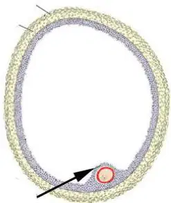
Choose the two correct statements! Identify the arrowed structure in the follicle diagram.
The structure is a Graafian/vesicular/mature/tertier follicle, the arrow shows the cumulus oophorus.
The structure is a Graafian/vesicular/tertier follicle, the arrow shows the cumulus oophorus, the innermost cells of which form the corona radiata.
The structure is a Graafian/vesicular/tertier follicle, the arrow shows the theca interna.
The structure is a secondary follicle, the arrow shows the theca interna.
The structure is a Graafian/vesicular/tertier follicle, the arrow shows the cumulus oophorus, which contains the tertiary oocyte.
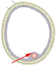
Choose the two correct statements! Identify the red arrowed structure in the follicle diagram.
The structure is a Graafian/vesicular/tertier follicle, the red arrow shows the cumulus oophorus.
The structure is a Graafian/vesicular/tertier follicle; the red arrow shows the cumulus oophorus, which contains the primary oocyte arrested in the prophase of first meiotic division.
The structure is a Graafian/vesicular/tertier follicle, the red arrow shows the cumulus oophorus, which contains the primary oocyte in the prophase of first mitotic division.
The structure is a secondary follicle, the red arrow shows the cumulus oophorus, which protrudes into the cavity of follicle.
The structure is a Graafian/vesicular/tertier follicle, the red arrow shows the theca interna, which contains the primary oocyte in the prophase of first mitotic division.
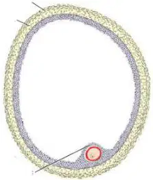
Choose the two correct statements! Identify the red-circled structure in the follicle diagram.
The structure is a Graafian/vesicular/tertier follicle, the red circle represents the zona pellucida.
During the ovulation, the zona pellucida is extruded from the Graafian/vesicular/tertier follicle.
The structure is a Graafian/vesicular/tertier follicle, the red circle represents the cell membrane of the oocyte.
During the ovulation, the zona pellucida is not extruded from the Graafian/vesicular/tertier follicle.
The zona pellucida is composed of squamous cells.
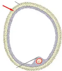
Choose the two correct statements! Identify the red arrowed structure in the follicle wall.
The structure is a Graafian/vesicular/tertier follicle, the red arrow shows the theca interna.
The structure is a Graafian/vesicular/tertier follicle, the red arrow shows the theca interna, which is located between the membrana granulosa and theca externa.
The structure is a Graafian/vesicular/tertier follicle, the red arrow shows the theca externa.
The structure is a Graafian/vesicular/tertier follicle, the red arrow shows the membrana granulosa, which is bordered by the theca interna.
The structure is a Graafian/vesicular/tertier follicle, the red arrow shows the corona radiata.
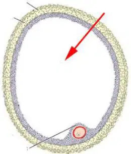
Choose the two correct statements! Identify the red arrowed space in the follicle diagram.
The red arrow shows the cavity/antrum of a Graafian/vesicular/tertier follicle, which is filled with follicular fluid.
The red arrow shows the cavity/antrum of a Graafian/vesicular/tertier follicle, which is bordered by the membrana granulosa.
The red arrow shows the cavity/antrum of a Graafian/vesicular/tertier follicle, which is filled with oxygenated blood.
The red arrow shows the cavity/antrum of a Graafian/vesicular/tertier follicle, which is bordered by the theca interna.
The red arrow shows the cavity/antrum of a Graafian/vesicular/tertier follicle, which appears in the primary follicle.

Choose the three correct statements related to the red line segment!
The structure is the head of the sperm/spermatozoon, which contains the sperm nucleus.
The structure is the head of the sperm/spermatozoon, which contains the sperm nucleus and the acrosome.
The structure is the head of the sperm/spermatozoon, which contains acrosomal enzymes.
The structure is the head of the sperm/spermatozoon, which is rich in mitochondria.
The structure is the head of the sperm/spermatozoon, which does not contain acrosomal enzymes.

Choose the only correct statement related to the red line segment!
The structure is the neck of the sperm/spermatozoon, which is located between the head and middle piece.
The structure is the neck of the sperm/spermatozoon, which is rich in mitochondria.
The structure is the neck of the sperm/spermatozoon, which is rich in proteolytic enzymes.
The structure is the neck of the spermatid, which is located between the head and middle piece.
The structure is the neck of the spermatid, which is located between the head and tail.

Choose the only correct statement related to the red line segment!
The structure is the middle piece of the sperm/spermatozoon, which is located between the neck and tail.
The structure is the middle piece of the sperm/spermatozoon, which does not contain mitochondria.
The structure is the middle piece of the sperm/spermatozoon, which contains proteolytic enzymes for the fertilization.
The structure is the middle piece of spermatid, which is rich in mitochondria.
The structure is the middle piece of the spermatid, which is located between the head and tail.
Choose the three correct statements!
Only the capacitated sperm cell is able to pass through the corona radiata
The acrosome reaction allows sperm to penetrate the zona pellucida
The acrosome reaction results in the release of enzymes
The acrosome reaction occurs in the testis
The acrosome reaction occurs in the cavity of uterus
Choose the only correct statement!
The acrosome reaction results in release of acrosin and trypsin-like substance from the sperm cell
The acrosome reaction results in release of acrosin and trypsin-like substance from the zona pellucida
The acrosome reaction results in release of trypsin-like substance from the oocyte
The acrosome reaction results in release of acrosin from the oocyte
The acrosome reaction results in release of cortical granules from the polar bodies
Which of the following processes prevents the polyspermy? One correct answer.
As a result of release of lysosomal enzymes from the cortical granules of oocyte, the oocyte membrane becomes impermeable to other sperm cells.
As soon as the sperm cell has entered the oocyte, it starts to produce proteolytic enzymes.
As soon as the sperm cell has entered the oocyte, the zona pellucida begins to secrete acrosin.
As a result of zona reaction, the oocyte begins to secrete acrosin.
As a result of zona reaction, the oocyte and sperm cell begin to secrete acrosin.
Choose the four correct statements!
The syncytiotrophoblast release proteolytic enzymes which help the implantation.
Lacunae of syncytiotrophoblast contain maternal blood.
The syncytiotrophoblast contributes to the formation of placenta
The syncytiotrophoblast is the outermost layer of the late blastocyst
The syncytiotrophoblast is the innermost layer of the late blastocyst
Choose the three correct statements!
The outer layer of the blastocyst is the trophoblast, which gives rise to the cytotrophoblast and syncytiotrophoblast layers.
The syncytiotrophoblast is the outer, multinucleated part of the blastocyst.
The cytotrophoblast is the inner, mononuclear part of the blastocyst.
The syncytiotrophoblast is the inner, multinucleated part of the blastocyst.
The cytotrophoblast is the outer, mononuclear part of the blastocyst.
Choose the two correct statements!
During the formation of morula, two groups of cells are formed: the inner one of the two groups of cells is called embryoblast cells.
During the formation of morula, two groups of cells are formed: the outer one of the two groups of cells is called trophoblast cells.
The developing blastocyst is surrounded by the extraembryonic mesoderm.
The cavity of blastocyst is the blastocoel, which contains amniotic fluid.
The cavity of blastocyst is the blastocoel, which is filled with fetal blood.
Choose the only correct statement!
During cleavage, the zygote divides with mitosis, and the subsequent mitoses results in the formation of morula and than the blastocyst.
During cleavage, the zygote divides with meiosis, and the subsequent meioses results in the formation of morula and than the blastocyst.
During cleavage, the secondary oocyte divides with mitosis, and the subsequent mitoses results in the formation of morula and than the blastocyst.
During cleavage, the zygote divides with mitosis, and the subsequent mitoses results in the formation of two polar bodies.
During cleavage, the secondary oocyte divides with mitosis, and the subsequent mitoses results in the formation of definitive oocyte and one polar body.
Choose the four correct statements!
The extraembryonic coelom is bordered by the extraembryonic somatic and splanchnic mesoderm
The extraembryonic coelom is found in the early extraembryonic mesoderm
The amniotic cavity is bordered by the epiblast layer and amnioblasts.
The chorionic cavity is located outer to the primitive yolk sac and the amniotic cavity
The primitive yolk sac is bordered by the epiblast layer and the exocoelomic (Heuser) membrane
Choose the three correct statements!
Gastrulation is the process whereby the three germ layers are established.
Gastrulation begins with formation of primitive streak.
The process of gastrulation occurs during the third week of gestation.
Gastrulation is the process whereby the stomach is established.
The first step of the gastrulation is the formation of primitive pit.
Choose the three correct statements!
Gastrulation is the process whereby the ectoderm, endoderm and the mesoderm are established.
The process of gastrulation, which occurs during the third week of gestation, results in the formation of the three germ layers.
Gastrulation begins with formation of primitive streak, which is formed on the surface of the epiblast.
Gastrulation is the process whereby the epiblast and hypoblast are formed.
Gastrulation is the process whereby the epiblast and amnion are formed.
Choose the three correct statements!
The primitive streak starts to form in the caudal region of the embryo.
All of the germ layers derive from the epiblast.
The primitive streak appears before the formation of the notochord.
The primitive streak plays a role in the establishment of mediolateral axis of the embryo.
The nervous system develops directly from the primitive streak.
Choose the only correct statement!
Mesenchyme refers to loosely organized embryonic connective tissue regardless of its origin.
Mesoderm is synonymous with mesenchyme.
Mesoderm develops from the hypoblast.
Mesoderm can develop either from the hypoblast or epiblast.
Mesenchyme develops exclusively from the mesoderm cells.
Choose the three correct statements!
The primitive node is the cephalic end of the primitive streak surrounding the primitive pit.
Detached cells of the epiblast give rise to the endoderm and mesoderm.
During the process of gastrulation, all of the germ layers derive from the epiblast.
During the process of gastrulation the endoderm develops by the transformation of hypoblast cells.
During the process of gastrulation the notochord develops from the mesoderm.
Choose the three correct statements!
The primitive streak is formed on the surface of the epiblast.
During the process of gastrulation, detached cells of the epiblast migrate along the primitive streak and through the node and appear between the epiblast and hypoblast.
Detached cells of epiblast, soon after they appear between the epiblast and hypoblast, begin to spread laterally and cranially and establish contact with the extraembryonic mesoderm.
During the process of gastrulation, which appears between the epiblast and hypoblast, migrate only in lateral direction.
The primitive streak is formed on the surface of ectoderm.
Choose the three correct statements!
During the formation of notochord, its cranial end develops first.
The notochord serves as the basis for the development of axial skeleton.
The neurenteric canal is a temporary connection between the amniotic and yolk sac cavities.
The neurenteric canal is involved in the formation of digestive system.
The neurenteric canal is involved in the formation of nervous system.
Choose the three correct statements!
The oropharyngeal membrane contains only ectoderm and endoderm without intervening mesoderm.
The oropharyngeal membrane is a temporary structure, which separates the stomodeum (= primitive oral cavity) from the primitive gut.
Rupture of the oropharyngeal membrane establishes connection between the amniotic cavity and primitive gut.
Rupture of the oropharyngeal membrane establishes connection between the amniotic and yolk sac cavity.
The oropharyngeal membrane is located in the caudal end of the embryo.
Choose the three correct statements!
The hindgut terminates temporarily at an ectodermal– endodermal membrane, the cloacal membrane, with no intervening mesoderm.
When the cloacal membrane appears, the allantois or allantoenteric diverticulum is also formed that extends into the connecting stalk.
The outpocketing of the yolk sac in the caudal part of embryo is the allantois or allantoenteric diverticulum, which later becomes obliterated.
Differentiation of three germ layers begin at the caudal part of the embryo
The outpocketing of the dorsal wall of neural tube is the allantois or allantoenteric diverticulum, which later becomes obliterated.
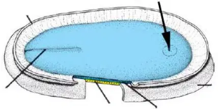
Choose the three correct statements. A dorsal view of the embryonic disc is shown; the blue area indicates the epiblast. A black arrow points to the cranial end of the disc.
The blue color represents the epiblast; the arrow shows the buccopharyngeal/oropharyngeal membrane.
The blue color represents the dorsal surface of the epiblast from the direction of opened amniotic cavity.
The arrow shows the buccopharyngeal/oropharyngeal membrane, which is located at the cranial/cephalic end of embryonic plate.
The arrow shows the cloacal membrane, which is located at the cranial/cephalic end of embryonic plate.
The blue color represents the dorsal surface of the epiblast from the direction of opened yolk sac.
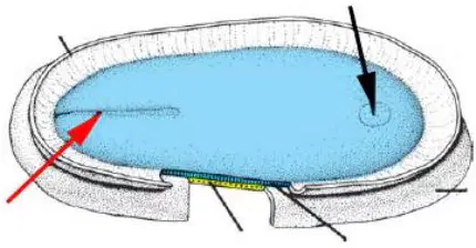
Choose the three correct statements. A dorsal view of the embryonic disc is shown; the blue area indicates the epiblast. A red arrow points to a structure on the left of the disc; a black arrow points to the cranial end.
The blue color represents the epiblast; the structure labeled with black arrow is the buccopharyngeal/oropharyngeal membrane.
The structure labeled with red arrow is the primitive streak; the structure labeled with black arrow is the buccopharyngeal/oropharyngeal membrane.
The blue color represents the dorsal surface of the epiblast from the direction of opened amniotic cavity.
The structure labeled with red arrow is the cloacal membrane; the structure labeled with black arrow is the buccopharyngeal/oropharyngeal membrane.
The blue color represents the epiblast; the structure labeled with red arrow is the cloacal membrane.
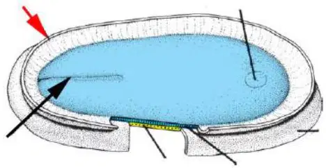
Choose the two correct statements. A dorsal view of the embryonic disc is shown; the blue area indicates the epiblast. A red arrow points to the cut edge at the amnion; a black arrow points to the primitive streak.
The structure labeled with black arrow is the primitive streak; the structure labeled with red arrow is the cut edge of the amnion.
The blue color represents the dorsal surface of the epiblast; the structure labeled with red arrow is the cut edge of the amnion.
The blue color represents the dorsal surface of the epiblast; the structure labeled with black arrow is the primitive node.
The blue color represents the dorsal surface of the hypoblast; the structure labeled with black arrow is the primitive streak.
The structure labeled with black arrow is the primitive streak; the structure labeled with red arrow is the cut edge of the yolk sac.
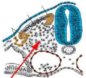
Choose the two correct statements. The picture is a cross section of the embryo; a red arrow marks a mesenchymal region near the paraxial mesoderm.
The picture is a cross section of the embryo; the cells around the arrow constitute the sclerotom.
The picture is a cross section of the embryo; the cells around the arrow give rise to the vertebrae.
The picture is a cross section of the embryo; the cells around the arrow give rise to the myotom.
The picture is a sagittal section of the embryo; the cells around the arrow constitute the myotom.
The picture is a sagittal section of the embryo; the cells around the arrow constitute the myotom.
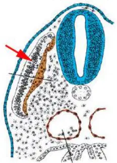
Choose the two correct statements. The picture is a cross section of the embryo; a red arrow marks the dorsolateral compartment of a somite.
The picture is a cross section of the embryo; the arrow shows the dermatome.
The picture is a cross section of the embryo; the arrow shows the dermatome which gives rise to the connective tissue (dermis) of skin on the dorsal side of the embryo.
The picture is a cross section of the embryo; the arrow shows the somite.
The picture is a sagittal section of the embryo; the arrow shows the sclerotom.
The picture is a sagittal section of the embryo; the arrow shows the intraembryonic mesenchyme.
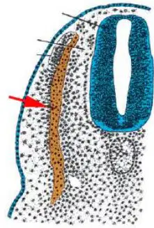
Choose the two correct statements. The picture is a cross section of the embryo; a red arrow marks the medial compartment of a somite.
The picture is a cross section of the embryo; the arrow shows the myotom.
The picture is a cross section of the embryo; the arrow shows the myotom which gives rise to muscles.
The picture is a cross section of the embryo; the arrow shows the sclerotom which gives rise to bones.
The picture is a cross section of the embryo; the arrow shows the somite.
The picture is a sagittal section of the embryo; the arrow shows the sclerotom.
Choose the only correct statement.
Neurulation is completed by day 28 of embryonic development.
Neurulation is completed by day 10 of embryonic development.
Neurulation is completed by day 38 of embryonic development.
Neurulation is completed by the end of second month of embryonic development.
Neurulation is completed by the end of third month of embryonic development.
Choose the only correct statement.
Placodes refer to ectodermal thickenings in the cephalic region of the embryo.
No permanent structures develop from the placodes.
Structures develop form placodes remain on the outer surface of the body.
Placode gives rise to the central nervous system.
Placodes refer to ectodermal thickenings in the posterior body wall.
Choose the only correct statement.
The notochord induces the overlying ectoderm to thicken and form the neural plate.
The notochord does not have any role in the development of nervous system, it only contributes to the development of axial skeleton.
The notochord gives rise to the spinal cord.
The notochord induces the overlying endoderm to thicken and form the neural plate.
The notochord induces the completion of neural development.
Choose the two correct statements.
The central nervous system develops from the neural tube.
Part of the peripheral nervous system develops from the neural crest.
Neurulation refers to the development of brain ventricles.
Neurulation refers to the development of peripheral nerves.
Neurulation refers to the development of the notochord and neural plate.
Choose the three correct statements.
The neural tube is derivative of the ectoderm.
Formation of neural tube is completed by the closure of anterior and posterior neuropore.
Formation of neural tube begins with the fusion of neural folds.
The cells of neural crest start to migrate from the wall of the neural tube.
The cavity of neural tube is continuous with the cavity of yolk sac through the anterior neuropore.
Choose the three correct statements.
The neural crest gives rise to neural and non neural structures.
There are ganglia which are derived from the neural crest.
The neural crest cells are formed at the crest of the neural fold and begin to dissociate from their neighbors and migrate away from this region after the closure of neural tube is completed.
The neural crest cells do not migrate to the head region.
The neural crest contribute to the formation of neural tube.
Which of the structures develop from the neural crest? Choose the three correct statements!
Some of the bones of skull
Melanocytes
Sympathetic ganglia
Enamel of the teeth
Hypophysis
Which of the structures develop from the ectoderm? Choose the four correct statements!
Epidermis of skin including its glands and nail
Neurons of central nervous system
Enamel of the teeth
Sensory epithelium of nose
Cartilage
Choose the three correct statements!
The first step in the differentiation of mesoderm is the appearance of paraxial, intermediate and lateral plate mesoderm at both sides of the notochord.
The intermediate mesoderm connects the paraxial mesoderm and lateral plate mesoderm.
The cavity which is formed within the lateral plate mesoderm is called intraembryonic coelom.
The bilateral intraembryonic coeloms communicate with each other.
The intraembryonic coelom opens to the amniotic cavity.
Choose the four correct statements!
In the lateral direction, the intraembryonic coelom is continued into the extraembryonic coelom.
Due to the formation of the intraembryonic coelom, the lateral plate mesoderm develops into the somatopleura and splanchnopleura.
The somatic mesoderm is the other name of the somatopleura.
The splanchnopleura is the other name of the splanchnic or visceral mesoderm.
The intraembryonic coelom appears in the paraxial part of the mesoderm.
Choose the four correct statements!
The somatopleura is continued into the part of extraembryonic mesoderm which covers the amnion.
The splanchnopleura is continued into the part of extraembryonic mesoderm which covers the yolk sac.
As a result of the segmentation of paraxial mesoderm, the newly formed structures in the head region are called somitomers, and caudally to the occipital region they are called somites.
In the lateral direction, the intraembryonic coelom is continued into the extraembryonic coelom.
The extraembryonic coelom appears in the paraxial mesoderm.
Choose the four correct statements!
Due to the formation of the intraembryonic coelom, the lateral plate mesoderm develops into a somatopleura and a splanchnopleura.
The first pairs of somites appear in the future occipital region, the new somites appear in craniocaudal sequence and at the end of fifth week, 42 to 44 pairs are present.
During the differentiation of somites, small cavity appears inside.
Cells at the ventral and medial walls of the somite migrate toward the neural tube and notochord and form the sclerotome.
Cells at the ventral and medial walls of the somite migrate toward the neural tube and notochord and form the myotome.
Choose the three correct statements!
Cells, migrate from the ventral and medial walls of the somite form the sclerotome.
The vertebrae and ribs develop from the sclerotome.
Cells at the dorsomedial and ventrolateral edges of the somite form the myotom.
Cells, migrate from the ventral and medial walls of the somite form the sclerotome and myotom.
Cells at the dorsomedial and ventrolateral edges of the somite form the dermatome, myotome and sclerotome.
Choose the only correct statement!
The extraembryonic mesoderm lining the cytotrophoblast and amnion is called the extraembryonic somatic mesoderm.
The extraembryonic mesoderm covering the yolk sac is not continuous with the somatic or parietal mesoderm.
The intermediate mesoderm connects the intra- and extraembryonic mesoderm.
The paraxial mesoderm connects the intra- and extraembryonic mesoderm.
The intraembryonic mesoderm differentiates directly into the sclerotom and lateral plate mesoderm.
Choose the only correct statement!
The extraembryonic mesoderm covering the yolk sac is continuous with the splanchnic or visceral mesoderm.
The extraembryonic mesoderm covering the amniotic cavity is continuous with the splanchnic or visceral mesoderm.
No permanent structures develop from the intermediate mesoderm.
The nephrotomes develop from the paraxial mesoderm.
All bones of the skull develop directly from the intermediate mesoderm.
Choose the four correct statements!
The splanchnic or visceral layer of lateral plate mesoderm is continuous with the visceral layer of extraembryonic mesoderm covering the yolk sac.
The parietal (somatic) layer of lateral plate mesoderm is continuous with the extraembryonic parietal mesoderm covering the amnion.
The gonads develop from the intermediate mesoderm.
The sclerotome derives from the paraxial mesoderm.
The myotom develops from the intermediate mesoderm
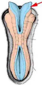
Choose the two correct statements in relation to the structure pointed by red arrow!
The structure is the neural fold, which develops from the neural plate.
The structure is the neural fold, which, by fusion of its contralateral pair, contributes to the closure of the neural tube.
The structure is the neural fold, which gives rise to the peripheral nervous system.
The structure is the neural fold, which gives rise to the red bone marrow.
The structure is the neural fold, which does not give rise to any permanent structures in the body.
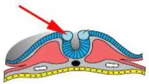
Choose the two correct statements in relation to the structure pointed by red arrow!
The arrow shows neural crest cells are formed at the crest of the neural fold and begin to dissociate from their neighbors and migrate away from this region after the closure of neural tube is completed.
The arrow shows the neural crest cells, which leave their original position by active migration and contribute to the development of different structures, e.g. some of the bones of the skull.
The arrow shows the neural crest cells, which migrate from the notochord.
The arrow shows the cells, which give rise to the somites.
The arrow shows the neural crest cells, which develop from the hypoblasts.
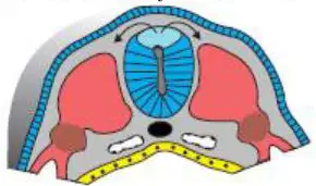
Choose the only one correct statement!
The arrows show the migration pathway of a group of cells, which give rise to the neural crest.
The arrows show the migration pathway of a group of cells, which give rise to the notochord.
The arrows show the migration pathway of a group of cells in the wall of the gut tube, which give rise to the neural crest.
The arrows show the migration pathway of a group of cells, which cells give rise to the sclerotome.
The arrows show the migration pathway of a group of cells in the wall of the gut tube, which give rise to the ganglia.
Choose the three correct statements!
During the folding of the embryo, the head and tail folds as well as the lateral (body wall) folds move ventrally.
As a result of longitudinal growth of the neural tube, the head and tail folds move ventrally.
Parallel to the development and growth of the brain vesicles, the embryonic disc begins to bulge into the amniotic cavity.
Parallel to the development and growth of the brain vesicles, the embryonic disc begins to bulge into the yolk sac.
As a result of ventral movement of the head and tail folds as well as the lateral (body wall) folds, the ventral body wall closes completely.
Choose the two correct statements!
As a result of ventral movement of the head and tail folds as well as the lateral (body wall) folds, the ventral body wall closes completely except for the region where the connecting stalk and yolk sac are found.
Initially, the allantois is found in the connecting stalk; later, its proximal part is incorporated into the body of the embryo.
As a result of ventral movement of the head and tail folds as well as the lateral (body wall) folds, the ventral body wall closes completely except for the region where the connecting stalk and neural tube are found.
Initially, the allantois is found in the connecting stalk; later, its distal part is incorporated into the body of the embryo.
The allantois and the vitelline duct open to the hindgut.
Choose the only correct statement!
The epithelium of primitive gut derives from the endoderm.
The allantois appears close to the oropharyngeal (buccopharyngeal) membrane.
The allantois appears in the anterior wall of the yolk sac and does not grow into the connecting stalk.
The allantois appears in the anterior wall of the yolk sac and then grows into the connecting stalk.
The epithelium of primitive gut derives from the ectoderm.
Choose the only correct statement!
The vitelline duct connects the yolk sac and midgut.
The vitelline duct connects the allantois and midgut.
The vitelline duct is not accompanied by blood vessels.
The vitelline duct connects the hindgut and the yolk sac.
The vitelline duct is not present in the primitive umbilical cord.
Choose the only correct statement!
Epithelium of the respiratory system develops from the endoderm.
Epithelium of gut tube develops from the ectoderm; cells of the endoderm migrate to the gut tube in a later stage of development.
Parenchyma of the liver develops from the mesoderm.
Parenchyma of the liver develops from the ectoderm.
Epithelium of urinary bladder develops from the mesoderm.
Choose the two correct statements!
As a result of cephalocaudal and lateral folding of the embryo, the epithelium of primitive gut develops from the endoderm.
The primitive gut is a blind-ending tube, which is divided into foregut, midgut and hindgut.
The hindgut is not present in the primitive gut it develops in a later stage of development.
The epithelium of primitive gut develops from the mesoderm and endoderm.
The epithelium of primitive gut develops from the endoderm and ectoderm.
Choose the two correct statements!
The ectodermally lined primitive oral cavity is called stomodeum.
The ectodermally lined primitive oral cavity is separated from the endodermally lined pharynx by the oropharyngeal (buccopharyngeal) membrane.
The endodermally lined primitive oral cavity is called stomodeum.
The endodermally lined primitive oral cavity is separated from the endodermally lined pharynx by the oropharyngeal (buccopharyngeal) membrane.
The ectodermally lined primitive oral cavity is separated from the ectodermally lined pharynx by the oropharyngeal (buccopharyngeal) membrane.
Choose the three correct statements!
Proctodeum, which is the lower part of the anal canal, originates from the ectoderm.
Opening for the anus is created by the rupture of cloacal membrane.
The upper and lower parts of the anal canal are separated by the cloacal membrane.
Proctodeum, which is the lower part of the anal canal, originates from the endoderm.
The cloacal membrane develops from the mesoderm.
Choose the two correct statements!
The hindgut terminates temporarily at an ectodermal–endodermal membrane, the cloacal membrane.
The oropharyngeal (buccopharyngeal) membrane breaks down earlier than the cloacal membrane.
The cloacal membrane consists up the ectoderm, mesoderm and endoderm.
The oropharyngeal (buccopharyngeal) membrane breaks down later than the cloacal membrane.
The oropharyngeal (buccopharyngeal) and the cloacal membrane break down simultaneously.
Choose the only correct statement!
The cloacal membrane appears in the embryonic disc.
The cloacal membrane appears after folding of the embryo.
The cloacal membrane appears in the embryonic disc, the oropharyngeal (buccopharyngeal) membrane develops during later stages.
The oropharyngeal (buccopharyngeal) membrane appears in the embryonic disc, the cloacal membrane develops during later stages.
The cloacal membrane contains mesoderm.
Choose the only correct statement!
The cloacal membrane is present in the embryonic disc.
The oropharyngeal (buccopharyngeal) membrane appears after completion of the folding of the embryo.
The cloacal membrane is found below the proctodeum.
The oropharyngeal (buccopharyngeal) membrane is found at the junction of stomodeum and midgut.
The cloacal membrane consists up the mesoderm and endoderm.
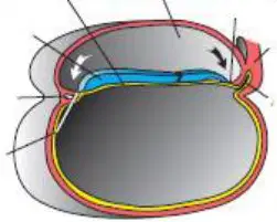
Choose the three correct statements!
The picture is the midsagittal section of an embryo, the white curved arrow shows the head fold.
The picture is the midsagittal section of an embryo and the black curved arrow shows the tail fold.
The picture is the midsagittal section of an embryo, the white and black curved arrows are located in the amniotic cavity.
The picture is the midsagittal section of an embryo, the white and black curved arrows show the lateral (body wall) fold.
The picture is the midsagittal section of an embryo, the white and black curved arrows are located in the extraembryonic cavity.
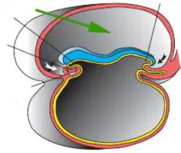
Choose the two correct statements!
The picture is the midsagittal section of an embryo, the arrow shows the amniotic cavity.
The picture is the midsagittal section of an embryo, the arrow shows the amniotic cavity which is filled with amniotic fluid.
The picture is the cross section of an embryo, the arrow shows the amniotic cavity which is filled with amniotic fluid.
The picture is the midsagittal section of an embryo; the arrow shows the yolk sac.
The picture is the cross section of an embryo; the arrow shows the yolk sac.
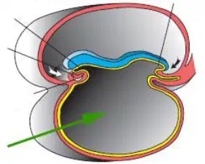
Choose the three correct statements!
The picture is the midsagittal section of an embryo, the arrow shows the yolk sac.
The arrow shows a cavity which, in a later stage of development, is connected to the midgut with the vitelline duct.
No permanent structure develops from the cavity which is labeled with arrow.
The arrow shows a cavity which develops into the foregut.
The small intestines develop from the vitelline duct.

Choose the three correct statements!
The picture is the midsagittal section of an embryo; the straight arrow shows the yellow colored allantois.
The allantois is found in the primitive umbilical cord.
In a later stage of development the lumen of the allantois is obliterated.
The picture is the midsagittal section of an embryo; the straight arrow shows the vitelline duct.
The picture is the cross section of an embryo; the straight arrow shows the allantois.
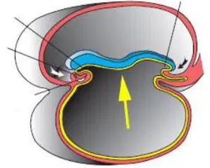
Choose the three correct statements!
The picture is the midsagittal section of an embryo, the blue color structure is the ectoderm.
The nervous system develops from the structure shown in blue color.
The epithelium of the skin develops from the structure shown in blue color.
The cavity above the blue color structure is the yolk sac.
The picture is the cross section of an embryo; the blue color structure is the ectoderm.
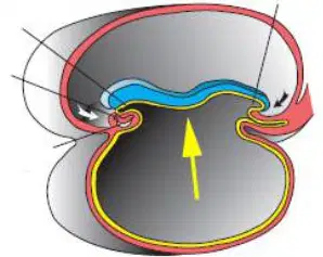
Choose the three correct statements!
The picture is the midsagittal section of an embryo, the straight arrow shows the endoderm seen in yellow color.
The picture is the midsagittal section of an embryo, the straight arrow shows the endoderm seen in yellow color; the endoderm gives rise to the epithelium of primitive gut.
The structure shown in yellow color derives from the epiblast.
The cavity above the blue color structure is the yolk sac.
The straight arrow shows a yellow color structure which derives from the notochord.
Choose the two correct statements.
The amnion and chorion are fetal membranes.
The amnion and chorion develop from embryonic tissue.
Both the amnion and chorion develop from maternal tissue.
Both the amnion and chorion are in direct contact with the amniotic fluid
The fetal membranes start to develop only in the last month of pregnancy
Choose the three correct statements.
The amniotic cavity is filled with a clear, watery fluid.
The amount of amniotic fluid increases from 30 mL at 10 weeks of gestation to 1000 mL at 37 weeks.
The amniotic fluid serves as a protective cushion.
The amniotic cavity is filled with amniotic fluid; its volume remains unchanged during the pregnancy.
The fetal urine is present in the amniotic fluid during the total length of pregnancy. The amniotic fluid prevents the adherence of the embryo to the uterus.
Choose the three correct statements.
The fetal period begins in the ninth week of pregnancy and lasts until birth.
The age of the embryo may be indicated with the crown-rump length (CRL, sitting height).
The age of the embryo may be indicated with the crown-heel length (CHL, standing height).
The age of the embryo may be indicated with the crown rump length (CRL, sitting height), which is measured from the face to the buttocks.
The age of the embryo may be indicated with the crown-heel length (CHL, standing height), which is measured from the vertex of the skull to the midpoint of sole of the foot.
Choose the four correct statements.
The length of pregnancy is considered to be 280 days, or 40 weeks after the onset of the last normal menstrual period.
The length of pregnancy is considered to be 266 days or 38 weeks after fertilization.
During the third month of pregnancy, the length of the head is approximately half of the crown-rump length (CRL, sitting height).
During the fifth month of pregnancy, movements of the fetus can be felt by the mother.
During the third month of pregnancy, the length of the head is approximately one fourth of the crown-rump length (CRL, sitting height).
Choose the three correct statements.
During the third month of pregnancy, the eyes move to the ventral aspect of the face.
It is not before of the 12th week of pregnancy when the external genitalia develop such a degree that the sex of the fetus can be determined by ultrasound.
Primary ossification centers are present in the long bones and skull by the 12th week of pregnancy.
By the 20th week of pregnancy, the external genitalia develop such a degree that the sex of the fetus can be determined by ultrasound.
During the seventh month of pregnancy, the eyes move to the ventral aspect of the face.
Choose the three correct statements.
At birth, the testes of mature male newborn are in the scrotum.
At birth, the body weight of mature newborn is at least 2500 g.
At birth, the border between the head hair and forehead of the mature newborn is sharp.
At birth, the labia majora of mature female newborn do not cover the labia minora.
At birth, the nails of the mature newborn do not exceed the fingertips.
Choose the three correct statements.
By the end of intrauterine life, the skin of fetus is covered by the vernix caseosa.
The lanugo hair is a fine hair which covers the fetus.
The age of the fetus can be determined with the biparietal diameter of the skull.
At the beginning of pregnancy, the body of the fetus is covered by the vernix caseosa, which disappears by the middle of the sixth month.
The lanugo is a fatty substance, which covers the body of the fetus.
Choose the three correct statements.
The primitive umbilical ring is an oval line of reflection between the amnion and body wall.
The connecting stalk along with the allantois and umbilical vessels passes through the primitive umbilical ring.
The vitelline duct and its vessels pass through the primitive umbilical ring.
The only structure which passes through the primitive umbilical ring is the connecting stalk
The primitive umbilical ring is found between the amnion and the midgut.
Choose the three correct statements.
The umbilical cord, surrounded by the amnion, contains the blood vessels which form connection between the placenta and fetus.
The blood vessels in the umbilical cord contain fetal blood; the umbilical vein carries the oxygenated blood.
Structures in the umbilical cord are surrounded by the Wharton's jelly.
The blood vessels in the umbilical cord contain maternal blood; the umbilical arteries carry the deoxygenated blood.
The blood vessels in the umbilical cord contain maternal and fetal blood; the umbilical vein carries the oxygenated blood.
Choose the three correct statements.
Approximately two thirds of twins are dizygotic.
Dizygotic twins develop from simultaneous release of two oocytes which are fertilized by different spermatozoa.
Monozygotic twins develop from a single zygote.
Dizygotic twins develop from a single oocyte which is fertilized by two spermatozoa
Monozygotic twins develop from two oocytes which are fused after the fertilization.
Choose the four correct statements.
Dizygotic twins usually have separate chorion and amnion.
Dizygotic twins usually have separate placenta, but sometimes the placentas are fused.
Dizygotic twins usually have separate chorionic sac (cavity), but sometimes the walls of sacs fuse.
The monozygotic twins always have common placenta.
If the separation of zygote occurs at the two-cells stage, the blastocysts implants separately and each embryo has its own placenta and chorionic sac (cavity).
Choose the one correct statement.
Monozygotic twins are of the same sex and they are genetically identical.
Dizygotic twins are genetically identical and they may or may not be of different sex.
Monozygotic twins can develop by the separation of inner cell mass at a late stage of development; each embryo has its own placenta.
Monozygotic twins can develop by the separation of inner cell mass at a late stage of development; each embryo has its own amniotic cavity.
The monozygotic twins always have common placenta.
Choose the two correct statements!
Monozygotic twins can develop by splitting of inner cell mass at a later stage of development; the two embryos have a common placenta.
Monozygotic conjoined twins can develop by partial or complete splitting of the primitive streak and node.
Monozygotic twins always have common placenta, chorionic sac (cavity), and amniotic cavity
Monozygotic twins can develop by the separation of zygote at the late blastocyst stage (bilaminar germ disk stage); the two embryos have a common placenta, but separate chorionic sac (cavity) and amniotic cavity.
Monozygotic twins have common umbilical cord.
Choose the two correct statements!
Monozygotic twins develop from a single zygote
Approximately one third of twins are monozygotic.
The only mechanism of monozygotic twins formation is the splitting of embryo in a two-cell stage.
The only mechanism of monozygotic twins formation is the splitting and separation of the embryoblast cells.
Approximately 90% of twins are monozygotic.
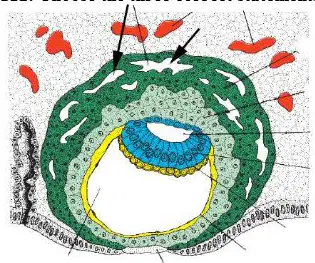
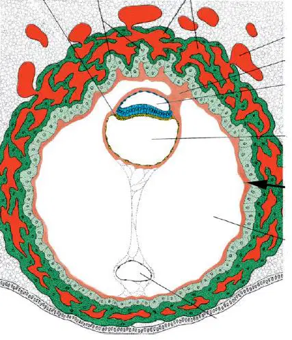
Choose the three correct statements. The diagram shows early implantation with arrows pointing to lacunae in the syncytiotrophoblast.
The arrows show lacunae in the syncytiotrophoblast; in a later stage of development maternal blood vessels will open into these lacunae.
The arrows show lacunae in the syncytiotrophoblast; in a later stage of development the lacunae will communicate with each other.
The arrows show lacunae in the syncytiotrophoblast, which, in a later stage of development, will be filled with maternal blood establishing the uteroplacental circulation.
The arrows show lacunae in the syncytiotrophoblast, which are filled with amniotic fluid.
The arrows show the lacunae in the syncytiotrophoblast, and in a later stage of development, fetal blood vessels will open into the lacunae.
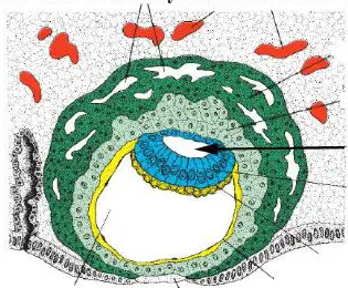
Choose the only one correct statement in relation to the cavity pointed by arrow. The diagram shows early implantation with an arrow indicating a small cavity within the embryonic disc region.
It is the amniotic cavity filled with amniotic fluid and bordered by amnioblasts and epiblast cells.
It is the amniotic cavity filled with amniotic fluid and bordered by amnioblasts and hypoblast cells.
It is the blastocyst cavity filled with amniotic fluid and bordered by amnioblasts and epiblast cells.
It is the extraembryonic coelom filled with amniotic fluid and bordered by amnioblasts and epiblast cells.
It is the amniotic cavity, which is bordered by amnioblasts and epiblast cells and in a later stage of development, will receive fetal blood vessels.
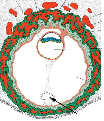
Choose the only one correct statement in relation to the cavity pointed by arrow!
The cavity is an exocoelomic cyst, which is located in the extraembryonic cavity (chorionic cavity).
The cavity is the secondary yolk sac bordered by amnioblast cells.
The cavity is the secondary yolk sac containing amniotic fluid
The cavity is an exocoelomic cyst, which is the remnant of the secondary yolk sac.
The cavity is an exocoelomic cyst, which is the remnant of the blastocyst cavity.
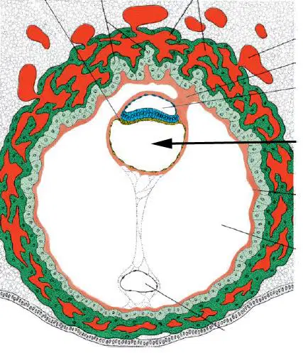
115. Choose the one correct statement in relation to the cavity pointed by arrow!
The cavity is the secondary/definitive yolk sac bordered by cells migrated from the hypoblast.
The cavity is the secondary/definitive yolk sac which appears within the exocoelomic cavity
The cavity is the secondary/definitive yolk sac which is bordered by epiblast cells.
It is the exocoelomic cavity which is the remnant of the secondary/definitive yolk sac.
The cavity is the exocoelomic cyst sac which is bordered by hypoblast cells.
116. Choose the three correct statements in relation to the structure pointed by arrow!
The structure is the connecting stalk, which is the part of the extraembryonic mesoderm.
The structure is the connecting stalk, which contributes to the formation of the umbilical cord.
The structure is the connecting stalk, which in the chorionic cavity.
The structure is the connecting stalk, which is not the part of the extraembryonic mesoderm
The structure is the connecting stalk, which becomes the part of the notochord.
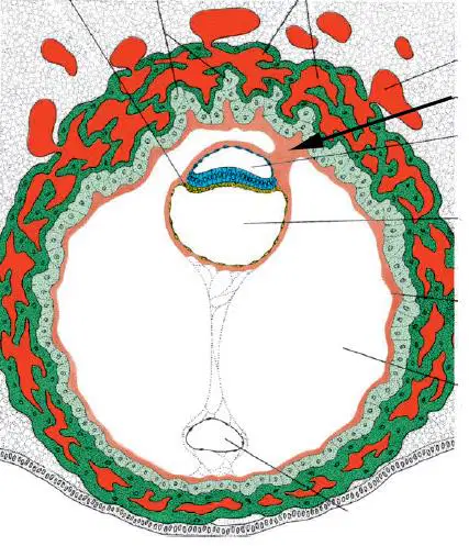
117. Choose the three correct statements in relation to the structure pointed by arrow!
The structure is the chorionic plate (extraembryonic somatic/somatopleuric mesoderm), which is the part of the extraembryonic mesoderm.
The structure is the chorionic plate (extraembryonic somatic mesoderm), which is surrounded by the cytotrophoblast cells.
The structure is the chorionic plate (extraembryonic somatic mesoderm), which participates in the formation of placental villi.
The structure is the chorionic plate (extraembryonic somatic mesoderm), which is not the part of the extraembryonic mesoderm
The structure is the chorionic plate (extraembryonic somatic mesoderm) which gives rise to the notochord.
118. Choose the only correct statement!
All types of formed elements of the blood originate from the myeloid progenitor cells (CFUM) with the exception of lymphocytes.
All types of formed elements of the blood originate from the myeloid progenitor cells (CFUM).
All types of formed elements of the blood originate from the myeloid progenitor cells (CFUM) except the red blood cells.
Only the white blood cells originate from the myeloid stem cell (CFU-M).
The myeloid stem cells (CFU-M) give rise to formed elements of the blood only in the intrauterine life
119. Choose the only correct statement!
In one phase of embryonic development the hemopoietic pluripotent stem cells (CFU-S) appear in the extraembryonic mesoderm of yolk sac.
The lymphoid progenitor cells (CFU-L) appear in the extraembryonic mesoderm of yolk sac.
The myeloid progenitor cells (CFU-M) appear in the extraembryonic mesoderm of yolk sac
The pluripotent myeloid stem cells appear in the extraembryonic mesoderm of yolk sac.
The pluripotent myeloid and lymphoid stem cells appear in the extraembryonic mesoderm of yolk sac.
120. Choose the only correct statement
The pluripotent hemopoietic stem cells (CFU-S) are the only cells within the hemopoietic system that possess the potential for self-renewal.
The pluripotent hemopoietic stem cells (CFU-S) and myeloid progenitor stem cells (CFU-M) possess the potential for self-renewal
The pluripotent hemopoietic stem cells (CFU-S) and lymphoid progenitor stem cells (CFU-L) possess the potential for self-renewal.
The lymphoid progenitor stem cells (CFU-L) are the only cells within the hemopoietic system that possess the potential for self-renewal.
The myeloid progenitor stem cells (CFU-M) are the only cells within the hemopoietic system that possess the potential for self-renewal
121. Choose the three correct statements!
The pluripotent hemopoietic stem cells (CFU-S) can give rise to myeloid (CFU-M) and lymphoid progenitor cells (CFU-L).
The pluripotent hemopoietic stem cells (CFU-S) have self-renewal capacity, which is the ability to give rise to identical daughter CFU-S.
The pluripotent hemopoietic stem cells (CFU-S) have self-renewal capacity throughout the life.
The pluripotent hemopoietic stem cells (CFU-S) have self-renewal capacity in the intrauterine life.
The pluripotent hemopoietic stem cells (CFU-S) have self-renewal capacity till the beginning of puberty.
122. Choose the three correct statements!
In the intrauterine life the hemopoiesis occurs in the yolk sac, liver, spleen, and red bone marrow.
After birth, the hemopoiesis occurs in the red bone marrow.
In the intrauterine life the shortest period of hemopoiesis occurs in the spleen.
In the intrauterine life the shortest period of hemopoiesis occurs in the liver.
In the intrauterine life the shortest period of hemopoiesis occurs in the red bone marrow
123. Choose the three correct statements!
The pluripotent hemopoietic stem cells (CFU-S) are found both in the peripheral blood and red bone marrow.
The shape and size of pluripotent hemopoietic stem cell (CFU-S) is similar to the small lymphocytes.
The pluripotent hemopoietic stem cells (CFU-S) can be identified by detecting their specific markers.
The pluripotent hemopoietic stem cells (CFU-S) are found only in the red bone marrow.
The shape and size of pluripotent hemopoietic stem cell (CFU-S) is similar to the monocyte.
124. Choose the only correct statement!
During the eryhropoiesis the first morphologically identifiable cell is the proerythroblast
The acidophilic cytoplasm of proerythroblast refers to the appearance of hemoglobin
The acidophilic cytoplasm of proerythroblast refers to the appearance of the hem component of hemoglobin.
During the eryhropoiesis the last cell which sill has nucleus is the polychromatophil erythroblast.
During the eryhropoiesis the last cell which sill has nucleus is the reticulocyte.
125. Choose the only correct statement!
One of the factors that control the erythropoiesis is the erythropoietin, which is produced in the kidney.
One of the factors that control the erythropoiesis is the erythropoietin, which is produced in the red bone marrow.
During the erythropoiesis the diameter of cells increases from the younger to mature erythrocytes.
During the erythropoiesis the diameter of cells remains constant.
During the erythropoiesis the diameter of cells increase in the younger forms followed by a decrease in the mature cells
126. Choose the two correct statements!
During the granulopoiesis the first morphologically identifiable cell is the myeloblast
During the granulopoiesis the nonspecific granules appear in the promyelocytes.
During the granulopoiesis the specific granules appear in the promyelocytes.
During the granulopoiesis the nonspecific granules appear in the myelocytes.
During the granulopoiesis the nonspecific granules appear in the myeloblasts.
Choose the cells of granulopoiesis in sequence! Only one correct answer!
Myeloblast, promyelocyte, myelocyte, metamyelocyte or juvenile, stab (band), granulocyte
Promyelocyte, myeloblast, myelocyte, metamyelocyte or juvenile, stab (band), granulocyte
Myelocyte, promyelocyte, myeloblast, metamyelocyte or juvenile, stab (band), granulocyte
Promyeloblast, promyelocyte, myelocyte, metamyelocyte or juvenile, stab (band), granulocyte
Myeloblast, promyelocyte, metamyelocyte or juvenile, stab (band), myelocyte, granulocyte
Choose the cells of erythropoiesis in sequence! Only one correct answer!
Proerythroblast, basophil erythroblast, orthochromatophil erythroblast, oxyphil erythroblast or normoblast, reticulocyte, erythrocyte.
Basophil erythroblast, proerythroblast, orthochromatophil erythroblast, oxyphil erythroblast or normoblast, reticulocyte, erythrocyte.
Basophil erythroblast, oxyphil erythroblast or normoblast, proerythroblast, orthochromatophil erythroblast, reticulocyte, erythrocyte.
Basophil erythroblast, orthochromatophil erythroblast, oxyphil erythroblast or normoblast, proerythroblast, reticulocyte, erythrocyte.
Orthochromatophil erythroblast, basophil erythroblast, oxyphil erythroblast or normoblast, proerythroblast, reticulocyta, erythrocyte.
Choose the only correct statement!
Progenitor stem cell of granulocytes (CFU-G) derives from the common granulocytemonocyte progenitor cell (CFU-GM).
Progenitor stem cell of granulocytes (CFU-G) derives directly from the pluripotent hemopoietic stem cell (CFU-S).
Progenitor stem cell of monocytes (CFU-Mo) derives directly from the pluripotent hemopoietic stem cell (CFU-S).
All the progenitor stem cells of formed elements of the blood derive directly from the pluripotent hemopoietic stem cell (CFU-S).
Progenitor stem cell of erythrocytes (CFU-E) derives directly from the pluripotent hemopoietic stem cell (CFU-S).
Which of the followings best describes the early stages of the skull in a developing embryo?
Solid bone plates fused together
Mesenchymal sheets that will eventually ossify
Cartilage structures that later transform into bone
Fully formed individual bones separated by sutures
Facial skeleton is a derivative of the endoderm
The anterior fontanelle in a newborn is at the junction of …. sutures:
Coronal and sagittal
Sagittal and lambdoid
Lambdoid and squamous
Lambdoid and coronal
Sagittal and Squamous
What is the main advantage of having non-ossified sutures between bones of calvaria in a newborn?
Provides flexibility demanded in the delivery canal.
Allows for facial expressions.
Enables muscle growth.
Provides attachment points for muscles.
Sutures remain elastic also in adulthood.
Fontanels are formed by…
Overlapping bone plates
Unfused areas between developing bone plates
Lack of calcium in specific areas
Absence of muscle tissue in the head
Excess amount of cerebrospinal fluid
Select the TWO FALSE statements about skull development!
The facial bones fuse together earlier than bones of neurocranium.
Sutures allow for some movement during childbirth.
All skull bones are completely fused by adulthood via synostosis
The final shape of the skull is influenced by genetics and environment.
Calvaria develops with enchondral ossificaion.
Choose the three correct statements!
The membranous portion of the neurocranium is derived from neural crest cells and paraxial mesoderm.
The frontal bone develops from the neural crest cells by intramembranous ossification.
The parietal bone develops from the paraxial mesoderm by intramembranous ossification.
The frontal bone develops from the neural crest cells by endochondral ossification.
The parietal bone develops from the paraxial mesoderm by endochondral ossification.
Choose the four correct statements!
The sutures of the newborn skull are formed by connective tissue, which derive from the neural crest and paraxial mesoderm.
At the newborn calvaria (vault of the skull) where more than two bones meet, the sutures are wide and are called fontanelles.
The anterior (major) fontanelle, which is a rhomboid or diamond shape, is found where the two frontal and two parietal bones meet.
The anterior (major) fontanelle becomes ossified by the 18th postnatal month, whereas the posterior (minor) fontanelle by 3rd month.
The posterior (minor) fontanelle becomes ossified by the 18th postnatal month, whereas the anterior (major) fontanelle by 1-2 year of child.
Choose the four correct statements!
The cartilaginous neurocranium consists of prechordal and chordal chondrocranium.
The lesser wing and anterior part of greater wing of sphenoid bone develops from the prechordal chondrocranium, cells of which derive from the neural crest.
In the first few years after birth, palpation of the anterior fontanelle may give valuable information as to whether ossification of the skull is proceeding normally.
In the first few years after birth, palpation of the anterior fontanelle may give valuable information as to whether ossification of the skull is proceeding normally.
The desmocranium consists of prechordal and chordal parts.
Choose the three correct statements!
Petrous part of temporal bone develops from the chondrocranium, whereas the squamous part derives from the viscerocranium.
The Meckel cartilage, which is found in the ventral portion of the first pharyngeal arch, disappears except in the sphenomandibular ligament.
Mesenchyme around the Meckel cartilage gives rise to the mandible by intramembranous ossification.
Mesenchyme around the Meckel cartilage gives rise to the mandible by endochondral ossification
The entire temporal bone develops from the chondrocranium.
Choose the three correct statements!
The viscerocranium develops mainly from the first two pharyngeal arches.
Mesenchyme for formation of the bones of the face derives from neural crest cells.
Flat bones which surround the brain develop with intramembraneous ossification.
Mesenchyme for formation of the bones of the face derives from the endoderm.
Bones of the face develop by endochondral ossification.

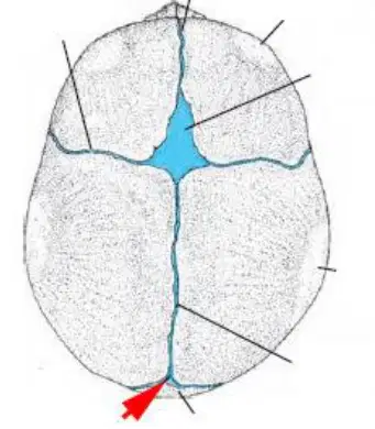
Choose the three correct statements!
The arrow shows the anterior fontanelle, which anterior fontanelle usually closes by 18 months of age.
The arrow shows the anterior fontanelle, which is bordered by the two frontal and two parietal bones.
At birth, the structure labeled with arrow consists of connective tissue
The arrow shows the anterior fontanelle, which anterior fontanelle closes by 36 months of age.
At birth, the structure labeled with arrow does not contain connective tissue.
Choose the two correct statements!
The arrow shows the posterior (minor) fontanelle, which closes by 1 to 2 months of age.
The arrow shows the posterior (minor) fontanelle, which is bordered by the occipital and two parietal bones.
The arrow shows the posterior (minor) fontanelle, which closes later than the anterior (major) fontanelle.
The structure labeled with arrow is closed before birth
The structure labeled with arrow does not contain dense connective tissue.
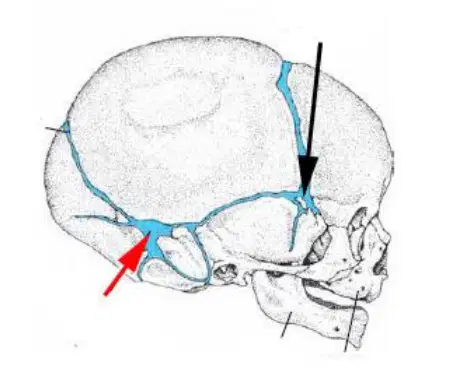
Choose the three correct statements!
The red arrow shows the posterolateral or mastoid fontanelle.
The black arrow shows the anterolateral or sphenoid fontanelle.
The structures labeled with black and red arrow contain connective tissue.
The red arrow shows the anterior (major) fontanelle.
The black arrow shows the posterior (minor) fontanelle.

