
 Worksheets
Vetscience2
Total questions: 99
Worksheet time: 50mins
What are the five basic functions of the musculoskeletal system? Fill in the blanks: Bones furnish five basic functions: _______, _______, _______, _______, and _______.
structure, leverage, protection, mineral reserves, blood cell production
movement, digestion, protection, mineral reserves, blood cell production
structure, leverage, protection, fat storage, blood cell production
structure, leverage, protection, mineral reserves, hormone secretion
Which of the following is NOT a function of the musculoskeletal system?
B) Leverage
A) Structure
C) Digestion
E) Mineral reserves
D) Protection
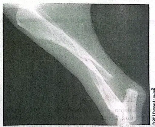
The radiograph in Figure 3-2 shows a fracture in which bone of Missy's leg?
Femur
Tibia
Fibula
Humerus
What is a radiograph?
A) A type of surgery
B) An X-ray image
C) A broken bone
D) A veterinarian
An orthopedic surgeon is a veterinarian who specializes in surgery of the bones.
True
False
Fill in the blank: Missy was not able to ______ weight on her injured leg when she arrived at the office.
bear
bare
bar
beer
Describe the most visible function of bones in the animal body as mentioned in the passage.
The most visible function of bones is structure. The collection of bones in the animal forms the skeleton, which provides the framework that defines an animal's shape and size.
The most visible function of bones is digestion. Bones help break down food in the animal body.
The most visible function of bones is respiration. Bones assist in the breathing process of animals.
The most visible function of bones is circulation. Bones pump blood throughout the animal body.
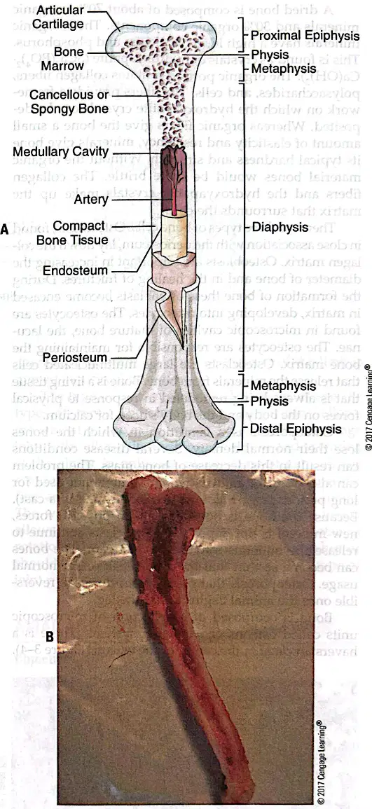
Match the following bone parts with their correct locations:
Articular Cartilage
Top end of bone covering joint surface
Bone Marrow
Center hollow region
Cancellous (Spongy) Bone
Area beneath compact bone at ends
Medullary Cavity
Central cavity
Compact Bone Tissue
Outer shell of shaft
What is the function of the rib cage in the musculoskeletal system?
Protects the heart and lungs
Produces blood cells
Stores minerals
Provides movement at joints
Fill in the blank: The outer shell of a long bone is composed of ________ bone.
compact
spongy
cancellous
trabecular
Which type of bone is found inside the ends of long bones and is made up of tiny spicules and plates?
Compact bone
Spongy bone
Cortical bone
Periosteum
The periosteum is a thin connective tissue that covers the bone and blends into tendons and ligaments.
True
False
Fill in the blank: Bone marrow is made in the ________ center, the medullary cavity.
hollow
solid
dense
outer
Which tissue provides protection as bones move against one another within a joint?
Periosteum
Endosteum
Articular cartilage
Spongy bone
What percentage of a dried bone is composed of inorganic minerals?
70%
30%
50%
90%
Which of the following is NOT a component of the organic portion of bone?
Collagen fibers
Polysaccharides
Hydroxyapatite
Cells
Name the three types of bone cells described in the passage.
Osteoblasts, osteocytes, osteoclasts
Chondrocytes, fibroblasts, erythrocytes
Neutrophils, lymphocytes, monocytes
Adipocytes, melanocytes, keratinocytes
Osteoporosis is a condition in which bones lose their normal density.
True
False
What is the function of osteoclasts in bone tissue?
Osteoclasts release minerals from bone into the bloodstream.
Osteoclasts produce new bone matrix.
Osteoclasts store calcium in bone tissue.
Osteoclasts aid in the formation of cartilage.
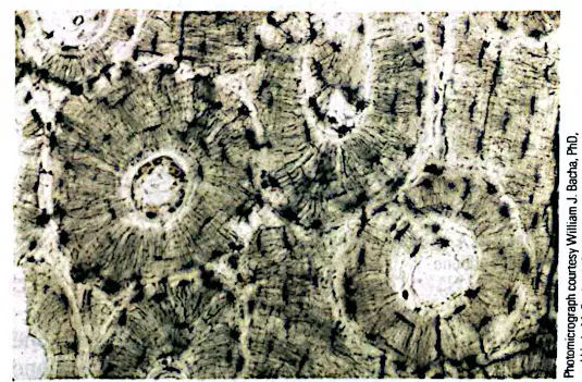
The central canal in the osteon is called the ________ canal.
haversian
volkmann
medullary
epiphyseal
Which type of joint allows little movement and is found in the skull?
Fibrous joint
Cartilage joint
Synovial joint
Haversian joint
Cartilage joints are connected with cartilage and allow for growth.
True
False
What is the function of synovial fluid in synovial joints?
Synovial fluid provides lubrication to the joint and carries nutrients to the cartilage.
Synovial fluid strengthens the bones and increases joint rigidity.
Synovial fluid produces red blood cells for the joint.
Synovial fluid connects muscles to bones.

In the diagram comparing the skeletons of a horse and a cat, what bone is labeled as the 'Atlas (C1)' in the horse?
Atlas (C1) is the first cervical vertebra in the horse.
Atlas (C1) is the last thoracic vertebra in the horse.
Atlas (C1) is the first lumbar vertebra in the horse.
Atlas (C1) is the last sacral vertebra in the horse.

Which joint in the horse's leg is identified as a 'Ball and Socket Joint'?
The joint between the pelvis and femur is a Ball and Socket Joint.
The joint between the femur and tibia is a Ball and Socket Joint.
The joint between the radius and ulna is a Ball and Socket Joint.
The joint between the cannon bone and pastern is a Ball and Socket Joint.

In the diagram, which bone is referred to as the 'Cannon Bone' in the horse?
The III Metatarsal is referred to as the Cannon Bone in the horse.
The Tibia is referred to as the Cannon Bone in the horse.
The Femur is referred to as the Cannon Bone in the horse.
The Calcaneus is referred to as the Cannon Bone in the horse.

Which of the following bones is present in both the horse and the cat?
Femur
Ulna
Patella
All of the above

What is the function of the 'Patella' as labeled in both the horse and cat diagrams?
The Patella functions as the kneecap, aiding in leg movement and protection of the knee joint.
The Patella is responsible for digestion in the animal.
The Patella helps in the animal's sense of smell.
The Patella is a bone in the tail that aids in balance.

The horse has more coccygeal (caudal) vertebrae than the cat.
True
False

Which bone is labeled as the 'Mandible' in both the horse and cat diagrams?
The Mandible is the lower jaw bone in both the horse and cat.
The Mandible is the upper jaw bone in both the horse and cat.
The Mandible is the cheek bone in both the horse and cat.
The Mandible is the nasal bone in both the horse and cat.

In the diagram, which animal has a clavicle bone?
The cat has a clavicle bone.
The dog has a clavicle bone.
The horse has a clavicle bone.
The cow has a clavicle bone.

Which joint type is found at the horse's hock (tarsus)?
Hinge Joint
Ball and Socket Joint
Pivot Joint
Saddle Joint
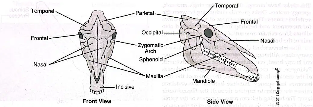
Refer to the diagram labeled 'Skull structure of a horse (Front View)'. Fill in the blank: The bone labeled at the top of the skull is called the _________.
Temporal
Frontal
Occipital
Parietal
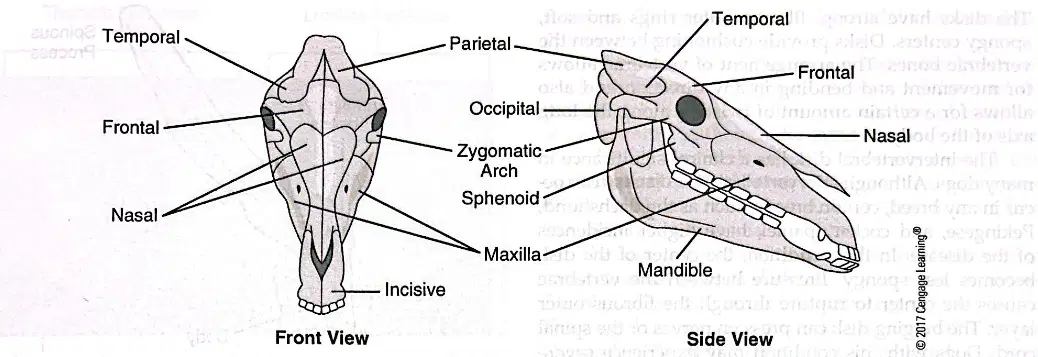
Refer to the diagram labeled 'Skull structure of a horse (Side View)'. Fill in the blank: The bone forming the lower jaw is called the _________.
Mandible
Maxilla
Frontal
Occipital
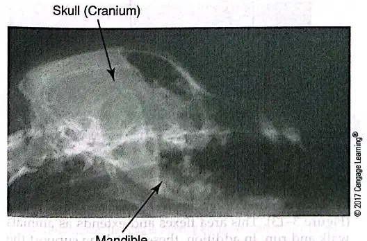
Refer to the radiograph labeled 'Skull (Cranium)'. Fill in the blank: The arrow is pointing to the _________
Mandible
Maxilla
Zygomatic bone
Frontal bone
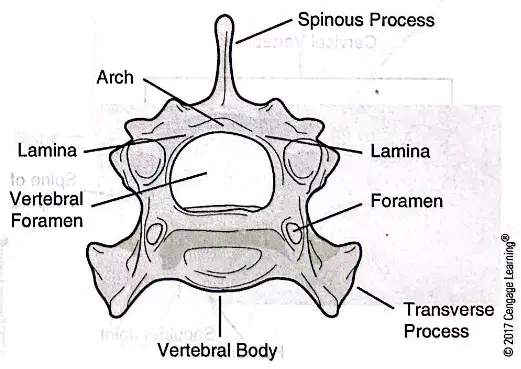
Refer to the diagram labeled 'The structure of a vertebra'. Fill in the blank: The central part of the vertebra is called the _________.
Vertebral Body
Spinous Process
Transverse Process
Vertebral Arch
Which bone in the skull is most notably responsible for protecting the brain?
Cranium
Mandible
Maxilla
Zygomatic
The mandible is a synovial joint that allows animals to secure and chew food.
True
False
Match the following bones to their functions:
Protects the brain
3
Holds the teeth
1
Allows animals to chew food
2
The function of the vertebral column in animals is:
to support and protect the spinal cord
to aid in digestion
to produce hormones
to filter waste from blood
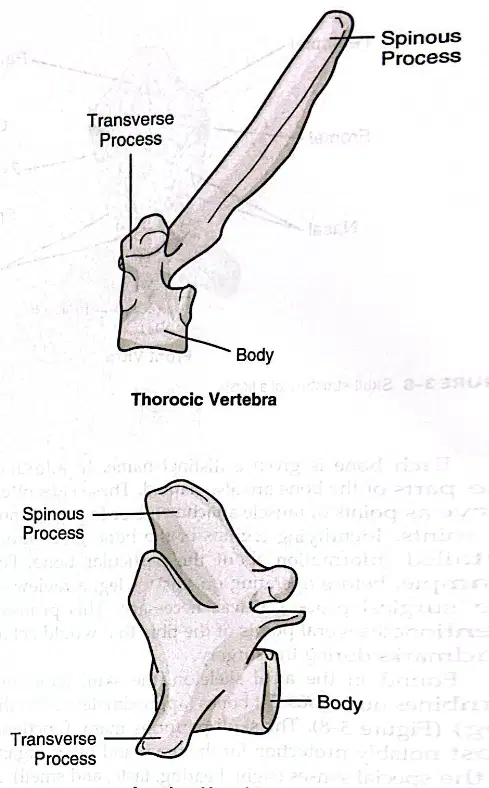
Refer to the diagram labeled 'Thoracic Vertebra.' Which part is labeled as the 'Spinous Process'?
The uppermost projection is labeled as the 'Spinous Process.'
The rounded area at the side is labeled as the 'Spinous Process.'
The lower curved section is labeled as the 'Spinous Process.'
The central hollow region is labeled as the 'Spinous Process.'
According to the passage, how many cervical vertebrae do mammals such as cats, horses, and giraffes have?
Seven cervical vertebrae.
Five cervical vertebrae.
Nine cervical vertebrae.
Twelve cervical vertebrae.
Which vertebra allows up-and-down motion of the head, as in nodding to signify yes?
The atlas (first cervical vertebra).
The axis (second cervical vertebra).
The thoracic vertebra.
The lumbar vertebra.
Dogs, cats, cattle, goats, and sheep all have how many pairs of ribs?
13 pairs of ribs.
10 pairs of ribs.
15 pairs of ribs.
12 pairs of ribs.
Horses have how many thoracic vertebrae?
18 thoracic vertebrae.
16 thoracic vertebrae.
20 thoracic vertebrae.
14 thoracic vertebrae.
The cartilage joining the ribs to the sternum in older animals can be replaced with bone.
True
False
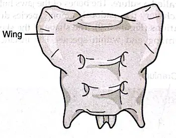
Refer to the diagram labeled 'Sacrum, Ventral View.' Which part is labeled as the 'Wing'?
The lateral projections are labeled as the 'Wing.'
The central ridge is labeled as the 'Wing.'
The lower tip is labeled as the 'Wing.'
The upper notch is labeled as the 'Wing.'
What is the clinical significance of intervertebral disk disease in dogs?
It can cause pain, paralysis, and may require medical or surgical treatment.
It only affects the skin and causes mild itching.
It improves mobility and increases energy levels.
It is a minor condition that resolves without intervention.
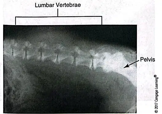
Which vertebrae are labeled in the radiograph of a cat?
A) Thoracic Vertebrae
B) Lumbar Vertebrae
C) Sacral Vertebrae
D) Caudal Vertebrae
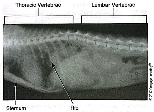
What age-related change is shown in the radiograph of the lumbar spine of a dog?
Spondylosis
Intervertebral disc herniation
Osteosarcoma
Hip dysplasia
Luxation
What is the name of the bone labeled in the diagram?
Scapula
Femur
Humerus
Clavicle
The sacrum is a group of three sacral vertebrae that fuses to support the pelvis.
True
False
Which part of the skeleton includes the bones of the forelimbs and hind limbs?
Axial skeleton
Appendicular skeleton
Pelvic skeleton
Thoracic skeleton
In cats, the scapula does not have a bony connection to the axial skeleton.
True
False
What is the clinical significance of the cat's ability to rotate its scapula and extend its front legs when falling from heights?
It allows the cat to land gracefully and rarely break a leg, but it may break its lower jaw due to the high speed of the fall.
It helps the cat to fly short distances and escape predators.
It enables the cat to swim efficiently when falling into water.
It causes the cat to lose balance and increases the risk of spinal injury.
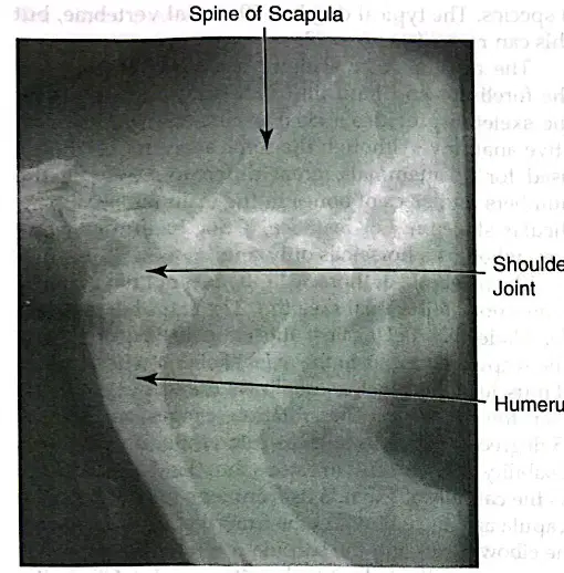
What is the upper bone of the forelimb called?
Humerus
Femur
Radius
Tibia
How many carpal bones does a dog have?
7
5
8
6
How many carpal bones do ruminants have?
6
7
8
5
How many carpal bones does a horse have?
7 or 8
5 or 6
9 or 10
3 or 4
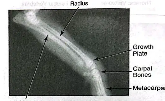
Which bone is labeled as running parallel to the radius in the forelimb of a dog?
Ulna
Humerus
Metacarpals
Carpal Bones
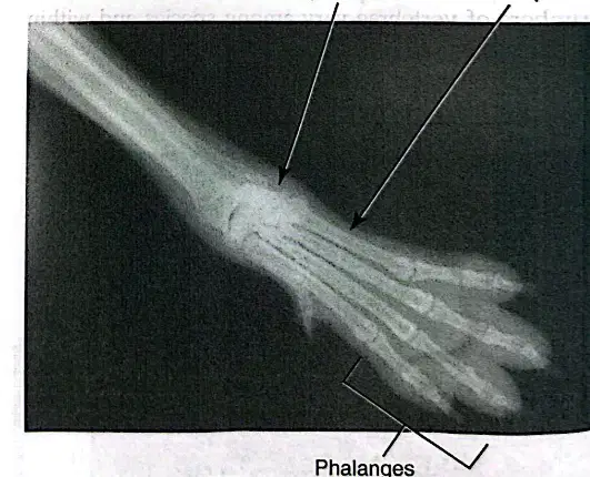
Which bones are found distal to the metacarpals in a dog's forelimb?
Carpal Bones
Phalanges
Ulna
Radius
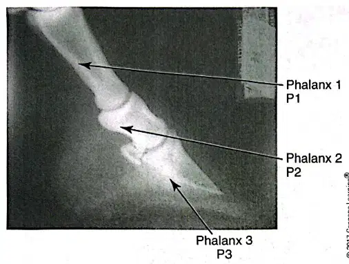
Refer to the radiograph of the foot of a horse (Figure 3-19). Which bone is labeled as Phalanx 1 (P1)?
The most distal bone
The middle bone
The most proximal bone
The bone covered by the nail or hoof
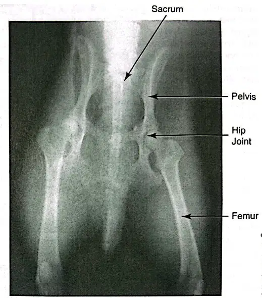
Refer to the radiograph of the pelvis. Match the following anatomical terms to their locations:
Uppermost part of the radiograph
Sacrum
Central region
Pelvis
Joint connecting pelvis and femur
Hip Joint
Lower part of the radiograph
Femur
Fill in the blank: The last phalanx is covered by the ______ or hoof.
nail
skin
bone
cartilage
Which bones fuse into one structure as the ruminant embryo develops?
First and second metacarpal bones
Third and fourth metacarpal bones
Phalanx 1 and Phalanx 2
Ilium and ischium
The pelvis is made of two halves. Each half divides into regions named the ______, ______, and ______.
ilium, ischium, pubis
ilium, sacrum, coccyx
ischium, femur, tibia
pubis, sacrum, femur
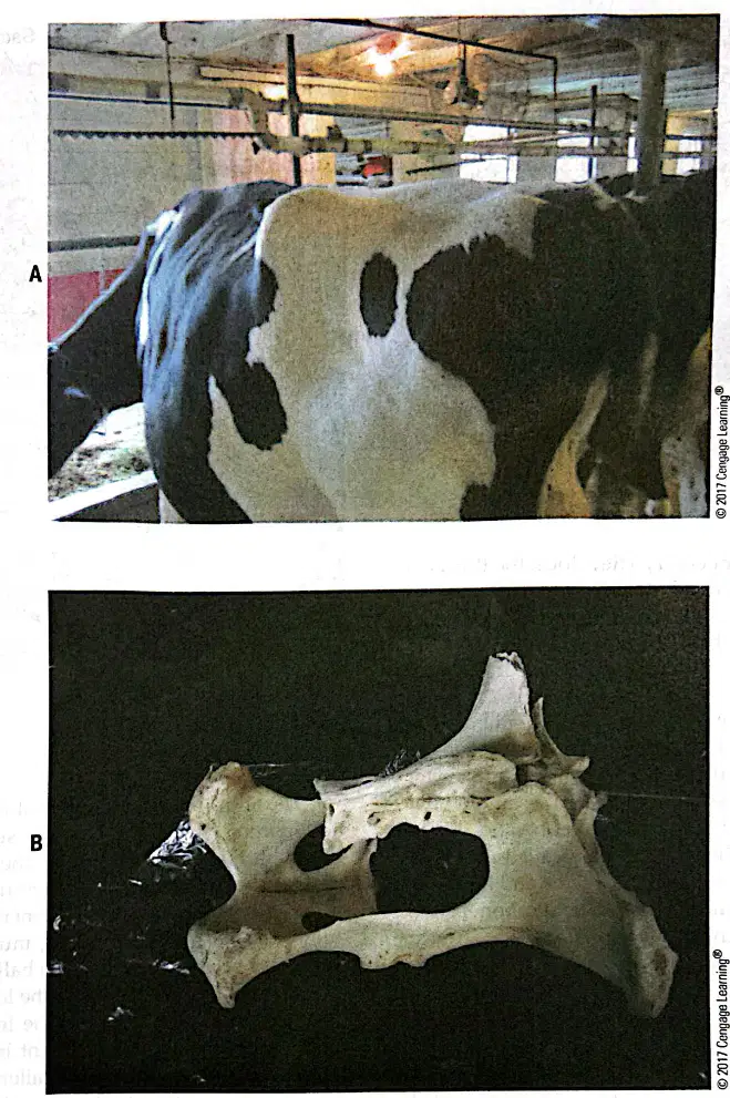
FIGURE 3-21. The wings of the ilium (hooks) and the ischium (pins) can be observed on each side of the tail in the rump of a cow. Which bones are being identified in this description?
Ilium and ischium
Femur and tibia
Scapula and humerus
Radius and ulna
Refer to the radiograph of the hind limb (Figure 3-22). Which bone is labeled as 'Femur'?
A) The uppermost bone
B) The lowermost bone
C) The bone on the right
D) The bone on the left.
Refer to the photograph of the internal structure of the knee (Figure 3-23). What is identified by the probe in the image?
The two cruciate ligaments
The meniscus
The patellar tendon
The lateral collateral ligament
Bone growth and remodeling in immature animals occurs through which process?
Endochondral ossification and bone resorption
Osteoarthritis development and cartilage breakdown
Muscle hypertrophy and fat deposition
Ligament stretching and joint fusion
What is the function of the growth plate in young animals?
The growth plate is the site where bone elongation occurs, allowing bones to grow longer as the animal matures.
The growth plate is responsible for producing red blood cells in young animals.
The growth plate helps in the absorption of nutrients from food in young animals.
The growth plate acts as a protective layer against infections in young animals.
What happens when the growth plate of the ulna closes prematurely in a basset hound?
A) The radius grows straight
B) The radius curves to fit between the other bones
C) The humerus shortens
D) The ulna lengthens
What is the correct term used to describe the picture produced when a stream of x-rays is passed through a body part and exposes a piece of photographic film?
Radiograph
Photograph
X-ray
Radiology
Which of the following tissues appear light on a radiograph because they are radiopaque?
Fat
Soft tissue
Teeth and bone
Mineral
Arrange the following in order from most radiolucent to most radiopaque as seen in radiographs: air, fat, soft tissue or muscle, bone, mineral.
Most radiolucent to most radiopaque: air, fat, soft tissue or muscle, bone, mineral.
Most radiolucent to most radiopaque: mineral, bone, soft tissue or muscle, fat, air.
Most radiolucent to most radiopaque: bone, mineral, soft tissue or muscle, fat, air.
Most radiolucent to most radiopaque: fat, air, soft tissue or muscle, bone, mineral.
What is the function of specially designed lead aprons and gloves during x-ray procedures?
To minimize exposure to humans and prevent x-ray penetration.
To enhance the clarity of x-ray images.
To keep the patient warm during the procedure.
To improve the comfort of the medical staff.
What is the advantage of having a radiographic image as a computer file compared to old film methods?
It allows for alterations in brightness, contrast, and size, and easy transfer of the image to a specialty clinic or radiologist for consultation.
It makes the image less clear and harder to interpret.
It increases the risk of losing the image due to physical damage.
It requires more space for storage than traditional film.
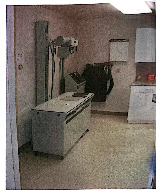
Refer to the image labeled 'FIGURE 3-25 Radiograph machine'. What is shown in the image?
A radiograph machine used for taking x-rays.
An ultrasound machine used for imaging soft tissues.
A centrifuge used for separating blood components.
A microscope used for viewing slides.
Objective: Relate Bone and Muscle Groups to Movement. Bones and muscles work together to provide mobility to the body by:
Muscles contract and pull on bones, causing movement at the joints.
Bones contract and pull on muscles, causing movement at the joints.
Muscles and bones both contract to create movement.
Bones move independently to create mobility.
What is a simple fracture?
A simple fracture is when the bone is broken into two pieces, a clean break.
A simple fracture is when the bone is shattered into many small pieces.
A simple fracture is when the bone breaks and pierces through the skin.
A simple fracture is when the bone bends but does not break.
What is a comminuted fracture?
A comminuted fracture results in several fragments of bone.
A comminuted fracture is a break that does not involve bone fragments.
A comminuted fracture is a simple crack in the bone.
A comminuted fracture is a fracture that heals without intervention.
What is a compound (open) fracture?
A compound (open) fracture results when one of the bone ends punctures through the skin.
A compound (open) fracture is a fracture that heals without medical intervention.
A compound (open) fracture is a fracture where the bone bends but does not break.
A compound (open) fracture is a fracture that only occurs in children.
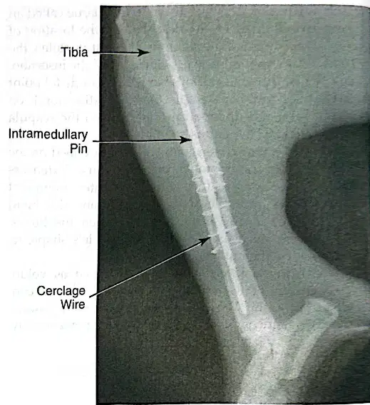
Which surgical method involves driving a stainless steel pin into the center of the broken tibia?
Bone plate and screws
Intramedullary pin
Cerclage wire
Cast or splint
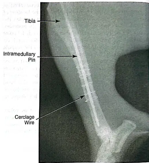
What is the purpose of cerclage wires in bone fracture repair?
Cerclage wires are used to stabilize the pieces of bone by wrapping around the bone fragments.
Cerclage wires are used to promote bone growth by releasing growth factors.
Cerclage wires are used to prevent infection by acting as a barrier.
Cerclage wires are used to replace missing bone tissue.
What is the initial structure that forms at the fracture site and helps stabilize it during bone healing?
Callus
Osteon
Ligament
Cartilage
After approximately two weeks, the callus at the fracture site is gradually replaced by what type of cell?
Osteoblasts
Chondrocytes
Fibroblasts
Adipocytes
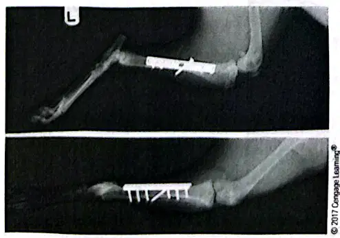
What method was used to repair the tibial fracture shown in the radiograph?
Bone plate and screws
Intramedullary pin
Cerclage wire
Cast or splint
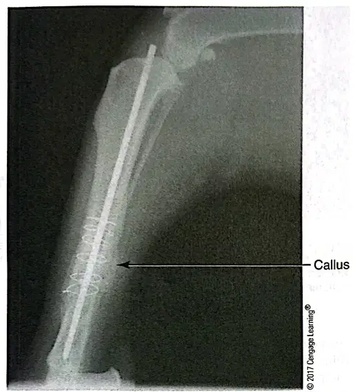
What is the name of the formation surrounding the site of the fracture in the radiograph of healing bone two months following surgery?
Callus
Osteoma
Granuloma
Fibroma
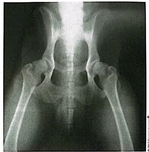
What condition is shown in the radiograph of a dog, characterized by a shallow acetabulum and subluxation (partial dislocation) of the femur?
Hip dysplasia
Osteosarcoma
Patellar luxation
Intervertebral disc disease
What is the most common treatment for mild cases of hip dysplasia in dogs?
Anti-inflammatory medications
Surgical hip replacement
Physical therapy only
High-protein diet
Which of the following is NOT a reason that fractures do not heal properly?
Bone infection
Proper alignment and immobilization
Open fracture
Movement after surgery
Degenerative joint disease in dogs is characterized by:
A) Pain and lameness
B) Trouble rising
C) Both A and B
D) None of the above
Hip dysplasia is generally associated with large- and giant-breed dogs, but any dog can have it.
True
False
What is the condition called that involves damage or degeneration of the navicular bone in horses, leading to lameness?
Navicular syndrome
Laminitis
Colic
Ringbone
Aspirin can be used to treat hip dysplasia.
True
False
The hinge joint allows rotation.
True
False
Dried bone consists of _____% inorganic and _____% organic material.
70% inorganic and 30% organic material
60% inorganic and 40% organic material
50% inorganic and 50% organic material
80% inorganic and 20% organic material
The appendicular skeleton contains the bones of the _______.
limbs
skull
vertebral column
rib cage
The smallest metacarpal bone in a dog is called the _______.
dewclaw
carpal pad
metacarpal tubercle
phalange

