
 Worksheets
CM-12
Total questions: 122
Worksheet time: 1hrs 1mins
Name
Class
Date
1.
Normal value is 0-2 or 0-3 per HPF
a)
RBC
b)
WBCs
c)
Squamous epithelial cells
d)
Transitional epithelial cells
2.
Sources of error = yeasts, oil droplets, air bubbles, monohydrate calcium oxalate crystals
a)
RBC
b)
WBCs
c)
Squamous epithelial cells
d)
Transitional epithelial cells
3.
Remedy is to add 2% acetic acid to lyse it.
a)
RBC
b)
WBCs
c)
Squamous epithelial cells
d)
Transitional epithelial cells
4.
Which concentration of acetic acid is added to lyse RBCs?
a)
1%
b)
2%
c)
3%
d)
4%
5.
What is the normal value of eosinophil in urine?
a)
1%
b)
2%
c)
3%
d)
4%
6.
Eosinophil value is considered significant if it exceeds... ___ since this is usually seen in acute interstitial nephritis
a)
>1%
b)
>2%
c)
>3%
d)
>4%
7.
Most predominant WBC seen in urine
a)
Neutrophil
b)
Eosinophil
c)
Basophil
d)
Monocyte
e)
Lymphocyte
8.
Increased in renal transplant rejection
a)
Neutrophil
b)
Eosinophil
c)
Basophil
d)
Monocyte
e)
Lymphocyte
9.
Which of the ff. mononuclear cells is increased in chronic inflammation and radiation therapy?
a)
Lymphocytes
b)
Monocytes
c)
Macrophages
d)
Histiocytes
10.
Glitter cells color when Sternheimer-Malbin stain is used
a)
Pale red
b)
Pale pink
c)
Pale yellow
d)
Pale blue
11.
Leukocytes color when Sternheimer-Malbin stain is used
a)
Pale red
b)
Pale pink
c)
Pale yellow
d)
Pale blue
12.
Which WBC is granulated and multilobed?
a)
Neutrophil
b)
Eosinophil
c)
Basophil
d)
Monocyte
e)
Lymphocyte
13.
Which WBC swell in hypotonic urine, and exhibits Brownian movement thereby producing a sparkling appearance?
a)
Neutrophil
b)
Eosinophil
c)
Basophil
d)
Monocyte
e)
Lymphocyte
14.
Which WBC form blebs and finger-like projections (myelin forms) when dying?
a)
Neutrophil
b)
Eosinophil
c)
Basophil
d)
Monocyte
e)
Lymphocyte
15.
Normal value of this WBC is 1%
a)
Neutrophil
b)
Eosinophil
c)
Basophil
d)
Monocyte
e)
Lymphocyte
16.
Glitter cells
a)
Neutrophil
b)
Eosinophil
c)
Basophil
d)
Monocyte
e)
Lymphocyte
17.
Smooth, non-nucleated, biconcave disks
a)
RBC
b)
WBCs
c)
Squamous epithelial cells
d)
Transitional epithelial cells
18.
Increased number indicates presence of infection or inflammation
a)
RBC
b)
WBCs
c)
Squamous epithelial cells
d)
Transitional epithelial cells
19.
What is the cell seen in cases of pyuria?
a)
RBC
b)
WBCs
c)
Squamous epithelial cells
d)
Transitional epithelial cells
20.
What is the cell seen in cases of leukocyturia?
a)
RBC
b)
WBCs
c)
Squamous epithelial cells
d)
Transitional epithelial cells
21.
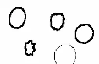
Identify the cell in the image
a)
RBC
b)
WBCs
c)
Squamous epithelial cells
d)
Transitional epithelial cells
22.
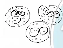
Identify the cell in the image
a)
RBC
b)
WBCs
c)
Squamous epithelial cells
d)
Transitional epithelial cells
23.
In which type of spx/condition would crenated or shrinked RBCs will be seen?
a)
Hypertonic urine
b)
Hypotonic urine
c)
Glomerular membrane damage
24.
In which type of spx/condition would swollen or lysed RBCs will be seen?
a)
Hypertonic urine
b)
Hypotonic urine
c)
Glomerular membrane damage
25.
In which type of spx/condition would shadow cells-like RBCs will be seen?
a)
Hypertonic urine
b)
Hypotonic urine
c)
Glomerular membrane damage
26.
In which type of spx/condition would dysmorphic with projections and fragmented RBCs will be seen?
a)
Hypertonic urine
b)
Hypotonic urine
c)
Glomerular membrane damage
27.
Normal value is 0-5 or 0-8 per HPF
a)
RBC
b)
WBCs
c)
Squamous epithelial cells
d)
Transitional epithelial cells
28.
Largest cell with abundant, irregular cytoplasm and prominent nucleus
a)
RBC
b)
WBCs
c)
Squamous epithelial cells
d)
Transitional epithelial cells
29.
Cell size is about 30 to 50 um, which is 5 to 7x the size of an RBC
a)
RBC
b)
WBCs
c)
Squamous epithelial cells
d)
Transitional epithelial cells
30.
The nucleus is about the size of an RBC
a)
RBC
b)
WBCs
c)
Squamous epithelial cells
d)
Transitional epithelial cells
31.
May occasionally appear folded, resembling a cast
a)
RBC
b)
WBCs
c)
Squamous epithelial cells
d)
Transitional epithelial cells
32.
Comes from linings of vagina, female urethra and lower male urethra
a)
RBC
b)
WBCs
c)
Squamous epithelial cells
d)
Transitional epithelial cells
33.
Also known as urothelial cell or bladder cell
a)
RBC
b)
WBCs
c)
Squamous epithelial cells
d)
Transitional epithelial cells
34.
Cell size is about 20 to 30 um which is 4 to 6x the size of an RBC; N:C ratio is DECREASED
a)
RBC
b)
WBCs
c)
Squamous epithelial cells
d)
Transitional epithelial cells
35.
Spherical, polyhedral, or caudate with CENTRALLY located nucleus
a)
RBC
b)
WBCs
c)
Squamous epithelial cells
d)
Transitional epithelial cells
36.
They tend to take on water and can look like large round balloons
a)
RBC
b)
WBCs
c)
Squamous epithelial cells
d)
Transitional epithelial cells
37.
From renal pelvis, calyces, ureter, urinary bladder and upper male urethra
a)
RBC
b)
WBCs
c)
Squamous epithelial cells
d)
Transitional epithelial cells
38.
True UTI is characterized by bacteria + ______
a)
RBC
b)
WBCs
c)
Squamous epithelial cells
d)
Transitional epithelial cells
39.
True yeast infection is characterized by yeast + ______
a)
RBC
b)
WBCs
c)
Squamous epithelial cells
d)
Transitional epithelial cells
40.
Increased following catheterization and appears either singly, in pairs, or in clumps (synctia)
a)
RBC
b)
WBCs
c)
Squamous epithelial cells
d)
Transitional epithelial cells
41.
If this cell exhibits an abnormal morphology, it implies malignancy or viral infection
a)
RBC
b)
WBCs
c)
Squamous epithelial cells
d)
Transitional epithelial cells
42.

Identify the cell in the image
a)
RBC
b)
WBCs
c)
Squamous epithelial cells
d)
Transitional epithelial cells
43.
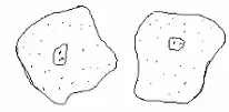
Identify the cell in the image
a)
RBC
b)
WBCs
c)
Squamous epithelial cells
d)
Transitional epithelial cells
44.
Variation of squamous epithelial cell covered with G. vaginalis; a bacteria associated with bacterial vaginosis
a)
Glitter cell
b)
Ghost cell
c)
Clue cell
d)
Bubble cell
45.
Clue cells are associated with presence of which of the ff.?
a)
Trichomonas vaginalis
b)
Lactobacillus acidophilus
c)
Gardnerella vaginalis
d)
Schistosoma haematobium
46.
What is the most frequently encountered parasite in urine?
a)
Trichomonas vaginalis
b)
Lactobacillus acidophilus
c)
Gardnerella vaginalis
d)
Schistosoma haematobium
47.
Pear-shaped flagellate with undulating membrane and jerky motility
a)
Trichomonas vaginalis
b)
Lactobacillus acidophilus
c)
Gardnerella vaginalis
d)
Schistosoma haematobium
48.
If not moving, may resemble WBC/TEC/RTE cell
a)
Trichomonas vaginalis
b)
Lactobacillus acidophilus
c)
Gardnerella vaginalis
d)
Schistosoma haematobium
49.
Blood fluke with terminal spine
a)
Trichomonas vaginalis
b)
Lactobacillus acidophilus
c)
Gardnerella vaginalis
d)
Schistosoma haematobium
50.
Causes hematuria
a)
Trichomonas vaginalis
b)
Lactobacillus acidophilus
c)
Gardnerella vaginalis
d)
Schistosoma haematobium
51.
Associated with bladder cancer
a)
Trichomonas vaginalis
b)
Lactobacillus acidophilus
c)
Gardnerella vaginalis
d)
Schistosoma haematobium
52.
What is the most common fecal contaminant?
a)
Ascaris lumbricoides
b)
Trichuris trichiura
c)
Enterobius vermicularis
d)
Strongyloides stercoralis
53.
Which of the following are urinary bladder cancer markers?
a)
CEA
b)
CD125
c)
NMP
d)
BTA
54.
Reported as rare, few, moderate, or many per HPF
a)
Trichomonas vaginalis
b)
Lactobacillus acidophilus
c)
Gardnerella vaginalis
d)
Schistosoma haematobium
55.
Causative agent of ping pong disease
a)
Trichomonas vaginalis
b)
Lactobacillus acidophilus
c)
Gardnerella vaginalis
d)
Schistosoma haematobium
56.
What is the most clinically significant epithelial cell?
a)
Renal tubular epithelial cell
b)
Transitional epithelial cell
c)
Squamous epithelial cell
57.
Location of nucleus of TEC
a)
Terminal
b)
Central
c)
Eccentric
58.
Location of nucleus of RTE cell
a)
Terminal
b)
Central
c)
Eccentric
59.
Origin is nephron
a)
Renal tubular epithelial cell
b)
Bacteria
c)
Yeasts
d)
Parasites
60.
Motility differentiates them from amorphous urates and phosphates
a)
Renal tubular epithelial cell
b)
Bacteria
c)
Yeasts
d)
Parasites
61.
Small refractile oval structure that may or may not bud
a)
Renal tubular epithelial cell
b)
Bacteria
c)
Yeasts
d)
Parasites
62.
Branched, mycelial forms are seen in severe infections
a)
Renal tubular epithelial cell
b)
Bacteria
c)
Yeasts
d)
Parasites
63.
Size is 3 to 5x the size of RBCs
a)
Renal tubular epithelial cell
b)
Bacteria
c)
Yeasts
d)
Parasites
64.
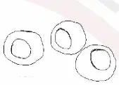
Identify the cell in the image
a)
Renal tubular epithelial cell
b)
Bacteria
c)
Yeasts
d)
Parasites
65.

Identify the cell in the image
a)
Renal tubular epithelial cell
b)
Bacteria
c)
Yeasts
d)
Parasites
66.
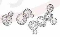
Identify the cell in the image
a)
Renal tubular epithelial cell
b)
Bacteria
c)
Yeasts
d)
Parasites
67.
Sometimes bilirubin-stained or hemosiderin-laden
a)
Renal tubular epithelial cell
b)
Bacteria
c)
Yeasts
d)
Parasites
68.
If RTE is from ___, it appears large, oblong, or cigar-shaped with grainy cytoplasm
a)
PCT
b)
DCT
c)
CD
d)
Nephron
e)
Ascending loop of Henle
69.
If RTE is from ___, it appears smaller, round to oval and may be mistaken for WBCs
a)
PCT
b)
DCT
c)
CD
d)
Nephron
e)
Ascending loop of Henle
70.
If RTE is from ___, it appears columnar, polygonal, or cuboidal with flat edge
a)
PCT
b)
DCT
c)
CD
d)
Nephron
e)
Ascending loop of Henle
71.
If RTE exceeds this amount per HPF, it is indicative of tubular injury
a)
>1
b)
>2
c)
>3
d)
>4
72.
N:C ratio of TEC
a)
Increased
b)
Decreased
c)
Normal
d)
Either
73.
N:C ratio of RTE cell
a)
Increased
b)
Decreased
c)
Normal
d)
Either
74.
Renal tubular fat bodies
a)
Oval fat body
b)
Bubble cell
c)
Clue cell
d)
Glitter cell
75.
Lipid containing RTE cell which may also be a monocyte or macrophage
a)
Oval fat body
b)
Bubble cell
c)
Clue cell
d)
Glitter cell
76.
Highly refractile RTE cell
a)
Oval fat body
b)
Bubble cell
c)
Clue cell
d)
Glitter cell
77.
Seen in lipiduria such as nephrotic syndrome
a)
Oval fat body
b)
Bubble cell
c)
Clue cell
d)
Glitter cell
78.
Identified by lipid stains (TAG and neutral fats)
a)
Oval fat body
b)
Bubble cell
c)
Clue cell
d)
Glitter cell
79.
RTE cell with non-lipid vacuoles
a)
Oval fat body
b)
Bubble cell
c)
Clue cell
d)
Glitter cell
80.
Injured cells in which the endoplasmic reticulum has dilated prior to cell death
a)
Oval fat body
b)
Bubble cell
c)
Clue cell
d)
Glitter cell
81.
Seen in acute tubular necrosis
a)
Oval fat body
b)
Bubble cell
c)
Clue cell
d)
Glitter cell
82.
In polarizing microscope, this exhibits maltese cross formation
a)
VLDL
b)
Babesia spp.
c)
Cholesterol
d)
HDL
e)
Triglycerides
83.
What is the most common cause of UTI?
a)
S. saprophyticus
b)
S. aureus
c)
E. coli
d)
P. vulgaris
84.
seen in DM and vaginal monoliasis
a)
P. immitis
b)
C. albicans
c)
C. botulinum
d)
C. difficile
85.
Oval, slightly tapered head; long, flagella-like tail and seen after sexual intercourse
a)
Spermatozoa
b)
Mucus threads
c)
Crystals
d)
Casts
e)
T. vaginalis
86.
Has low refractive index and its major constituent is uromodulin (Tam-Horsfall protein)
a)
Spermatozoa
b)
Mucus threads
c)
Crystals
d)
Casts
e)
T. vaginalis
87.
Squamous epithelial cells are reported as
a)
Rare, few, moderate, or many per LPF
b)
Rare, few, moderate, or many per HPF
c)
Average number per 10 HPFs
d)
Average number per HPF
e)
Average number per LPF
88.
Transitional epithelial cells are reported as
a)
Rare, few, moderate, or many per LPF
b)
Rare, few, moderate, or many per HPF
c)
Average number per 10 HPFs
d)
Average number per HPF
e)
Average number per LPF
89.
Yeasts are reported as
a)
Rare, few, moderate, or many per LPF
b)
Rare, few, moderate, or many per HPF
c)
Average number per 10 HPFs
d)
Average number per HPF
e)
Average number per LPF
90.
Renal tubular epithelial cells are reported as
a)
Rare, few, moderate, or many per LPF
b)
Rare, few, moderate, or many per HPF
c)
Average number per 10 HPFs
d)
Average number per HPF
e)
Average number per LPF
91.
Oval fat bodies are reported as
a)
Rare, few, moderate, or many per LPF
b)
Rare, few, moderate, or many per HPF
c)
Average number per 10 HPFs
d)
Average number per HPF
e)
Average number per LPF
92.
Abnormal crystals are reported as
a)
Rare, few, moderate, or many per LPF
b)
Rare, few, moderate, or many per HPF
c)
Average number per 10 HPFs
d)
Average number per HPF
e)
Average number per LPF
93.
Casts are reported as
a)
Rare, few, moderate, or many per LPF
b)
Rare, few, moderate, or many per HPF
c)
Average number per 10 HPFs
d)
Average number per HPF
e)
Average number per LPF
94.
Epithelial cells
a)
quantitated per LPF
b)
quantitated per HPF
95.
Mucus threads
a)
quantitated per LPF
b)
quantitated per HPF
96.
Casts
a)
quantitated per LPF
b)
quantitated per HPF
97.
Normal crystals
a)
quantitated per LPF
b)
quantitated per HPF
98.
Bacteria
a)
quantitated per LPF
b)
quantitated per HPF
99.
RBCs
a)
quantitated per LPF
b)
quantitated per HPF
100.
WBCs
a)
quantitated per LPF
b)
quantitated per HPF
101.
Epithelial cells: 0 to 5
a)
Rare
b)
Few
c)
Moderate
d)
Many
102.
Epithelial cells: 5 to 20
a)
Rare
b)
Few
c)
Moderate
d)
Many
103.
Epithelial cells: 20 to 100
a)
Rare
b)
Few
c)
Moderate
d)
Many
104.
Epithelial cells: >100
a)
Rare
b)
Few
c)
Moderate
d)
Many
105.
Normal crystals: 0 to 2
a)
Rare
b)
Few
c)
Moderate
d)
Many
106.
Normal crystals: 2 to 5
a)
Rare
b)
Few
c)
Moderate
d)
Many
107.
Normal crystals: 5 to 20
a)
Rare
b)
Few
c)
Moderate
d)
Many
108.
Normal crystals: >20
a)
Rare
b)
Few
c)
Moderate
d)
Many
109.
Bacteria: >200
a)
Rare
b)
Few
c)
Moderate
d)
Many
110.
Bacteria: 0 to 10
a)
Rare
b)
Few
c)
Moderate
d)
Many
111.
Bacteria: 10 to 50
a)
Rare
b)
Few
c)
Moderate
d)
Many
112.
Bacteria: 50 to 200
a)
Rare
b)
Few
c)
Moderate
d)
Many
113.
Mucus threads: 0-1
a)
Rare
b)
Few
c)
Moderate
d)
Many
114.
Mucus threads: 1 to 3
a)
Rare
b)
Few
c)
Moderate
d)
Many
115.
Mucus threads: 3 to 10
a)
Rare
b)
Few
c)
Moderate
d)
Many
116.
Mucus threads: >10
a)
Rare
b)
Few
c)
Moderate
d)
Many
117.
Numerical ranges: 0 to 2, 2 to 5, 5 to 10, >10
a)
Casts
b)
RBCs
c)
WBCs
118.
Numerical ranges: 0 to 2, 2 to 5, 5 to 10, 10 to 25, 25 to 50, 50 to 100, >100
a)
Casts
b)
RBCs
c)
WBCs
119.
Rare: 0 to 5
Few: 5 to 20
Moderate: 20 to 100
Many: >100
a)
Epithelial cells
b)
Normal crystals
c)
Bacteria
d)
Mucus threads
120.
Rare: 0 to 2
Few: 2 to 5
Moderate: 5 to 20
Many: >20
a)
Epithelial cells
b)
Normal crystals
c)
Bacteria
d)
Mucus threads
121.
Rare: 0 to 10
Few: 10 to 50
Moderate: 50 to 200
Many: >200
a)
Epithelial cells
b)
Normal crystals
c)
Bacteria
d)
Mucus threads
122.
Rare: 0 to 1
Few: 1 to 3
Moderate: 3 to 10
Many: >10
a)
Epithelial cells
b)
Normal crystals
c)
Bacteria
d)
Mucus threads
100 %

