
 Worksheets
BIOLOGY FINAL QUIZ
Total questions: 145
Worksheet time: 1hrs 16mins
HOW MANY BONES DO WE HAVE
AROUND 206
AROUND 400
AROUND 300
AROUND 304
HOW MANY MUSCLES DO WE HAVE
AROUND 600
AROUND 500
AROUND 400
AROUND 300
FUNCTION OF BONES
Supporter and provider of body shape
Inner organ protector
Place to form red blood cells
Muscles adherence place
As the passive motion organ
TYPES OF BONES BASED ON COMPOSITION
CARTILAGE
HARD BONES
LONG BONES
FLAT BONES
IRREGULAR BONES
WHAT IS THE ONLY CELL TYPE THAT IS FOUND IN CARTILAGE?
CHONDROCYTE
CHONDRINE
CHONDRIUM
WHAT IS THE MATRIX (the extracellular, structural material that surrounds the chondrocyte (cartilage cells)) IN CARTILAGE
CHONDROCYTE
CHONDRINE
CHONDRIUM
WHAT IS THE PROTECTIVE LAYER IN CARTILAGE
CHONDROCYTE
CHONDRINE
CHONDRIUM
Types of Cartilage
Hyaline
Elastic
Fibrous
Osteocyte
Zombie Cartilage 🧟♀️🧟♂️🧟
Hyaline Cartilage
Smooth and shiny glasslike structure
End of leg, arm, trachea, ribs, breastbone
Flexible, elastic, easily back into shape
Yellow appearance
Auricle, epiglottis, nose
Absorb shock and reduce friction
Withstand tension and pressure
intervertebral disks of the spine and
knee
Man-made cartilage, used for transplants
Made of iron and silver, can adapt to the body and withstand high pressure and doesn't break
Elastic Cartilage
Smooth and shiny glasslike structure
End of leg, arm, trachea, ribs, breastbone
Flexible, elastic, easily back into shape
Yellow appearance
Auricle, epiglottis, nose
Absorb shock and reduce friction
Withstand tension and pressure
intervertebral disks of the spine and
knee
Man-made cartilage, used for transplants
Made of iron and silver, can adapt to the body and withstand high pressure and doesn't break
Fibrous Cartilage
Smooth and shiny glasslike structure
End of leg, arm, trachea, ribs, breastbone
Flexible, elastic, easily back into shape
Yellow appearance
Auricle, epiglottis, nose
Absorb shock and reduce friction
Withstand tension and pressure
intervertebral disks of the spine and
knee
Man-made cartilage, used for transplants
Made of iron and silver, can adapt to the body and withstand high pressure and doesn't break
Bones (Osteon)
More calcium, less collagen
Less calcium, more collagen
the cells are osteocytes
(a bone cell, formed when an osteoblast becomes embedded in the matrix it has secreted.)
the protective layer is periosteum
(a fibrous membrane covering the outer surface of most bones that contains blood vessels and nerves and plays a crucial role in bone growth and repair)
Produce white blood cells and thrombocytes
periosteum?
Attached firmly
Muscle attachment place
Red Blood cell formation
Fat disposal
red marrow
Attached firmly
Muscle attachment place
Red Blood cell formation
Fat disposal
yellow marrow
Attached firmly
Muscle attachment place
Red Blood cell formation
Fat disposal
Red marrow in children!!!
skull
backbones
ribs
eyeball
Red marrow in adults!!!
skull
backbones
ribs
eyeball
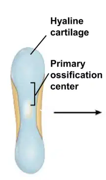
What stage of bone formation is this
1 : Formation of bone collar around hyaline cartilage model.
2 : Cavitation of the hyaline cartilage within the cartilage model.
3 : Invasion of internal cavities by the periosteal bud and spongy bone formation
4 : Formation of medullary cavity as ossification continues; appearance of secondary ossification centers in the epiphyses.
5 : Ossification of the epiphyses; when completed, hyaline cartilage remains only in the epiphyseal plates and articular cartilages
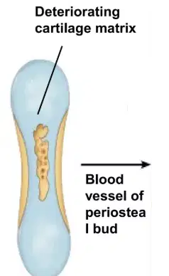
What stage of bone formation is this
1 : Formation of bone collar around hyaline cartilage model.
2 : Cavitation of the hyaline cartilage within the cartilage model.
3 : Invasion of internal cavities by the periosteal bud and spongy bone formation
4 : Formation of medullary cavity as ossification continues; appearance of secondary ossification centers in the epiphyses.
5 : Ossification of the epiphyses; when completed, hyaline cartilage remains only in the epiphyseal plates and articular cartilages
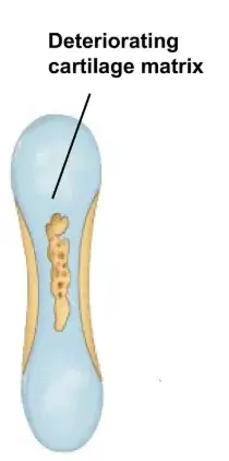
What stage of bone formation is this
1 : Formation of bone collar around hyaline cartilage model.
2 : Cavitation of the hyaline cartilage within the cartilage model.
3 : Invasion of internal cavities by the periosteal bud and spongy bone formation
4 : Formation of medullary cavity as ossification continues; appearance of secondary ossification centers in the epiphyses.
5 : Ossification of the epiphyses; when completed, hyaline cartilage remains only in the epiphyseal plates and articular cartilages
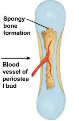
What stage of bone formation is this
1 : Formation of bone collar around hyaline cartilage model.
2 : Cavitation of the hyaline cartilage within the cartilage model.
3 : Invasion of internal cavities by the periosteal bud and spongy bone formation
4 : Formation of medullary cavity as ossification continues; appearance of secondary ossification centers in the epiphyses.
5 : Ossification of the epiphyses; when completed, hyaline cartilage remains only in the epiphyseal plates and articular cartilages
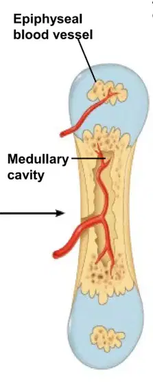
What stage of bone formation is this
1 : Formation of bone collar around hyaline cartilage model.
2 : Cavitation of the hyaline cartilage within the cartilage model.
3 : Invasion of internal cavities by the periosteal bud and spongy bone formation
4 : Formation of medullary cavity as ossification continues; appearance of secondary ossification centers in the epiphyses.
5 : Ossification of the epiphyses; when completed, hyaline cartilage remains only in the epiphyseal plates and articular cartilages
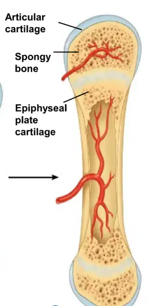
What stage of bone formation is this
1 : Formation of bone collar around hyaline cartilage model.
2 : Cavitation of the hyaline cartilage within the cartilage model.
3 : Invasion of internal cavities by the periosteal bud and spongy bone formation
4 : Formation of medullary cavity as ossification continues; appearance of secondary ossification centers in the epiphyses.
5 : Ossification of the epiphyses; when completed, hyaline cartilage remains only in the epiphyseal plates and articular cartilages
What is the function of calcium in bone formation?
provides the mineral component that gives bones their strength and structure
to combine with calcium to form hydroxyapatite, the mineral that provides bones with strength and rigidity
promoting the absorption of calcium and phosphate, stimulating osteoblast activity, and controlling bone mineralization
decrease bone resorption by inhibiting osteoclast activity, which in turn helps to maintain blood calcium levels
stimulates bone formation mainly when administered intermittently, a process also known as anabolism
What is the function of phosphate in bone formation?
provides the mineral component that gives bones their strength and structure
to combine with calcium to form hydroxyapatite, the mineral that provides bones with strength and rigidity
promoting the absorption of calcium and phosphate, stimulating osteoblast activity, and controlling bone mineralization
decrease bone resorption by inhibiting osteoclast activity, which in turn helps to maintain blood calcium levels
stimulates bone formation mainly when administered intermittently, a process also known as anabolism
What is the function of vitamin d in bone formation?
provides the mineral component that gives bones their strength and structure
to combine with calcium to form hydroxyapatite, the mineral that provides bones with strength and rigidity
promoting the absorption of calcium and phosphate, stimulating osteoblast activity, and controlling bone mineralization
decrease bone resorption by inhibiting osteoclast activity, which in turn helps to maintain blood calcium levels
stimulates bone formation mainly when administered intermittently, a process also known as anabolism
What is the function of Calcitonin hormone in bone formation?
provides the mineral component that gives bones their strength and structure
to combine with calcium to form hydroxyapatite, the mineral that provides bones with strength and rigidity
promoting the absorption of calcium and phosphate, stimulating osteoblast activity, and controlling bone mineralization
decrease bone resorption by inhibiting osteoclast activity, which in turn helps to maintain blood calcium levels
stimulates bone formation mainly when administered intermittently, a process also known as anabolism
What is the function of Paratyroid hormone in bone formation?
provides the mineral component that gives bones their strength and structure
to combine with calcium to form hydroxyapatite, the mineral that provides bones with strength and rigidity
promoting the absorption of calcium and phosphate, stimulating osteoblast activity, and controlling bone mineralization
decrease bone resorption by inhibiting osteoclast activity, which in turn helps to maintain blood calcium levels
stimulates bone formation mainly when administered intermittently, a process also known as anabolism
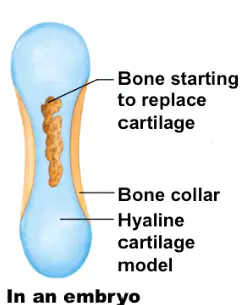
This is bone formation in......
EMBRYO
FETUS
CHILD
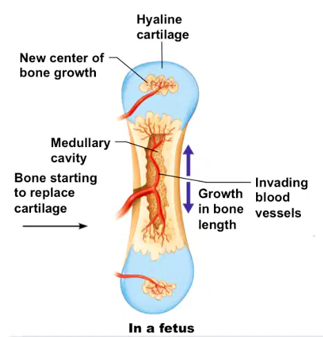
This is bone formation in......
EMBRYO
FETUS
CHILD
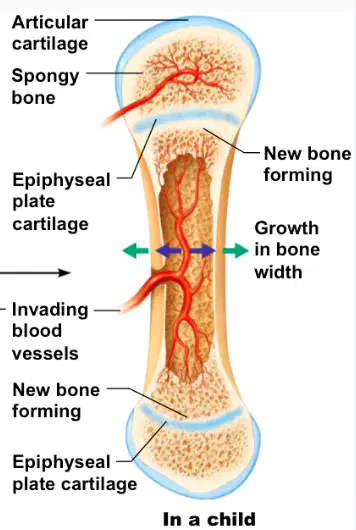
This is bone formation in......
EMBRYO
FETUS
CHILD
These are the types of bones based on the shape EXCEPT
Lamellar bones
Long/pipe bones
Short bones
Irregular bones
Circle bones
skull (cranial bones), ribs, breastbone, scapula
Lamellar bones
Long/pipe bones
Short bones
Irregular bones
arms, femur, tibia, fibula, finger, clavicle
Lamellar bones
Long/pipe bones
Short bones
Irregular bones
carpals, tarsals
Lamellar bones
Long/pipe bones
Short bones
Irregular bones
mandible, sacrum, vertebrae, facial bones
Lamellar bones
Long/pipe bones
Short bones
Irregular bones
Flat shaped, consist red bone marrow
Lamellar bones
Long/pipe bones
Short bones
Irregular bones
Long cylindrical with hump tip, yellow marrow
Lamellar bones
Long/pipe bones
Short bones
Irregular bones
as long as they are wide, provide stability and some movement
Lamellar bones
Long/pipe bones
Short bones
Irregular bones
Irregular shape
Lamellar bones
Long/pipe bones
Short bones
Irregular bones
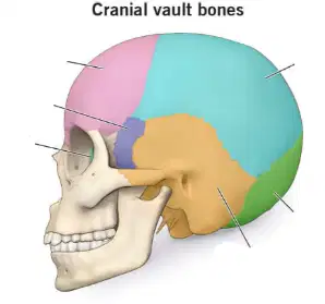
What is the light blue one
Parietal
Occipital
Temporal
Frontal
Sphenoid
What is the green one
Ethmoid
Occipital
Temporal
Frontal
Sphenoid
What is the yellow one
Ethmoid
Occipital
Temporal
Frontal
Sphenoid
What is the purple one
Ethmoid
Occipital
Temporal
Frontal
Sphenoid
What is the pink one
Ethmoid
Occipital
Temporal
Frontal
Sphenoid
What is the one inside the eye place (dark green)
Ethmoid
Occipital
Temporal
Frontal
Sphenoid
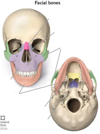
What is the pink one
Nasal
Lacrimal
Zygomatic
Maxilla
Mandible
What is the light blue one (it's inside the eye place near the pink one)
Nasal
Lacrimal
Zygomatic
Maxilla
Mandible
What is the green one
Nasal
Lacrimal
Zygomatic
Maxilla
Mandible
What is the light purple one
Nasal
Lacrimal
Zygomatic
Maxilla
Mandible
What is the orange one
Nasal
Lacrimal
Zygomatic
Maxilla
Mandible
What is the yellow one
Palatine
Vomer
Zygomatic
Mandible
What is the dark purple one
Palatine
Vomer
Zygomatic
Mandible
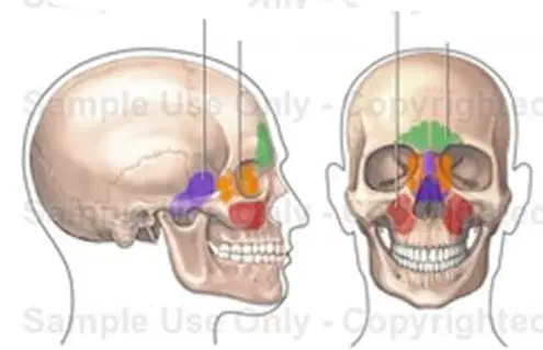
What is the purple one
Sphenoidal Sinus
Ethmoidal sinus
Maxillary Sinus
Frontal Sinus
What is the orange one
Sphenoidal Sinus
Ethmoidal sinus
Maxillary Sinus
Frontal Sinus
What is the red one
Sphenoidal Sinus
Ethmoidal sinus
Maxillary Sinus
Frontal Sinus
What is the green one
Sphenoidal Sinus
Ethmoidal sinus
Maxillary Sinus
Frontal Sinus
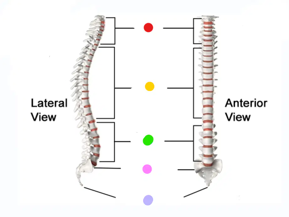
What is the red one
Cervical
C1-C7
Thoracic
T1-T12
Lumbar
L1-L5
Sacrum
S1-S5
(FUSED)
Coccyx
Co1-Co4
(FUSED)
What is the yellow one
Cervical
C1-C7
Thoracic
T1-T12
Lumbar
L1-L5
Sacrum
S1-S5
(FUSED)
Coccyx
Co1-Co4
(FUSED)
What is the green one
Cervical
C1-C7
Thoracic
T1-T12
Lumbar
L1-L5
Sacrum
S1-S5
(FUSED)
Coccyx
Co1-Co4
(FUSED)
What is the pink one
Cervical
C1-C7
Thoracic
T1-T12
Lumbar
L1-L5
Sacrum
S1-S5
(FUSED)
Coccyx
Co1-Co4
(FUSED)
What is the light purple one
Cervical
C1-C7
Thoracic
T1-T12
Lumbar
L1-L5
Sacrum
S1-S5
(FUSED)
Coccyx
Co1-Co4
(FUSED)
What is the light purple one
Cervical
C1-C7
Thoracic
T1-T12
Lumbar
L1-L5
Sacrum
S1-S5
(FUSED)
Coccyx
Co1-Co4
(FUSED)
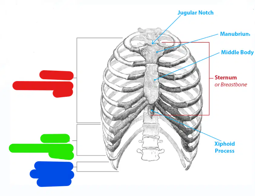
What is the red one
True ribs
(vertebrostenal ribs)
1-7
False ribs
(vertebrochondral ribs)
8-10
Floating ribs
(vertebral ribs)
11 and 12
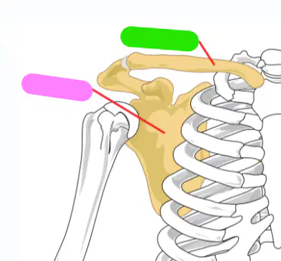
What is the pink one
Scapula
Clavicle
What is the green one
Scapula
Clavicle
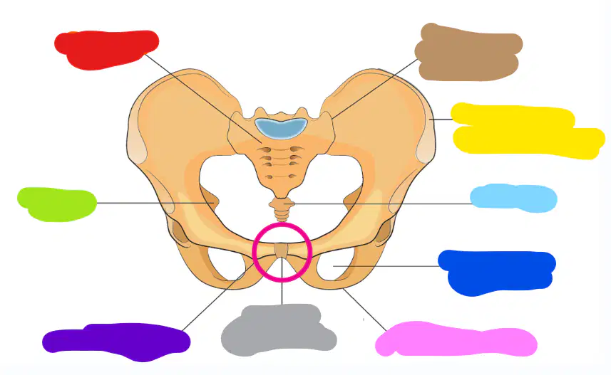
what is the red one
sacrum
ischium
pubic tubercle
pubic symphsis
what is the green one
sacrum
ischium
pubic tubercle
pubic symphsis
what is the purple one
sacrum
ischium
pubic tubercle
pubic symphsis
what is the gray one
sacrum
ischium
pubic tubercle
pubic symphsis
what is the brown one
iliac-sacral junction
the upper front iliac bone
coccyx
the orbutrator foramen
ischial tuberosity
what is the yellow one
iliac-sacral junction
the upper front iliac bone
coccyx
the orbutrator foramen
ischial tuberosity
what is the light blue one
iliac-sacral junction
the upper front iliac bone
coccyx
the orbutrator foramen
ischial tuberosity
what is the dark blue one
iliac-sacral junction
the upper front iliac bone
coccyx
the orbutrator foramen
ischial tuberosity
what is the pink one
iliac-sacral junction
the upper front iliac bone
coccyx
the orbutrator foramen
ischial tuberosity
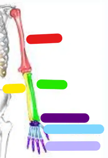
red
Humerus
Ulna
Radius
Carpals (8)
Metacarpals (5)
green
Humerus
Ulna
Radius
Carpals (8)
Metacarpals (5)
yellow
Humerus
Ulna
Radius
Carpals (8)
Metacarpals (5)
dark purple
Phalanges (14)
Ulna
Radius
Carpals (8)
Metacarpals (5)
blue
Phalanges (14)
Ulna
Radius
Carpals (8)
Metacarpals (5)
light purple
Phalanges (14)
Ulna
Radius
Carpals (8)
Metacarpals (5)
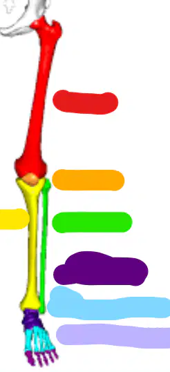
red
Femur
Patella
Tibia
Fibula
orange
Femur
Patella
Tibia
Fibula
yellow
Femur
Patella
Tibia
Fibula
green
Femur
Patella
Tibia
Fibula
dark purple
Tarsals (7)
Metatarsals (5)
Phalanges (14)
blue
Tarsals (7)
Metatarsals (5)
Phalanges (14)
light purple
Tarsals (7)
Metatarsals (5)
Phalanges (14)
What are joints?
Between bone and another bone
allow for movement and provide instability to the body
rigid, living organs that form the skeleton, providing structural support, protecting vital organs, and enabling movement
What are the type of joints
Synarthrosis
Amphiarthrosis
Diarthrosis
Ms Devi joints
Robot joints
SYNARTHROSIS
IMMOVABLE
LITTLE MOVEMENT
FULL MOVEMENT
AMPHIARTHROSIS
IMMOVABLE
LITTLE MOVEMENT
FULL MOVEMENT
DIARTHROSIS
IMMOVABLE
LITTLE MOVEMENT
FULL MOVEMENT
SYNARTHROSIS
Fibrous joint
Connected by fibrous connective tissue
No movement
Cartilagenous joint
Connected by hyaline cartilage
Slight movement
Synovial joint
Lubricating fluid in joint cavity
Free movement
AMPHIARTHROSIS
Fibrous joint
Connected by fibrous connective tissue
No movement
Cartilagenous joint
Connected by hyaline cartilage
Slight movement
Synovial joint
Lubricating fluid in joint cavity
Free movement
Types of synovial joints are.....
Hinge Joint
Saddle Joint
Ball and socket joint
Pivot joint
Gliding joint
HINGE JOINT
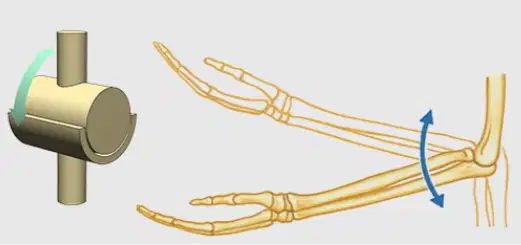
One direction
One direction
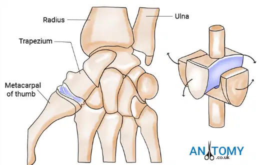
Two direction
Two direction
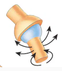
All direction
All direction

One bone rotate around another bone
One bone rotate around another bone
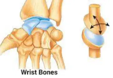
between the surfaces of two flat bones
between the surfaces of two flat bones
SADDLE JOINT
One direction
One direction
Two direction
Two direction
All direction
All direction
One bone rotate around another bone
One bone rotate around another bone
between the surfaces of two flat bones
between the surfaces of two flat bones
BALL AND SOCKET JOINT
One direction
One direction
Two direction
Two direction
All direction
All direction
One bone rotate around another bone
One bone rotate around another bone
between the surfaces of two flat bones
between the surfaces of two flat bones
PIVOT JOINT
One direction
One direction
Two direction
Two direction
All direction
All direction
One bone rotate around another bone
One bone rotate around another bone
between the surfaces of two flat bones
between the surfaces of two flat bones
GLIDING JOINT
One direction
One direction
Two direction
Two direction
All direction
All direction
One bone rotate around another bone
One bone rotate around another bone
between the surfaces of two flat bones
between the surfaces of two flat bones
MUSCLES
35-40% of body mass
Responsible for body movement
Contract and relax
As the passive motion organ
Skeletal muscle
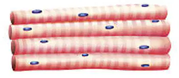
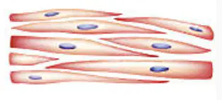
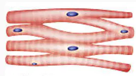
Smooth muscle
Cardiac muscle
Skeletal muscle
Dark and light area
Parallel - elongated cell
Voluntary movement
Many nuclei at the edge
Attached to bone
One nucleus at center
Have branches
Dark-light area
Involuntary movement
70 times/min
Internal organs and skin
Single centrally located nucleus (uninucleated)
Non-striated
Spindle/tapered
Involuntary
Cardiac muscle
Dark and light area
Parallel - elongated cell
Voluntary movement
Many nuclei at the edge
Attached to bone
One nucleus at center
Have branches
Dark-light area
Involuntary movement
70 times/min
Internal organs and skin
Single centrally located nucleus (uninucleated)
Non-striated
Spindle/tapered
Involuntary
Smooth muscle
Dark and light area
Parallel - elongated cell
Voluntary movement
Many nuclei at the edge
Attached to bone
One nucleus at center
Have branches
Dark-light area
Involuntary movement
70 times/min
Internal organs and skin
Single centrally located nucleus (uninucleated)
Non-striated
Spindle/tapered
Involuntary
Tendon : Edge of muscle stucks to bone
TRUE
FALSE
I give up 🥀😢
the attachment point of a muscle's tendon that is more stable and has little to no movement during muscle contraction
(immoveable bones)
ORIGO
INSERTIO
the attachment point that moves during contraction
(moveable bones)
ORIGO
INSERTIO
Interaction of skeletal muscle :
SYNERGIST
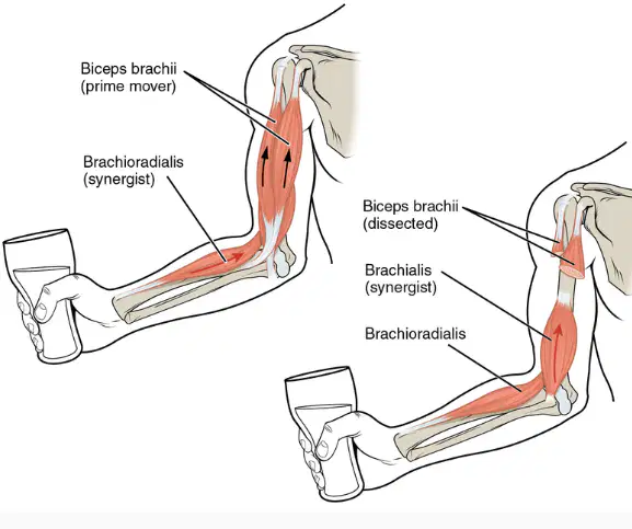
Work together with main muscle
Work together with main muscle
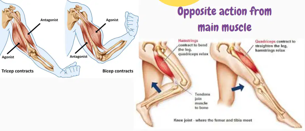
Opposite action from main muscle
Opposite action from main muscle
Interaction of skeletal muscle :
ANTAGONIST
Work together with main muscle
Work together with main muscle
Opposite action from main muscle
Opposite action from main muscle
flexion
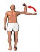

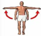
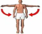
extension
abduction
adduction
Medial rotation
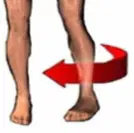
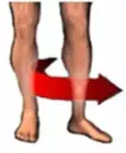
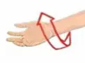
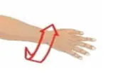
Lateral rotation
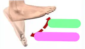
Green = dorsiflexion
Green = Plantar flexion
Pink = Plantar flexion
Pink = dorsiflexion
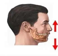
up = depression
up = elevation
down = depression
down = elevation
cracked bone accident only can be seen through X ray
FISSURE
FRACTURE
broken bone open and closed fractured
FISSURE
FRACTURE
Hydrocephalus (megalocephalus)
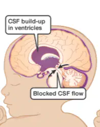
coagulation of spinal fluid inside skull and head enlargement
coagulation of spinal fluid inside skull and head enlargement
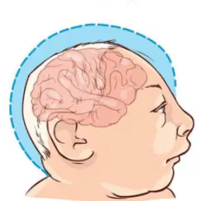
condition in which a person's head is significantly smaller than normal. This happens because lack of calcium when baby
condition in which a person's head is significantly smaller than normal. This happens because lack of calcium when baby
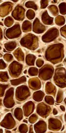
abnormal loss of bony tissues resulting in fragile porous. Caused by lack of estrogen (in woman) and calcium.
abnormal loss of bony tissues resulting in fragile porous. Caused by lack of estrogen (in woman) and calcium.
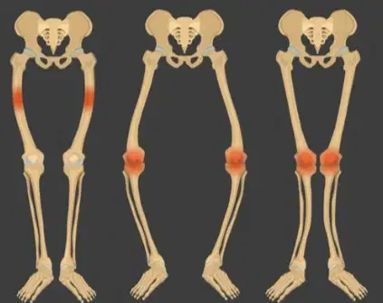
lack of vitamin D so disturb bone growth and caused rickets & softening of bones. Result --> bowed legs (X and O shaped)
lack of vitamin D so disturb bone growth and caused rickets & softening of bones. Result --> bowed legs (X and O shaped)
Microcephalus
coagulation of spinal fluid inside skull and head enlargement
coagulation of spinal fluid inside skull and head enlargement
condition in which a person's head is significantly smaller than normal. This happens because lack of calcium when baby
condition in which a person's head is significantly smaller than normal. This happens because lack of calcium when baby
abnormal loss of bony tissues resulting in fragile porous. Caused by lack of estrogen (in woman) and calcium.
abnormal loss of bony tissues resulting in fragile porous. Caused by lack of estrogen (in woman) and calcium.
lack of vitamin D so disturb bone growth and caused rickets & softening of bones. Result --> bowed legs (X and O shaped)
lack of vitamin D so disturb bone growth and caused rickets & softening of bones. Result --> bowed legs (X and O shaped)
Osteoporosis
coagulation of spinal fluid inside skull and head enlargement
coagulation of spinal fluid inside skull and head enlargement
condition in which a person's head is significantly smaller than normal. This happens because lack of calcium when baby
condition in which a person's head is significantly smaller than normal. This happens because lack of calcium when baby
abnormal loss of bony tissues resulting in fragile porous. Caused by lack of estrogen (in woman) and calcium.
abnormal loss of bony tissues resulting in fragile porous. Caused by lack of estrogen (in woman) and calcium.
lack of vitamin D so disturb bone growth and caused rickets & softening of bones. Result --> bowed legs (X and O shaped)
lack of vitamin D so disturb bone growth and caused rickets & softening of bones. Result --> bowed legs (X and O shaped)
Rickets
coagulation of spinal fluid inside skull and head enlargement
coagulation of spinal fluid inside skull and head enlargement
condition in which a person's head is significantly smaller than normal. This happens because lack of calcium when baby
condition in which a person's head is significantly smaller than normal. This happens because lack of calcium when baby
abnormal loss of bony tissues resulting in fragile porous. Caused by lack of estrogen (in woman) and calcium.
abnormal loss of bony tissues resulting in fragile porous. Caused by lack of estrogen (in woman) and calcium.
lack of vitamin D so disturb bone growth and caused rickets & softening of bones. Result --> bowed legs (X and O shaped)
lack of vitamin D so disturb bone growth and caused rickets & softening of bones. Result --> bowed legs (X and O shaped)
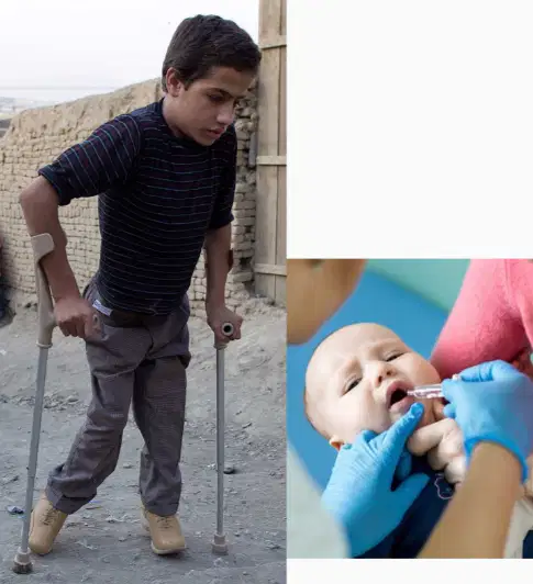
Polio
Caused by virus
Paralysis
Prevent by immunization
is a variant of covid 100
Scoliosis
condition in which a person's spine is curved from side to side
curving of the spine that causes a bowing or rounding of the back
curving of spine that causes a slanted frontwards shaped.
Kyphosis
condition in which a person's spine is curved from side to side
curving of the spine that causes a bowing or rounding of the back
curving of spine that causes a slanted frontwards shaped.
Lordosis
condition in which a person's spine is curved from side to side
curving of the spine that causes a bowing or rounding of the back
curving of spine that causes a slanted frontwards shaped.
Ankylosis
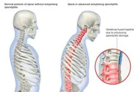
The consolidation of bones or their parts to form a single unit.
The consolidation of bones or their parts to form a single unit.
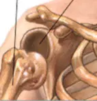
joints move from its original motion
joints move from its original motion
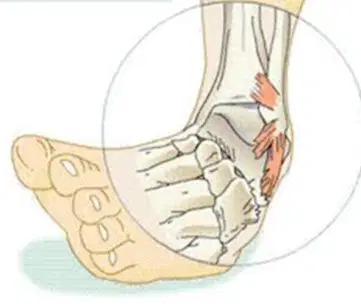
ligament is stretched too far or tears, the joint will become painful and swell.
ligament is stretched too far or tears, the joint will become painful and swell.
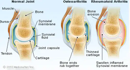
is a form of joint disorder that involves inflammation of one or more joints.
is a form of joint disorder that involves inflammation of one or more joints.
Dislocation
The consolidation of bones or their parts to form a single unit.
The consolidation of bones or their parts to form a single unit.
joints move from its original motion
joints move from its original motion
ligament is stretched too far or tears, the joint will become painful and swell.
ligament is stretched too far or tears, the joint will become painful and swell.
is a form of joint disorder that involves inflammation of one or more joints.
is a form of joint disorder that involves inflammation of one or more joints.
Sprained joint
The consolidation of bones or their parts to form a single unit.
The consolidation of bones or their parts to form a single unit.
joints move from its original motion
joints move from its original motion
ligament is stretched too far or tears, the joint will become painful and swell.
ligament is stretched too far or tears, the joint will become painful and swell.
is a form of joint disorder that involves inflammation of one or more joints.
is a form of joint disorder that involves inflammation of one or more joints.
Arthritis
The consolidation of bones or their parts to form a single unit.
The consolidation of bones or their parts to form a single unit.
joints move from its original motion
joints move from its original motion
ligament is stretched too far or tears, the joint will become painful and swell.
ligament is stretched too far or tears, the joint will become painful and swell.
is a form of joint disorder that involves inflammation of one or more joints.
is a form of joint disorder that involves inflammation of one or more joints.
Joint infection :
Exudative arthritis
joint inflammation that has pus inside
joint inflammation so synovial fluid will dried
unenergetic condition in joints because break in cakraephyphisis/metaphyseal
break in cakraephyphisis that causes death of bone
Joint infection :
Sika arthritis
joint inflammation that has pus inside
joint inflammation so synovial fluid will dried
unenergetic condition in joints because break in cakraephyphisis/metaphyseal
break in cakraephyphisis that causes death of bone
Joint infection :
Weak joints
joint inflammation that has pus inside
joint inflammation so synovial fluid will dried
unenergetic condition in joints because break in cakraephyphisis/metaphyseal
break in cakraephyphisis that causes death of bone
Joint infection :
Necrosis
joint inflammation that has pus inside
joint inflammation so synovial fluid will dried
unenergetic condition in joints because break in cakraephyphisis/metaphyseal
break in cakraephyphisis that causes death of bone
Atrophy
decrease in the mass of the muscle
increase in size of skeletal muscle
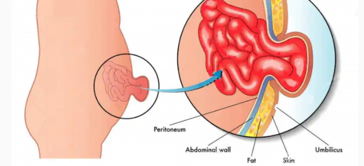
abdominal muscle is tore so intestine is going down to stomach cavity
abdominal muscle is tore so intestine is going down to stomach cavity
sudden, involuntary contractions or spasms in one or more of your muscles
Hypertrophy
decrease in the mass of the muscle
increase in size of skeletal muscle
abdominal muscle is tore so intestine is going down to stomach cavity
abdominal muscle is tore so intestine is going down to stomach cavity
sudden, involuntary contractions or spasms in one or more of your muscles
Hernia abdominal
decrease in the mass of the muscle
increase in size of skeletal muscle
abdominal muscle is tore so intestine is going down to stomach cavity
abdominal muscle is tore so intestine is going down to stomach cavity
sudden, involuntary contractions or spasms in one or more of your muscles
Cramps
decrease in the mass of the muscle
increase in size of skeletal muscle
abdominal muscle is tore so intestine is going down to stomach cavity
abdominal muscle is tore so intestine is going down to stomach cavity
sudden, involuntary contractions or spasms in one or more of your muscles
Tetanus
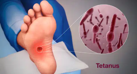
infection of the nervous system with the potentially deadly bacteria Clostridium tetani. This cause spasms in your muscle.
infection of the nervous system with the potentially deadly bacteria Clostridium tetani. This cause spasms in your muscle.
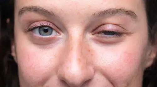
chronic autoimmune neuromuscular disease
Weakness of the muscle gradually
Can cause paralysis and death
chronic autoimmune neuromuscular disease
Weakness of the muscle gradually
Can cause paralysis and death
Myasthenia Gravis
infection of the nervous system with the potentially deadly bacteria Clostridium tetani. This cause spasms in your muscle.
infection of the nervous system with the potentially deadly bacteria Clostridium tetani. This cause spasms in your muscle.
chronic autoimmune neuromuscular disease
Weakness of the muscle gradually
Can cause paralysis and death
chronic autoimmune neuromuscular disease
Weakness of the muscle gradually
Can cause paralysis and death

