
 Worksheets
Medical A & P First Semester Final Review
Total questions: 224
Worksheet time: 3hrs 35mins

Which structure is the dorsal body cavity?
7
1
b
3

Which structure is the pericardial cavity?
d
c
1
a

Which structure is the cranial cavity?
1
b
3
c

Which structure is the abdominal cavity?
3
c
e
1

Which structure is the diaphragm?
e
b
5
3

Which structure is the pelvic cavity?
4
3
6
c

Which structure is the ventral body cavity?
5
6
2
1
Which statement is false regarding the anatomical position?
the palms face front
the thumbs point away from body
the head is turned to the left side
the posture is standing erect
Which term means that a structure is near the surface?
superficial
medial
deep
inferior
The hands are ____ to the feet.
superior
inferior
superficial
deep
The elbow is __ to the fingers.
distal
proximal
What is the term for the backside or "back" in the anatomical position?
anterior
ventral
caudal
posterior
away from the midline of the body
distal
posterior
anterior
lateral
A patient needs to have an intravenous line (IV) placed for medication in the emergency department.
The physician wants the line to be placed proximal to the elbow.
Where should the IV be placed?
In the ___.
hand
forearm
upper arm
femoral
Match the following terms to the definition
sagittal plane
divides the body into R/L
coronal plane
divides into dorsal & ventral
transverse plane
divides into superior & inferior
supine
facing up while laying down
prone
facing down while laying down
"The small intestine absorbs nutrients from our food and they are carried through the blood to the cells."
"The ________ system allows us to move around, but the _________ system controls our movement."
A process which has the ability to maintain the internal environment of the body regardless of changes inside or outside of the body.
Homeostasis
Negative Feedback Loop
Positive Feedback Loop
Feedback Loop

This is an example of __________.
Homeostasis
Negative Feedback Loop
Positive Feedback Loop
Feedback Loop

This is an example of __________.
Homeostasis
Negative Feedback Loop
Positive Feedback Loop
Feedback Loop
A process which increases (enhances) a response to an event in the body, which is not at a set-point.
Homeostasis
Negative Feedback Loop
Positive Feedback Loop
Feedback Loop
An organ such as the brain or glands in the endocrine system which receive information about changes in the body and then send signals to other organs to respond to the change.
Control Center
Receptor
Effector
Stimulus
A specialized cell in an organ which monitor the body for changes inside the body.
Control Center
Receptor
Effector
Stimulus
A specialized cell in an organ which respond to changes in the body's environment.
Control Center
Receptor
Effector
Stimulus
An event or change inside the body which signals a disturbance has happened and a response is required to maintain the health of the body.
Control Center
Receptor
Effector
Stimulus
Which of the following are examples of positive feedback loops (list in alphabetical order) (a) (b)
Which of the following are examples of Negative feedback loops (drag in alphabetical order) (a) (b) (c)

The bodies starting position is called what?
Sagittal
Coronal
Anatomical
Biological

on the smooth ER
In the nucleus
On the Rough ER only!
On the Rough ER and in the cytoplasm
The ____________ is a flexible structure that can self repair, and contain the organelles of a cell.
Cell wall
Cell membrane
Nucleus
Vacuole
The ______________ converts energy from food into a form of energy cells can use: ATP.
Cell membrane
Mitochondria
Nucleus
All of the above


What is the function of the cell membrane?
To control what comes in and out of the cell
To break down food inside the cell
to serve as a barrier
to eat the cells food

Which organelle produces the energy a cell needs to carry out its function
Mitochondria
Golgi Bodies
Cytoplasm
Nucleus
Function: Makes proteins.
Found in: All cells
Ribosomes
Nucleus
Rough ER
Golgi apparatus
Cells of organisms must go through cell division for?
growth
repair of damaged cells
replacement of dying cells.
All of these
During most of interphase, DNA is found in a relaxed form called
chromosome
centromere
centriole
chromatin
During mitosis, DNA is found in the condensed form called a
chromosome
centromere
centriole
chromatin
In prophase of mitosis, chromosomes
form and the nuclear envelope disappears.
are pulled toward opposite ends of the cell.
chromosomes line up along the equator of the cell
chromosomes begin to decondense and the two new nuclei begin to form.
In metaphase of mitosis, chromosomes
form and the nuclear envelope disappears.
are pulled toward opposite ends of the cell.
chromosomes line up along the equator of the cell
chromosomes begin to decondense and the two new nuclei begin to form.
In anaphase of mitosis, chromosomes
form and the nuclear envelope disappears.
are pulled toward opposite ends of the cell.
chromosomes line up along the equator of the cell
chromosomes begin to decondense and the two new nuclei begin to form.
In telophase of mitosis, chromosomes
form and the nuclear envelope disappears.
are pulled toward opposite ends of the cell.
chromosomes line up along the equator of the cell
chromosomes begin to decondense and the two new nuclei begin to form.


This fluid in the cell is where the organelles are found. It contains water and chemicals the cell needs.
Blood
Cytoplasm
Plasma
Vacuole


Match the organelle with its correct function.

Stores and Protects DNA

Builds Proteins

Generates energy for the cell

Controls what enters and leaves the cell

Packages up proteins to be shipped out of the cell
Identify the adipose CT:


What type of protein fibers are present in areolar CT?
Collagen
Reticular fibers
Elastic Fibers
All three
What type of protein fibers are present in elastic CT?
Collagen
Reticular fibers
Elastic Fibers
All three
What is the function of elastic CT?
Provides supportive framework.
Allows for stretching and recoiling.
Withstands stress.
Transport
Which is hyaline cartilage?


What is the function of blood CT?
Provides supportive framework.
Allows for stretching and recoiling.
Withstands stress.
Transport

Hyaline cartilage
Dense irregular tissue
Areolar tissue
Fibrocartilage

Compact (bone)
Elastic connective tissue
Fibrocartilage
Dense regular connective tissue

Blood cells
Areolar connective tissue
Reticule connective tissue
Hyaline cartilage

Hyline cartilage
Elastic cartilage
Elastic connective tissue
Adipose tissue
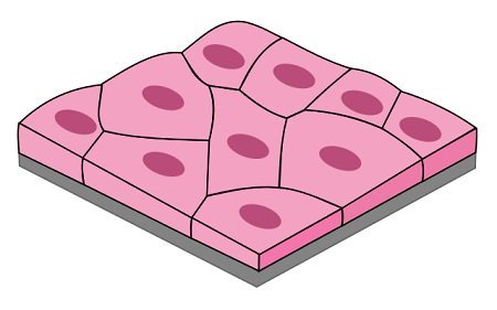
Name the type of epithelium
simple cuboidal
simple squamous
simple columnar
stratified squamous
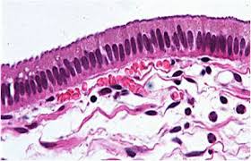
Name the type of epithelium
simple squamous
stratified squamous
simple cuboidal
simple columnar
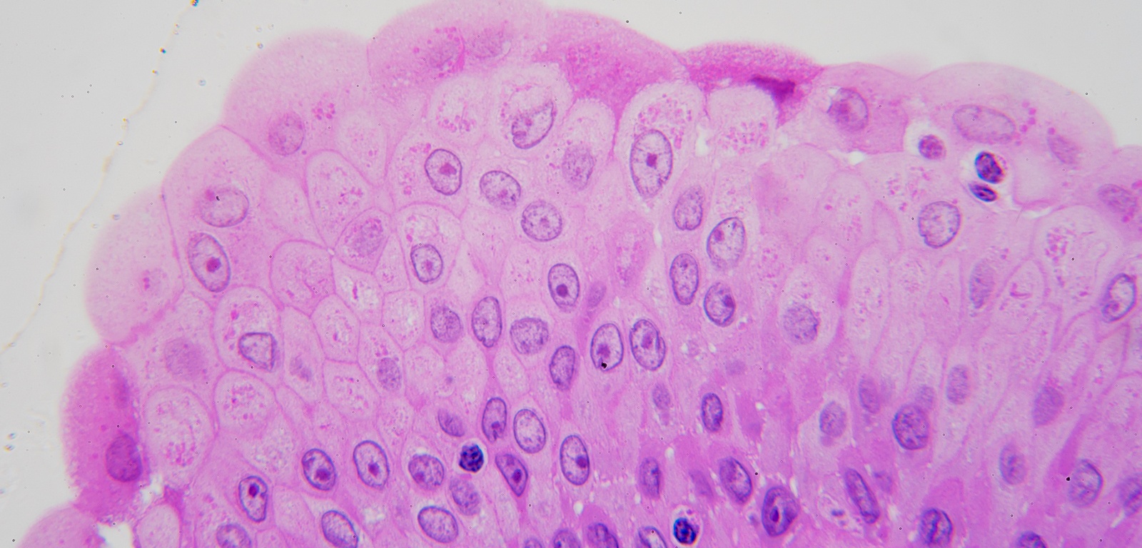
What type of epithelial tissue is this and where might you find it?
stratified cuboidal ; small intestine
simple squamous ; lung
pseudostratified columnar ; trachea
transitional ; bladder

Squamous

Cuboidal

Columnar

Blood

Bone

Loose Connective

Dense Connective

Hyaline Cartilage
The only fluid connective tissue
Blood
Adipose
Loose connective

What type of epithelium?
Non-Ciliated pseudostratified columnar epithelium.
Ciliated pseudostratified columnar epithelium.
Stratified columnar epithelium.
Simple columnar epithelium.

What type of epithelium?
Relaxed transitional epithelium.
Stratified columnar epithelium.
Simple columnar epithelium.
Distended transitional epithelium.

simple columnar
simple cuboidal
simple squamous
stratified cuboidal
Which type of connective tissue allows muscle to attach to bone?
adipose
cartilage
ligament
tendon
Which connective tissue stores fat in the cells?
adipose
cartilage
ligaments
tendons
Which of the following is NOT a main tissue type?
muscle
epithelial
cuboidal
connective
Where would you NOT find epithelial tissue?
skin
glands
heart
lining of hollow organs
How are epithelial tissue classified?
by shape
by size
by number of layers
by shape and number of layers.
Basement membranes are found in:
connective tissues
Epithelial tissues
nervous tissues
Muscle Tissue
Which of the following is not true for epithelial tissues?
they have a basement membraine
they provide protection
they reproduce slowly
they make up your glands.
Which of the following is connective tissue?
Bone
cartilage
blood
all of these
What do the connective tissues do? (their main job)
protect
connect tissues together
provide movement
provide impulses.
What cell type is most likely to be present in dense regular CT?
Fibroblasts
Adipocytes.
Mesenchymal cells.
Macrophages.
What is the function of dense irregular CT?
Binds skin and epithelia to deeper tissue.
Attach bone to bone or bone to muscle.
Withstands stress.
Provides supportive framework.
What is the cell type of hyaline cartilage?
Fibroblast
Chondrocytes
Osteocytes
Lacunae.
What is the cell type of bone CT?
Fibroblast
Chondrocytes
Osteocytes
Lacunae.
What are formed elements of blood CT?
Dissolved protein fibers.
Liquid nature of the ground substance.
A method of transporting nutrients.
Erythrocytes, leukocytes, and platelets.
What is the function of blood CT?
Provides supportive framework.
Allows for stretching and recoiling.
Withstands stress.
Transport
Which type of muscle tissue is responsible for voluntary muscle control?
Cardiac muscle tissue
Smooth muscle tissue
Connective tissue
Skeletal muscle tissue
Which type of muscle tissue is found in the heart?
cardiac muscle tissue
nervous tissue
smooth muscle tissue
skeletal muscle tissue
Which type of muscle tissue is found in the walls of blood vessels and digestive organs?
Cardiac muscle tissue
Skeletal muscle tissue
Adipose tissue
Smooth muscle tissue
Which type of muscle tissue is striated and multinucleated?
Skeletal muscle tissue
Smooth muscle tissue
Cardiac muscle tissue
Adipose tissue
Which type of muscle tissue is responsible for movement of bones?
Connective tissue
Cardiac muscle tissue
Smooth muscle tissue
Skeletal muscle tissue
Which type of muscle tissue is found in the walls of hollow organs?
Smooth muscle tissue
Adipose tissue
Skeletal muscle tissue
Cardiac muscle tissue

What type of Muscle tissue is this?
Smooth
Cardiac
Skeletal
Straight

Which type of muscle tissue has intercalated disks?
Smooth
Cardiac
Skeletal
Straight

Which type of muscle tissue has spindle shaped cells?
Smooth
Cardiac
Skeletal
Which pattern is Cardiac Muscle organized?

Spindle

Branched

Straight
Which pattern is skeletal muscle organized?
Spindle
Branched
Straight
Which muscle tissue(s) are striated?
Cardiac
Smooth
Skeletal


What type of connective tissue has a fluid-like ground substance?
bone
cartilage
blood
ligaments
What type of connective tissue has a hard ground substance of calcium and salts?
Bone
Adipose
Blood
Dense Regular CT

Which type of muscle tissue is this
Skeletal Muscle
Smooth Muscle
Cardiac Muscle
Unable to judge

nervous
muscle
epithelial
connective
Which cells produce the matrix of bone?
Adipocytes
Chondrocytes
Osteocytes
Fibroblasts
Which type of tissue would line the intestines?
epithelial
neural
muscle
connective
Which type of tissue is avascular (no blood vessels) and tightly packed?
Epithelial
Connective
Areolar
Adipose

B
Axon
Cell body
Dendrites
Schwann cell

A
Cell body
Nucleus
Axon
Dendrite
Schwann cell
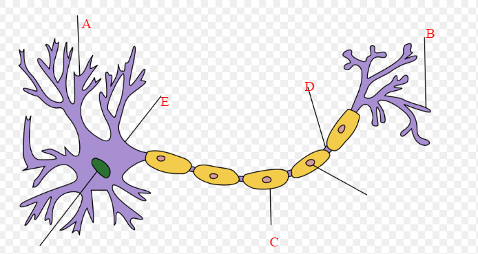
What structure is labeled B
Dendrite
Axon
Axon Terminal
Cell Body

What structure is labeled E
Synapse
Cell Body
Axon
Dendrite

What structure is labeled D
Synapse
Axon
Axon Terminal
Dendrite
What structure is labeled B
Dendrite
Axon
Axon Terminal
Cell Body
What is the primary function of osseous tissue?
Support and protect organs
Produce hormones
Store vitamins
Regulate temperature
Osseous tissue is also known as ______.
Muscle tissue
Bone tissue
Nerve tissue
Skin tissue
Which of the following is NOT a function of the skeletal system?
A) Support
B) Point of attachment for Movement
C) Hormone production
D) Blood cell production
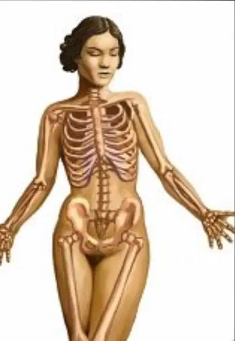
How many bones does an adult human have?
206
270
300
250
The skeletal system provides structure to the body.
True
False
The skeletal system stores minerals such as calcium.
True
False
Which of the following bones is part of the axial skeleton?
Femur
Humerus
Skull
Tibia
Fill in the blank: The _________ is a bone that connects the arm to the body.
Femur
Humerus
Clavicle
Tibia
Leverage in the skeletal system is related to ______.
Storage of lipids
Force of motion
Blood cell production
Protection of organs
What type of bones are classified by their shape and include the femur and humerus?
Long bones
Flat bones
Sutural bones
Irregular bones
Flat bones are found in the ______.
Skull
Femur
Tibia
Humerus
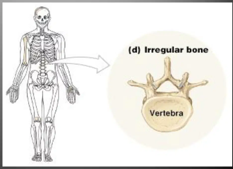
Which of the following is an example of an irregular bone?
Femur
Spinal vertebrae
Tibia
Radius
The compact bone is primarily composed of which of the following?
Osteons
Trabeculae
Cartilage
Marrow

Match the following
A
Haversian canal
B
Osteocyte
C
Canaliculi
D
Osteon
Mark the correct position of the diaphysis and main epiphysis

#7 in the diagram is: (a)

match the following regions of spongy and compact bone.
canaliculi
B
osteon
A
trabeculae
C
spongy bone
D
compact bone
E
Match the following
largest bone in the rear leg of an animal
femur
The enlarged end of a long bone
epiphysis
Substance that is primarily collagen, which gives bone high tensile strength
osteoid
These are typically thinner than others and are usually curved
flat
This makes up the lower jaw
mandible


What is the strongest bone in the body?
Femur
Clavicle
Ulna
Metacarpals


Which letter contains yellow marrow
G
H
C
A

How are lamellae arranged in an osteon?
Haversian canals are rings of bone tissue that surround central lamellae.
Lacunae are tiny channels that connect lamellae in separate rings of bone tissue.
Lamellae are rings of bone tissue that surround a central Haversian canal.
Lamellae are tiny channels that connect lacunae in separate rings of bone tissue.
In the bone development, primary ossification happens at the
joints
bone collar
shaft of the bone
the two epiphyses
In bone development, the secondary ossification takes place on the
collar bone
shaft of the bone
epiphyses plates
articular cartilage
Bone form first as
tendons
ligaments
hyaline cartilage
articular cartilage
A process when a cartilage gradually changes into bone tissue is called
osseus
ossification
resorption
bone modification
During endochondral ossification
hyaline cartilage is replaced by bone
hyaline cartilage turns into bone
bone replaced by hyaline cartilage
bone turns into hyaline cartilage

Name this joint
gliding
saddle
ball and socket
condyloid


Which is not a division of the skeletal system?
Axial
Central
Appendicular
What division of the skeleton would we find the Skull?
Axial
Appendicular

What is this an image of?
Pectoral Girdle
Pelvic Girdle
Femoral Girdle
Shoulder Girdle
Match the following common names of bones to their scientific names
Kneecap
Patella
Thigh Bone
Femur
Collar Bone
Clavicle
Shoulder blade
Scapula
Which of the following describes the joints in the skull and ribs?
movable
hinge
pivot
immovable
Which bone disease causes bones to become weak and brittle?
artereosclerosis
arthritis
fracture
osteoporosis
Where are red blood cells produced?
compact bone
red bone marrow
spongy bone
yellow bone marrow
An example of a pivot joint can be found in the forearm.
True
False
The carpals are your ankle bones.
True
False
The operation of the sodium-potassium "pump" moves:
sodium & potassium ions INTO the cell
sodium & potassium ions OUT of the cell
sodium ions OUT of the cell & potassium ions INTO the cell
sodium ions INTO the cell & potassium ions OUT of the cell


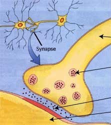
The location of neurotransmitters
axon terminals
dendrites
Schwann cells
cell body
Which of the following best describes a neuron?
Specialized cells that protects the body from any foreign particles
Specialized cell that transports oxygen throughout the cardiovascular system
Specialized cell that functions to transmit electrical impulses within the nervous system
Specialized cells that receive and read signals sent from the body
What are the 3 stages in action potential? Select all that apply.
Depolarization
Repolarization
Refractory period
Resting potential
Which type of neuron relays information from the (Afferent) body to the CNS?
sensory neuron
interneuron
motor neuron
myelinated neuron
Which chemical enters the synapse in order to transfer an impulse to another neuron?
hormone
plasma
neurotransmitter
cerebrospinal fluid
Where is the nucleus of a neuron located?
dendrite
synapse
axon
cell body
What part of a neuron carries an impulse toward the cell body?
axon
neurotransmitter
dendrite
synaptic cleft
A neuron that carries impulses from the brain and the spinal cord to a muscle or gland
sensory neuron
interneuron
motor neuron
myelinated neuron
A synapse occurs between
the axons of two different neurons.
the dendrites of two different neurons.
the axon of one neuron and the cell body of another neuron.
the axon of one neuron and the dendrite of another neuron.
What are the 2 MAJOR divisions of the peripheral nervous system (PNS)?
Sensory & Motor
(afferent and efferent)
Somatic & Autonomic
(SNS and ANS)

Which is the correct order/sequence of the parts of a neuron affected when a neural impulse occurs?

Schwann cells that make Myelin would be which color of structure?
the red one
the blue one
the green one
the purple one

What is the name of the blue structure
axon terminal
dendrite
schwann cell
axon

This is made up of the brain and spinal cord
Central Nervous System (CNS)
Peripheral Nervous System (PNS)
Autonomic nervous system
Somatic nervous system

All the parts of the nervous system except the brain and spinal cord.
Central Nervous System (CNS)
Peripheral nervous system (PNS)
Autonomic nervous system
Somatic nervous system
Which part of the peripheral nervous system deals with involuntary activities?
somatic
autonomic
I support neurons and help produce myelin what am I?
Axons
Dendrites
Glial cells
Cell bodies
Action Potential/Electrical Impulse

The tiny gap between neurons where nerve impulses are sent from one neuron to another.
synaptic cleft
axon terminal
impulse
myelin
The level of stimulation required to trigger a neural impulse.
Threshold
Action Potential
Resting Potential
Refractory Period
Type of neurons that carry information from the CNS through nerves to effectors such as muscles or glands.
Sensory Neurons
Interneurons
Motor Neurons
A neurons reaction of either firing (with a full length response) or not firing.
all-or-none response
refractory period
threshold
action potential
Place the following steps in the correct order to describe the firing of an ACTION POTENTIAL
Stimulus occurs changing the resting potential of the neuron
depolarization by 15 mVolts occurs (Threshold is reached)
Na+ channels begin to open up and sodium "whooshes" inside the neuron
More Na+ channels open and more "whooshing" to propagate the impulse
Neurotransmitters are released at the synapse by exocytosis...the receptor on the next neuron gets the chemical message...

