
 Worksheets
Cancer and Lymph Node Metastases Quiz
Total questions: 87
Worksheet time: 44mins
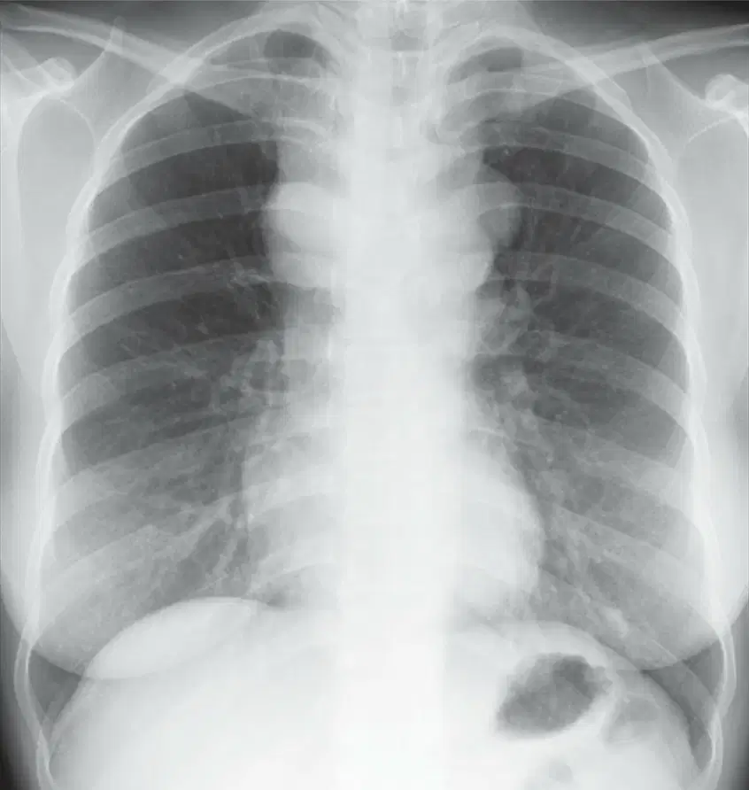
What type of cancer is associated with mediastinal lymph node metastases as shown in the image?
Papillary thyroid carcinoma
Testicular carcinoma
Breast cancer
Sarcoma
Which type of lymph node metastases is most typical of mucinous adenocarcinoma and sarcomas?
Calcified lymph node metastases
Necrotic lymph node metastases
Rim-enhancing lymph node metastases
Low-attenuation lymph node metastases
What type of lymph node metastases is commonly seen in testicular carcinoma, renal cell carcinoma, breast cancer, and lung cancer?
Necrotic, rim-enhancing, or low-attenuation lymph nodes
Calcified lymph node metastases
Enlarged lymph nodes without distinguishing characteristics
Superior mediastinal lymph nodes
Which cancers are associated with enhancing lymph nodes secondary to metastases?
Renal cell carcinoma, papillary thyroid carcinoma, lung cancer, sarcomas, and melanoma
Breast cancer, testicular carcinoma, and mucinous adenocarcinoma
Thyroid carcinoma, sarcomas, and adenocarcinoma
Lung cancer, renal cell carcinoma, and testicular carcinoma
What type of lymph node metastasis is shown in FIG. 5.23, and what is its characteristic feature?
Enhancing lymph node metastasis with metastatic paraganglioma; densely enhancing left paraaortic lymph node.
Calcified lymph node metastasis in thyroid carcinoma; faintly calcified pretracheal lymph node.
Enlarged lymph node metastasis in lung carcinoma; enlarged hilar lymph node.
Non-enhancing lymph node metastasis in breast carcinoma; non-enhancing axillary lymph node.
What type of lymph node metastasis is shown in FIG. 5.24, and what is its characteristic feature?
Calcified lymph node metastasis in thyroid carcinoma; faintly calcified pretracheal lymph node.
Enhancing lymph node metastasis with metastatic paraganglioma; densely enhancing left paraaortic lymph node.
Enlarged lymph node metastasis in lung carcinoma; enlarged hilar lymph node.
Non-enhancing lymph node metastasis in breast carcinoma; non-enhancing axillary lymph node.
What type of lymph nodes are visible following the treatment of metastatic gastric cancer?
Necrotic lymph nodes
Calcified lymph nodes
Enlarged lymph nodes
Inflamed lymph nodes
What characteristic is observed in the pretracheal lymph node in metastatic renal cell carcinoma?
High-attenuation center
Low-attenuation center and rim enhancement
Dense calcification
Uniform texture
Which type of lymph node involvement suggests a head and neck tumor?
Superior mediastinal lymph node involvement
Posterior mediastinal lymph node involvement
Retrocrural lymph node involvement
Paracardiac lymph node involvement
What does posterior mediastinal or paravertebral lymph node enlargement typically suggest about the primary tumor location?
Head and neck tumor
Abdominal tumor
Breast carcinoma
Colon carcinoma
Internal mammary lymph node metastases are most likely due to which type of cancer?
Lung cancer
Breast carcinoma
Colon carcinoma
Ovarian cancer
Paracardiac lymph node enlargement may occur as a result of metastasis from which types of tumors?
Head and neck tumors
Abdominal or thoracic tumors
Testicular carcinoma
Retroperitoneal tumors
What does the presence of multiple pleural nodules in a CT scan suggest?
Pleural effusion
Pleural metastases
Pneumothorax
Pulmonary embolism
Which type of cancer is most commonly associated with pleural metastases?
Adenocarcinoma
Squamous cell carcinoma
Small cell carcinoma
Basal cell carcinoma
What are the possible routes through which pleural metastases can spread?
Direct invasion, hematogenous spread, lymphatic spread
Airborne transmission, hematogenous spread, lymphatic spread
Direct invasion, airborne transmission, lymphatic spread
Hematogenous spread, lymphatic spread, airborne transmission
Which of the following is a finding that strongly suggests pleural metastasis in a patient with known malignancy?
Concentric pleural thickening
Pleural effusion, nonspecific
Nodular pleural thickening
Lymphatic obstruction
What is the most typical symptom of metastatic sarcoma involving the pleura?
Pleural effusion
Pneumothorax
Concentric pleural thickening
Lymphatic spread
Which mechanism is responsible for nonspecific pleural effusion in patients with neoplasm?
Local spread of tumor
Hematogenous spread
Lymphatic obstruction
Concentric pleural thickening
What is Kaposi's sarcoma (KS) primarily derived from?
Primitive vascular tissues
Bone marrow cells
Neural tissues
Muscular tissues
Which virus is associated with Kaposi's sarcoma (KS)?
Human herpesvirus 8 (HHV-8)
Epstein-Barr virus (EBV)
Cytomegalovirus (CMV)
Human papillomavirus (HPV)
What percentage of AIDS patients with Kaposi's sarcoma (KS) experience pulmonary involvement?
10%–30%
20%–50%
40%–60%
50%–70%
Which of the following is NOT a characteristic of pulmonary involvement in Kaposi's sarcoma (KS)?
Coarse reticular opacities at the lung bases
Pleural effusion
Interlobular septal thickening
Enlarged heart
What is the typical radiographic finding in Kaposi's sarcoma (KS) involving the lungs?
Bilateral diffuse abnormalities
Unilateral localized abnormalities
Normal lung appearance
Enlarged bronchi
What percentage of patients exhibit the chest radiographic appearance analogous to lymphangitic spread of carcinoma?
5%
10%
15%
20%
What radiographic features are typical of Kaposi Sarcoma (KS) in AIDS patients as seen in the perihilar regions and lower lobes?
Coarse, ill-defined opacities and consolidation
Streaky opacities and hilar lymph node enlargement
Both A and B
None of the above
Which radiographic feature is visible in the lower lobes of AIDS patients with Kaposi Sarcoma (KS)?
Coarse, ill-defined opacities
Streaky opacities
Consolidation
Enlarged hilar lymph nodes
What additional feature is visible in the radiographic appearance of Kaposi Sarcoma (KS) in AIDS patients besides opacities and nodules?
Pleural effusion
Hilar lymph node enlargement
Pneumothorax
Atelectasis
What is a typical CT feature of Kaposi Sarcoma (KS) in advanced cases?
Irregular and ill-defined spiculated nodules
Ground-glass opacity
Pleural effusion
Normal lung appearance
Which area of the lung is typically affected by early CT findings of KS in AIDS patients?
Lung apices
Lung bases
Middle lobes
Pleural cavity
What does the presence of ground-glass opacity in KS patients potentially represent?
Pulmonary hemorrhage
Pleural effusion
Normal lung tissue
Bronchial obstruction
Which feature allows KS to be distinguished from other thoracic complications in AIDS patients?
Perihilar distribution of irregular nodules larger than 1 cm
Pleural effusion
Ground-glass opacity
Interlobular septal thickening
What year did the World Health Organization (WHO) classify neoplasms of hematopoietic and lymphoid tissues, as mentioned in the text?
2005
2008
2010
2015
Which classification is included in the WHO classification of lymphomas primary to the lung, as mentioned in the text?
Lymphomas, leukemias, and lymphoproliferative diseases
Lymphomas and sarcomas
Leukemias and carcinomas
Lymphomas and melanomas
What percentage of non-Hodgkin lymphomas (NHL) are mature B-cell neoplasms?
50%
85%
15%
70%
Which type of lymphoma is associated with HHV-8 and multicentric Castleman disease?
Follicular lymphoma
Large B-cell lymphoma
Mantle cell lymphoma
Primary effusion lymphoma
Which of the following is a type of mature T-cell and NK-cell neoplasm?
Mantle cell lymphoma
Angioimmunoblastic T-cell lymphoma
Waldenström’s macroglobulinemia
Lymphoplasmacytic lymphoma
Which subtype of Hodgkin’s lymphoma is characterized by nodular lymphocyte predominance?
Classical Hodgkin’s lymphoma
Nodular lymphocyte-predominant Hodgkin’s lymphoma
Mixed cellularity classical Hodgkin’s lymphoma
Lymphocyte-depleted classical Hodgkin’s lymphoma
Which of the following is a histiocytic and dendritic cell neoplasm?
Lymphoplasmacytic lymphoma
Erdheim-Chester disease
Primary effusion lymphoma
Anaplastic large cell lymphoma
What does PTLD stand for in the context of posttransplantation lymphoproliferative disorders?
Posttransplantation lymphocyte disorder
Polymorphic T-cell lymphocyte disorder
Posttransplantation lymphoproliferative disorder
Primary T-cell lymphocyte disease
What is the primary basis for the classification of neoplasms in the WHO classification system?
Geographic location of the patient
Cell type and genetic differences
Patient's age and gender
Environmental factors
Which of the following infectious agents has been linked to the development of lymphomas and lymphoproliferative diseases?
Influenza virus
Epstein-Barr virus (EBV)
Rhinovirus
Streptococcus bacteria
What staging system is commonly used for lymphomas, including its modifications for NHLs?
TNM staging system
Ann Arbor staging system
Lugano staging system
Cotswolds staging system
In the Ann Arbor staging system, what does stage IV lymphoma indicate?
Single lymph node group involvement
Multiple lymph node groups, ipsilateral to the diaphragm
Involvement of lymph node groups above and below the diaphragm
Noncontiguous extranodal involvement, such as liver, lung, or bone marrow
What revisions to the staging system for lymphomas were recommended at the 11th International Conference on Malignant Lymphoma in Lugano, Switzerland?
Introduction of a new staging system
Recognition of extranodal involvement in staging
Removal of subcategories A and B
Inclusion of genetic mutations in staging criteria
What imaging technique is commonly used in the initial evaluation and staging of most lymphomas, improving accuracy compared to CT?
MRI
PET
Ultrasound
X-ray
Which lymphoma type is excluded from FDG-avid NHL in PET imaging?
Hodgkin lymphoma (HL)
Chronic lymphocytic leukemia (CLL)
Marginal zone NHL
Mycosis fungoides
What is the primary benefit of using PET-CT in staging lymphomas compared to CT alone?
Reduced imaging time
Improved staging accuracy
Lower radiation exposure
Enhanced patient comfort
In the Lugano Classification, what does Stage I indicate?
Involvement of lymph node regions on both sides of the diaphragm
Involvement of a single lymph node region or a single extralymphatic organ or site
Additional noncontiguous extralymphatic involvement
Involvement of two or more lymph node regions on the same side of the diaphragm
What distinguishes Stage II bulky from Stage II in the Lugano Classification?
Presence of fever and night sweats
Involvement of lymph node regions on both sides of the diaphragm
Presence of bulky disease
Additional noncontiguous extralymphatic involvement
What suffix is used in Hodgkin lymphoma (HL) to denote the absence or presence of systemic symptoms such as fever, night sweats, or unexplained weight loss?
X or Y
A or B
I or II
M or N
What is the current definition of "bulky" disease in Hodgkin's Lymphoma (HL)?
A mass greater than 5 cm in diameter
A mass greater than 10 cm or 1/3 of the transthoracic diameter
A mass visible on CT with modifiers X and A
A mass greater than 15 cm in diameter
What is the primary goal of the Lugano Classification in lymphoma follow-up and assessment?
To eliminate the use of CT scans in staging
To simplify and standardize response assessment and reporting
To focus solely on FDG-PET/CT for all lymphoma types
To replace the Deauville criteria with new guidelines
Which of the following is NOT a category of lymph node response using CT as defined in the Lugano Classification?
Complete radiologic response
Partial response
Progressive disease
Residual disease
What is the male-to-female ratio prevalence of Hodgkin's Lymphoma (HL)?
1:1
2:1
3:1
4:1
What is the significance of FDG uptake in response assessment for lymphoma?
It determines the size of the lymph nodes
It serves as a marker of metabolic activity
It eliminates the need for CT scans
It categorizes lymphomas into histological subtypes
What is the peak incidence age range for Hodgkin's Lymphoma?
First and second decades
Third and eighth decades
Fifth and sixth decades
Ninth and tenth decades
Which cell type is characteristic of Hodgkin's Lymphoma?
Reed-Sternberg cells
T-cells
B-cells
Plasma cells
What percentage of adult Hodgkin's Lymphoma cases are accounted for by the nodular sclerosis cell type?
20%-40%
50%-80%
10%-30%
70%-90%
Which staging system is used for Hodgkin's Lymphoma?
TNM system
Ann Arbor system
WHO classification system
Lugano system
What percentage of Hodgkin's Lymphoma cases show thoracic involvement at diagnosis?
50%
85%
70%
95%
What is the most common mediastinal node enlargement percentage in Hodgkin's Lymphoma cases?
75%
85%
98%
65%
What percentage of Hodgkin's Lymphoma cases show lung involvement at diagnosis?
5%
10%
15%
20%
What is the most common cause of pleural effusion in Hodgkin's Lymphoma cases?
Lung infection
Lymphatic obstruction
Thoracic injury
Pulmonary embolism
Which histologic type of Hodgkin's Lymphoma accounts for 50%-80% of adult cases?
Mixed cellularity HL
Nodular sclerosis HL
Lymphocyte-depleted HL
Lymphocyte-predominant HL
What percentage of patients with Hodgkin's Lymphoma have mediastinal lymph node enlargement at diagnosis?
75%
85%
98%
65%
Which lymph node groups are most often involved in Hodgkin lymphoma (HL)?
Prevascular, paratracheal, and aortopulmonary lymph nodes
Cervical, axillary, and inguinal lymph nodes
Mesenteric, iliac, and retroperitoneal lymph nodes
Submandibular, occipital, and supraclavicular lymph nodes
What percentage of patients with thoracic involvement in HL show abnormal lymph nodes in imaging studies?
75%
85%
98%
65%
If lymph nodes appear normal on CT imaging, what is unlikely to represent HL?
Intrathoracic adenopathy
Mediastinal mass
Pleural effusion
Pulmonary fibrosis
Which imaging technique shows prevascular anterior mediastinal lymph node enlargement and pretracheal lymph node enlargement in HL?
PA chest radiograph
Lateral view radiograph
Contrast-enhanced CT scan
MRI scan
What is typical of HL in the regions of superior mediastinal lymph nodes?
Lymph node enlargement
Mass formation
Fluid accumulation
Tissue calcification
Based on the diagram, which region has the highest percentage indicated for lymph node involvement?
Superior Mediastinal (Paratracheal)
Subcarinal
Hilar
Paracardiac
What is the percentage of lymph node involvement in the Subcarinal region as shown in the diagram?
5%
25%
35%
10%
Which region has the lowest percentage of lymph node involvement according to the diagram?
Paracardiac
Subcarinal
Posterior Mediastinal
Superior Mediastinal (AP Window)
What is the percentage of lymph node involvement in the Hilar region as indicated in the diagram?
35%
10%
5%
98%
Which lymph node group is involved in 98% of patients with thoracic disease according to the diagram?
Subcarinal nodes
Superior mediastinal (prevascular) nodes
Paracardiac nodes
Internal mammary nodes
What percentage of patients with thoracic disease have involvement of the subcarinal lymph nodes?
10%
25%
5%
35%
Which lymph node group is involved in 5% of patients with thoracic disease?
Internal mammary nodes
Subcarinal nodes
Paracardiac nodes
Hilar nodes
What is the percentage of involvement of paracardiac lymph nodes in patients with thoracic disease?
5%
10%
25%
35%
Which lymph node group is involved in 35% of patients with thoracic disease?
Hilar nodes
Subcarinal nodes
Posterior mediastinal nodes
Internal mammary nodes
What does the PA chest radiograph show in patients with HL involving multiple lymph node groups?
Superior mediastinal widening, hilar enlargement, and nodules in the left upper lobe
Normal lymph node appearance
Enlarged lymph nodes only in the prevascular anterior mediastinum
No visible abnormalities
Which lymph node groups are visible in the prevascular anterior mediastinum in HL patients?
Prevascular anterior mediastinum, pretracheal space, aortopulmonary window, subcarinal space, and both hila
Only pretracheal space and subcarinal space
Only aortopulmonary window and hila
None of the above
What percentage of HD patients with thoracic node involvement have multiple node groups affected?
85%
50%
70%
95%
What is visible at a lower level on CT in HL patients?
Paracardiac lymph node enlargement
Normal lymph nodes
Enlarged lymph nodes in the prevascular anterior mediastinum
Subcarinal space abnormalities
What is the most common lymph node group involved as a single group in nodular sclerosis HD?
Paratracheal lymph nodes
Prevascular lymph nodes
Aortopulmonary window nodes
Posterior mediastinal lymph nodes
What percentage of cases with nodular sclerosis HD involve enlargement of a single node group?
10%
15%
20%
25%
What does poor definition of mediastinal masses in HL indicate?
Benign growth
Invasion or extension into adjacent lung
Normal lymph node enlargement
Absence of abnormality
What is the typical contour of mediastinal masses in HL when multiple lymph nodes are involved?
Spherical
Elongated or lobulated
Flat and smooth
Irregular and spiked

