
 Worksheets
Abdomen Wk 1 book
Total questions: 83
Worksheet time: 1hrs 23mins
A cavity containing dead tissue and pus that forms owing to an infectious process
(a)
An accumulation of serous fluid in the peritoneal cavity
(a)
Redness of the skin owing to inflammation
(a)
Fibrous structure that runs down the midline of the abdomen from the xiphoid process to the symphis pubis separating the right and left rectus abdomens muscles
(a)
Rhythmic wavelike contraction of gastrointestinal tract that forces food through it
(a)
Collapsed lung that occurs when air leaks into the space between the chest wall and lung
(a)
The (a) cavity is completely encased in bone and is subdivided into the cranial and spinal cavity
The (a) abdominal wall is often described as a single unit
Extends from thoracic cage to the pelvis
(a)
Bound by cartilages of the 7th to 10th ribs and the xiphoid process superiorly
(a)
Inferiorly bound by the inginual ligament and iliac crests, pubic crests and pubic symphis
(a)
Attached to the skin and composed of connective tissue containing varying quantities of fat
(a)
Covers the muscles and partitions them into groups
(a)
Layers of flat tendinous fibrous sheets fused with strong connective tissue that attach muscles to fixed points, functioning like a tendon
(a)
Abdominal are primarily located in the (a) abdominal regions
Have minimal vascularity and nervation
(a)
Most readily known abdominal aponeurosis is the (a)
The skin attaches loosely to most of the subcutaneous tissues except at the (a) where attachment is firm
the subcutaneous tissue anterior to the muscle layers makes up the (a)
(a) fascia gives the body wall its rounded appearance
Consists of a combination of fat and fibrous tissue that blends with the deep fascia
(a)
The membraneous layer that continues into the perineal region as the superficial perineal fascia is called (a)
Internal to the endoabdominal fascia is the (a)
The (a) is the outer layer of the serous membrane lining the abdominopelvic cavity formed by a single layer of epithelial cells and supporting connective tissue
There are (a) bilaterally paired muscles in the anterolateral wall
Rectus abdominis is a (a) muscle
Internal and external oblique are (a) muscles
Transverse abdominis are (a) muscles
The pyramidal is a (a) muscle
A strong, dense connective tissue fascia that encases the rectus abdominis and pyramidal muscles as well as some arteries, veins, lymphatic vessels, and nerves
(a)
Is a midline, dense connective tissue structure that separates the right and left bellies of the rectus abdominis muscle.
(a)
Area where all layers of the anterolateral wall fuse.
(a)
It is an important canal where structures enter the abdominal cavity and exit and the entry pathways are potential sites for herniation
(a)
Composed of the lumbar vertebra, posterior abdominal wall muscles, diaphragm, fascia, lumbar plexus, fat, nerves, blood vessels, and lymphatic vessels
(a)
The (a) enclose the bilateral erector spine muscles or vertical deep back muscles in the same manner that the rectus sheath encloses the rectus abdominis
Psoas major, iliacus, quadratus luborum, psoas minor, Latissimus dorsi are all (a) muscles
The (a) is covered with a continuous layer of endoabdominal fascia. Located between the parietal peritoneum and the muscles
The (a) is a fibromuscular domed structure separating the thoracic cavity from the abdominal cavity
The right dome of diaphragm is slightly (a) than the left because of the presence of the liver
The (a) part of the diaphragm descends during inspiration and ascends during expiration
Musculotendinous bands that arise from the anterior surfaces of the bodies of the superior three lumbar vertebrae, the anterior longitudinal ligament, and intervertebral disks
(a)
The (a) allow several structures to pass between the thorax and abdomen -- esophagus, blood vessels, nerves, ETC
The IVC (a) during inspiration
The diaphragm appears (a) in children and adults
The diaphragm appears (a) in fetus
A __ __ linear array should be used for the abdominal wall
(a)
In some cases a (a) can be used to avoid the "main bang" artifact
A pocket of infection typically containing pus, blood, and degenerating tissue
(a)
Surface area of a. peritoneal organ devoid of peritoneum
(a)
A small pocket communicating with a larger cavity or tube
(a)
Fluid outside of vessels, bowel, and organs, seen in potential spaces
(a)
An extravasated collection of blood localized within a potential space or tissue
(a)
Area of the organ where blood vessels, lymph, and nerves enter and exit
(a)
treatment induced; may be intentional or unintentional
(a)
An extravasated collection of lymph
(a)
Two layers of fused peritoneum that conduct nerves, lymph, blood vessels between the small bowel and posterior peritoneal cavity wall
(a)
Peritoneum lining the walls of the peritoneal cavity
(a)
Solid organs and some portions of the bowel within the peritoneal cavity that are covered by visceral peritoneum
(a)
An empty fold where the peritoneal layer reflects between two organs or an organ and peritoneal wall, which may contain fluid and other materials when disease is present
(a)
Organs posterior to the parietal peritoneum that are typically covered on their anterior surface or fatty capsule by parietal peritoneum
(a)
Peritoneum encasing peritoneal organs
(a)
A thin sheet of tissues, called (a) divides the abdominal cavity into peritoneal and retroperitoneal compartments
largest division of the peritoneal cavity, the (a) contains the liver, spleen, stomach, digestion portion of duodenum, jejunum, ileum, cecum, transverse colon, sigmoid colon, and upper two thirds of the rectum
The (a) does not contain any organs and lies immediately posterior to the stomach
The splenorenal and gastrosplenic ligament create the left lateral wall of this pocket
(a)
The (a) also called the small momentum, is a fused double layer of peritoneum stretching between the lesser curvature of the stomach and the left sagittal fissure for the ligamentum venosum
Within the greater sac, the (a) is a large apron like double layered sheet of peritoneum that extends inferiorly from the greater curvature of the stomach and transverse colon
Is very mobile and moves to area of inflammation, surrounding the inflamed area by creating adhesions to wall off infection. Also acts as a cushion to the abdominal organs to prevent trauma and acts to prevent the loss of body heat from abdominal organs
(a)
The greater omentum subdivides the greater sac into a __ (above the colon) compartment and an __ (below the colon)
(a)
The ___ or __ is an extension of the greater sac between the diaphragm and the anterior superior liver leftward of the falciform ligament
(a)
The (a) is also called the superior recess of the lesser sac; this space is an extension of the lesser sac between the diaphragm and the posterior superior liver
The __ or __ is an extension of the greater sac between the right hemidiaphragm and the anterior superior liver rightward of the falciform ligament
(a)
The (a) is also referred to as the Morrisons pouch. This peritoneal space is created by the peritoneum reflecting from the liver over the right kidney and right posterior peritoneal wall
The (a) or lesser sac is sandwiched between the posterior stomach and parietal peritoneum covering the anterior pancreas and the splenorenal and gastrosplenic ligaments, and epiploic foramen
The right and left __ __ are potential spaces or grooves found along the lateral ascending and descending colons that conduct fluid between the supra colic compartment of the inferior abdomen and pelvis
(a)
The (a) or cul-de-sac in the male is the potential space created by the peritoneal reflection over the rectum and posterior bladder wall
The (a) is also called the rectovaginal pouch, pouch of Douglas, or posterior cul de sac in the female. This potential space is created by the parietal peritoneum draping over the anterior rectum, posterior vaginal wall, and posterior uterus
The (a) is also called the uterovesical pouch or anterior cul-de-sac in the female and is the space created by the peritoneal reflection over the uterine fundus
The (a) is also called the premedical or retropubic space and is an exztraperitoneal potential space located between the anterior wall of the urinary bladder and pubic symphis
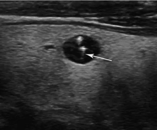
Caused by sound interacting with small air
bubbles causing the bubbles to vibrat
(a)
?
(a)
pancreas, liver, gallbladder, right kidney
(a)
The abdomen has several locations that are common abdominal fluidcollection points, also referred to as
(a)

